Downloaded 61 times
![Standarized uptake value:-
It is used for relative tissue uptake of FDG
tracer.
SUV threshold value used for malignancy is
(> 2.5) .
Formula:-
SUV= Tumor activity concentration [mCi/ml]
X body weight (g)/injected dose FDG [mCi ].](https://image.slidesharecdn.com/petct-150616140915-lva1-app6891/85/Pet-ct-technique-5-320.jpg)
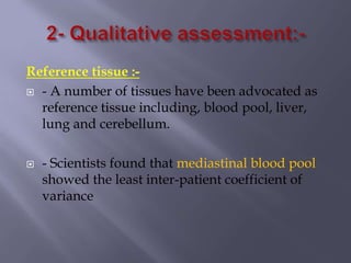
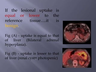
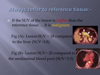


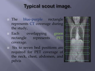
PET-CT combines functional PET imaging with anatomical CT imaging. A PET-CT scan involves fasting for 4-6 hours before injection of 18F-FDG tracer, followed by a whole body scan 40-60 minutes later lasting 13-17 minutes. Standardized uptake values (SUVs) are used to measure tracer uptake in tissues, with values over 2.5 suggesting malignancy. Dual time scanning one hour later can identify if SUV increases by 10% indicating malignancy or remains the same/decreases indicating benignity. Reference tissues like blood pools and livers are used to compare lesion uptake values to determine if they are higher, suggesting malignancy, or equal/lower, suggesting benignity.


![ONFH[AVN HIP] -TRIPLE REGIME -A NOVAL SURGICAL CONCEPT .pptx](https://cdn.slidesharecdn.com/ss_thumbnails/onfhavnhip2026koaconcalicutdrgokuldevdrmashraf-260210064517-213ec005-thumbnail.jpg?width=640&height=640&fit=bounds)


![PERI-PROSTHETIC FRACTURE NAIL-PLATE CONSTRUCT [NPC].pptx](https://cdn.slidesharecdn.com/ss_thumbnails/drarunkumardrmohamedashrafperiprostheticfrasturenail-plateconstructnpc-260209164459-7e9d15a1-thumbnail.jpg?width=640&height=640&fit=bounds)


