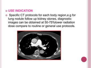
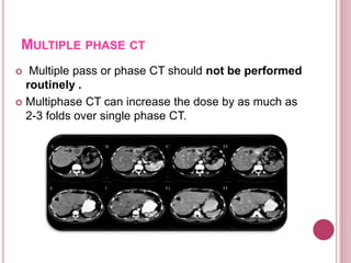
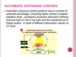
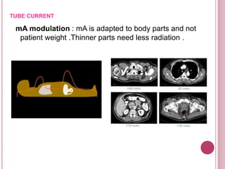
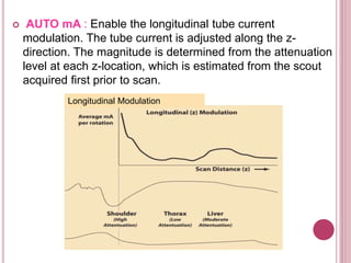
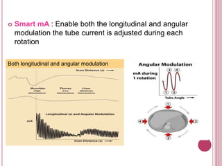
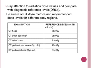
This document discusses various techniques for reducing radiation dose in computed tomography (CT) scans. It outlines strategies such as using automatic exposure control, adjusting scan parameters based on patient size, employing noise-tolerant images when possible, limiting scan lengths and phases, and utilizing newer reconstruction techniques. The goal is to lower radiation dose without compromising diagnostic image quality.
Basic physics of multidetector computed tomography ( CT Scan) - how ct scan works, different generations of ct, how image is generated and displayed and image artifacts related to CT Scan.
this power-point slide presentation includes lots of information like how MRI coil works. what is shimming, magnet, fringe, and design of mri coil and also magnet. this will help a lot for radiologist and technician radiographers.. thanks.
Recent advancements in modern x ray tubeSantosh Ojha
All the advancements in X-ray tubes till date are done to increase the Tube heat storage capacity thus increasing the lifetime of x -ray tubes. This slide explains about these recent advancements in x-ray tubes.
CT is one of the highest contributor for medical radiation exposure to patients. Some common CT dose descriptors and dose optimizations methods are briefly described in this presentation.
this slide sharer contents are basic principle of CT fluoroscopy , software and hardware parts of equipment and image aqua cation and radiation dose comparison and videos related to equipment .
CT Dose Issues.pptx on the factors to be considered on radiation protectionsanyengere
summary, mobile radiography allows for the diagnostic imaging of patients who are unable to be seen in the X-ray examination room. Therefore, mobile X-ray equipment is useful for patients who have difficulty with movement. However, staff are exposed to scattered radiation from the patient, and can receive potentially harmful radiation doses during radiography. The protection of staff is of utmost importance; therefore, we investigated the occupational radiation doses received by RTs, particularly eye doses, using phantom measurements. RTs can be located close to a patient (i.e., the source of scattered radiation) during mobile radiography. As eye doses can be significant, protective measures are essential for RTs. Protective aprons are important for protecting RTs, as is increasing the distance from the radiation source (i.e., the patient). Lead glasses may also be necessary for protecting the eyes of RTs. To reduce RT radiation exposure, RTs should remain distant from the patient if possible. However, because this distance may hinder verification of the patient’s condition, RTs sometimes work in close proximity to patients. This is a patient phantom study. In future, the data may need validation by comparison with personal RT dosimeter records. It is important to evaluate the radiation doses delivered to RTs during mobile radiography, as well as the scattered radiation distribution, to ensure adequate protection. Further comparison studies may be needed using the Monte Carlo method.
radiographers and nurses have a responsibility to ensure that no one is within the radiation field during the X-ray exposure of the patient. This is achieved by informing all persons in the immediate area that an X-ray exposure is about to be made and asking them to stand a safe distance from the radiation field area.
Shielding
Placing a barrier of lead or concrete between the radiation source and an individual provides protection from X-radiation (Jones and Taylor, 2006; Ehrlich and Coakes, 2017). During mobile radiography, anyone assisting in an examination and staying in the radiation field should wear a lead-rubber apron or stand behind a mobile lead screen. Generally, walls in special care units where ionising radiation is used are designed to contain the radiation produced by the mobile X-ray tube within a set of criteria and limits determined by relevant legislation (Hart et al, 2002).
Radiation protection during mobile radiography
Nurses' understanding and adherence to radiation protection control measures during mobile radiography is of paramount importance in protecting patients, themselves and members of the public visiting the ward/unit. However, some research studies have found limited awareness and non-adherence to radiation protection control measures among nurses during mobile radiography (Anim-Sampong et al, 2015; Luntsi et al, 2016; Azimi et al, 2018). This can be attributed to a lack of radiation protection awareness programmes for nurses working
Dear B.Sc MIT Students,
Attached is an essential document featuring comprehensive Questions & Answers for Nuclear medicine 3 marks questions and answers. We encourage you to utilize this resource to deepen your understanding and excel in your studies. Wishing you all the success in your academic endeavors and future careers.
Best regards,
MAGNETIC RESONANCE IMAGING QUESTION AND ANSWER 3 MARKS.pdfGanesan Yogananthem
Dear B.Sc MIT Students,
Attached is an essential document featuring comprehensive Questions & Answers for MAGNETIC RESONANCE IMAGING. We encourage you to utilize this resource to deepen your understanding and excel in your studies. Wishing you all the success in your academic endeavors and future careers.
Best regards,
QUESTIONS & ANSWERS FOR QUALITY ASSURANCE, RADIATIONBIOLOGY& RADIATION HAZARD...Ganesan Yogananthem
Dear B.Sc MIT Students,
Attached is an essential document featuring comprehensive Questions & Answers for Quality Assurance, Radiation Biology, and Radiation Hazards. We encourage you to utilize this resource to deepen your understanding and excel in your studies. Wishing you all the success in your academic endeavors and future careers.
Best regards,
Medical imaging technique is well advanced today and we are measuring signals from protons level in MRI imaging. But we left behind a error in 2-D measurement in digital X- ray in imaging. This we found out during imaging of Scanogram. In this presentation we are explaining that how we are controlling this error by applying small trick for given plane interest on the digital X-ray Scanogram image.
Ethanol (CH3CH2OH), or beverage alcohol, is a two-carbon alcohol
that is rapidly distributed in the body and brain. Ethanol alters many
neurochemical systems and has rewarding and addictive properties. It
is the oldest recreational drug and likely contributes to more morbidity,
mortality, and public health costs than all illicit drugs combined. The
5th edition of the Diagnostic and Statistical Manual of Mental Disorders
(DSM-5) integrates alcohol abuse and alcohol dependence into a single
disorder called alcohol use disorder (AUD), with mild, moderate,
and severe subclassifications (American Psychiatric Association, 2013).
In the DSM-5, all types of substance abuse and dependence have been
combined into a single substance use disorder (SUD) on a continuum
from mild to severe. A diagnosis of AUD requires that at least two of
the 11 DSM-5 behaviors be present within a 12-month period (mild
AUD: 2–3 criteria; moderate AUD: 4–5 criteria; severe AUD: 6–11 criteria).
The four main behavioral effects of AUD are impaired control over
drinking, negative social consequences, risky use, and altered physiological
effects (tolerance, withdrawal). This chapter presents an overview
of the prevalence and harmful consequences of AUD in the U.S.,
the systemic nature of the disease, neurocircuitry and stages of AUD,
comorbidities, fetal alcohol spectrum disorders, genetic risk factors, and
pharmacotherapies for AUD.
ARTIFICIAL INTELLIGENCE IN HEALTHCARE.pdfAnujkumaranit
Artificial intelligence (AI) refers to the simulation of human intelligence processes by machines, especially computer systems. It encompasses tasks such as learning, reasoning, problem-solving, perception, and language understanding. AI technologies are revolutionizing various fields, from healthcare to finance, by enabling machines to perform tasks that typically require human intelligence.
Lung Cancer: Artificial Intelligence, Synergetics, Complex System Analysis, S...Oleg Kshivets
RESULTS: Overall life span (LS) was 2252.1±1742.5 days and cumulative 5-year survival (5YS) reached 73.2%, 10 years – 64.8%, 20 years – 42.5%. 513 LCP lived more than 5 years (LS=3124.6±1525.6 days), 148 LCP – more than 10 years (LS=5054.4±1504.1 days).199 LCP died because of LC (LS=562.7±374.5 days). 5YS of LCP after bi/lobectomies was significantly superior in comparison with LCP after pneumonectomies (78.1% vs.63.7%, P=0.00001 by log-rank test). AT significantly improved 5YS (66.3% vs. 34.8%) (P=0.00000 by log-rank test) only for LCP with N1-2. Cox modeling displayed that 5YS of LCP significantly depended on: phase transition (PT) early-invasive LC in terms of synergetics, PT N0—N12, cell ratio factors (ratio between cancer cells- CC and blood cells subpopulations), G1-3, histology, glucose, AT, blood cell circuit, prothrombin index, heparin tolerance, recalcification time (P=0.000-0.038). Neural networks, genetic algorithm selection and bootstrap simulation revealed relationships between 5YS and PT early-invasive LC (rank=1), PT N0—N12 (rank=2), thrombocytes/CC (3), erythrocytes/CC (4), eosinophils/CC (5), healthy cells/CC (6), lymphocytes/CC (7), segmented neutrophils/CC (8), stick neutrophils/CC (9), monocytes/CC (10); leucocytes/CC (11). Correct prediction of 5YS was 100% by neural networks computing (area under ROC curve=1.0; error=0.0).
CONCLUSIONS: 5YS of LCP after radical procedures significantly depended on: 1) PT early-invasive cancer; 2) PT N0--N12; 3) cell ratio factors; 4) blood cell circuit; 5) biochemical factors; 6) hemostasis system; 7) AT; 8) LC characteristics; 9) LC cell dynamics; 10) surgery type: lobectomy/pneumonectomy; 11) anthropometric data. Optimal diagnosis and treatment strategies for LC are: 1) screening and early detection of LC; 2) availability of experienced thoracic surgeons because of complexity of radical procedures; 3) aggressive en block surgery and adequate lymph node dissection for completeness; 4) precise prediction; 5) adjuvant chemoimmunoradiotherapy for LCP with unfavorable prognosis.
These lecture slides, by Dr Sidra Arshad, offer a quick overview of physiological basis of a normal electrocardiogram.
Learning objectives:
1. Define an electrocardiogram (ECG) and electrocardiography
2. Describe how dipoles generated by the heart produce the waveforms of the ECG
3. Describe the components of a normal electrocardiogram of a typical bipolar leads (limb II)
4. Differentiate between intervals and segments
5. Enlist some common indications for obtaining an ECG
Study Resources:
1. Chapter 11, Guyton and Hall Textbook of Medical Physiology, 14th edition
2. Chapter 9, Human Physiology - From Cells to Systems, Lauralee Sherwood, 9th edition
3. Chapter 29, Ganong’s Review of Medical Physiology, 26th edition
4. Electrocardiogram, StatPearls - https://www.ncbi.nlm.nih.gov/books/NBK549803/
5. ECG in Medical Practice by ABM Abdullah, 4th edition
6. ECG Basics, http://www.nataliescasebook.com/tag/e-c-g-basics
Anti ulcer drugs and their Advance pharmacology ||
Anti-ulcer drugs are medications used to prevent and treat ulcers in the stomach and upper part of the small intestine (duodenal ulcers). These ulcers are often caused by an imbalance between stomach acid and the mucosal lining, which protects the stomach lining.
||Scope: Overview of various classes of anti-ulcer drugs, their mechanisms of action, indications, side effects, and clinical considerations.
Prix Galien International 2024 Forum ProgramLevi Shapiro
June 20, 2024, Prix Galien International and Jerusalem Ethics Forum in ROME. Detailed agenda including panels:
- ADVANCES IN CARDIOLOGY: A NEW PARADIGM IS COMING
- WOMEN’S HEALTH: FERTILITY PRESERVATION
- WHAT’S NEW IN THE TREATMENT OF INFECTIOUS,
ONCOLOGICAL AND INFLAMMATORY SKIN DISEASES?
- ARTIFICIAL INTELLIGENCE AND ETHICS
- GENE THERAPY
- BEYOND BORDERS: GLOBAL INITIATIVES FOR DEMOCRATIZING LIFE SCIENCE TECHNOLOGIES AND PROMOTING ACCESS TO HEALTHCARE
- ETHICAL CHALLENGES IN LIFE SCIENCES
- Prix Galien International Awards Ceremony
MANAGEMENT OF ATRIOVENTRICULAR CONDUCTION BLOCK.pdfJim Jacob Roy
Cardiac conduction defects can occur due to various causes.
Atrioventricular conduction blocks ( AV blocks ) are classified into 3 types.
This document describes the acute management of AV block.
Pulmonary Thromboembolism - etilogy, types, medical- Surgical and nursing man...VarunMahajani
Disruption of blood supply to lung alveoli due to blockage of one or more pulmonary blood vessels is called as Pulmonary thromboembolism. In this presentation we will discuss its causes, types and its management in depth.
Ozempic: Preoperative Management of Patients on GLP-1 Receptor Agonists Saeid Safari
Preoperative Management of Patients on GLP-1 Receptor Agonists like Ozempic and Semiglutide
ASA GUIDELINE
NYSORA Guideline
2 Case Reports of Gastric Ultrasound
The prostate is an exocrine gland of the male mammalian reproductive system
It is a walnut-sized gland that forms part of the male reproductive system and is located in front of the rectum and just below the urinary bladder
Function is to store and secrete a clear, slightly alkaline fluid that constitutes 10-30% of the volume of the seminal fluid that along with the spermatozoa, constitutes semen
A healthy human prostate measures (4cm-vertical, by 3cm-horizontal, 2cm ant-post ).
It surrounds the urethra just below the urinary bladder. It has anterior, median, posterior and two lateral lobes
It’s work is regulated by androgens which are responsible for male sex characteristics
Generalised disease of the prostate due to hormonal derangement which leads to non malignant enlargement of the gland (increase in the number of epithelial cells and stromal tissue)to cause compression of the urethra leading to symptoms (LUTS
- Video recording of this lecture in English language: https://youtu.be/lK81BzxMqdo
- Video recording of this lecture in Arabic language: https://youtu.be/Ve4P0COk9OI
- Link to download the book free: https://nephrotube.blogspot.com/p/nephrotube-nephrology-books.html
- Link to NephroTube website: www.NephroTube.com
- Link to NephroTube social media accounts: https://nephrotube.blogspot.com/p/join-nephrotube-on-social-media.html
Title: Sense of Smell
Presenter: Dr. Faiza, Assistant Professor of Physiology
Qualifications:
MBBS (Best Graduate, AIMC Lahore)
FCPS Physiology
ICMT, CHPE, DHPE (STMU)
MPH (GC University, Faisalabad)
MBA (Virtual University of Pakistan)
Learning Objectives:
Describe the primary categories of smells and the concept of odor blindness.
Explain the structure and location of the olfactory membrane and mucosa, including the types and roles of cells involved in olfaction.
Describe the pathway and mechanisms of olfactory signal transmission from the olfactory receptors to the brain.
Illustrate the biochemical cascade triggered by odorant binding to olfactory receptors, including the role of G-proteins and second messengers in generating an action potential.
Identify different types of olfactory disorders such as anosmia, hyposmia, hyperosmia, and dysosmia, including their potential causes.
Key Topics:
Olfactory Genes:
3% of the human genome accounts for olfactory genes.
400 genes for odorant receptors.
Olfactory Membrane:
Located in the superior part of the nasal cavity.
Medially: Folds downward along the superior septum.
Laterally: Folds over the superior turbinate and upper surface of the middle turbinate.
Total surface area: 5-10 square centimeters.
Olfactory Mucosa:
Olfactory Cells: Bipolar nerve cells derived from the CNS (100 million), with 4-25 olfactory cilia per cell.
Sustentacular Cells: Produce mucus and maintain ionic and molecular environment.
Basal Cells: Replace worn-out olfactory cells with an average lifespan of 1-2 months.
Bowman’s Gland: Secretes mucus.
Stimulation of Olfactory Cells:
Odorant dissolves in mucus and attaches to receptors on olfactory cilia.
Involves a cascade effect through G-proteins and second messengers, leading to depolarization and action potential generation in the olfactory nerve.
Quality of a Good Odorant:
Small (3-20 Carbon atoms), volatile, water-soluble, and lipid-soluble.
Facilitated by odorant-binding proteins in mucus.
Membrane Potential and Action Potential:
Resting membrane potential: -55mV.
Action potential frequency in the olfactory nerve increases with odorant strength.
Adaptation Towards the Sense of Smell:
Rapid adaptation within the first second, with further slow adaptation.
Psychological adaptation greater than receptor adaptation, involving feedback inhibition from the central nervous system.
Primary Sensations of Smell:
Camphoraceous, Musky, Floral, Pepperminty, Ethereal, Pungent, Putrid.
Odor Detection Threshold:
Examples: Hydrogen sulfide (0.0005 ppm), Methyl-mercaptan (0.002 ppm).
Some toxic substances are odorless at lethal concentrations.
Characteristics of Smell:
Odor blindness for single substances due to lack of appropriate receptor protein.
Behavioral and emotional influences of smell.
Transmission of Olfactory Signals:
From olfactory cells to glomeruli in the olfactory bulb, involving lateral inhibition.
Primitive, less old, and new olfactory systems with different path
These simplified slides by Dr. Sidra Arshad present an overview of the non-respiratory functions of the respiratory tract.
Learning objectives:
1. Enlist the non-respiratory functions of the respiratory tract
2. Briefly explain how these functions are carried out
3. Discuss the significance of dead space
4. Differentiate between minute ventilation and alveolar ventilation
5. Describe the cough and sneeze reflexes
Study Resources:
1. Chapter 39, Guyton and Hall Textbook of Medical Physiology, 14th edition
2. Chapter 34, Ganong’s Review of Medical Physiology, 26th edition
3. Chapter 17, Human Physiology by Lauralee Sherwood, 9th edition
4. Non-respiratory functions of the lungs https://academic.oup.com/bjaed/article/13/3/98/278874
1. RADIATION DOSE REDUCTION IN
COMPURIZED TOMOGRAPHY
BY
CH.Harisree
BSc. Radiology-3rd Year
Guided by Dr.I.Gurubharath. M.D., Ph.D.,
Sri Sathya Sai Medical College and Research Institute
3. Perform scan only if indicated.
It should be understood that performing more than
significant number of imaging examinations are
unnecessary. So, consultation between the
referring physician and radiologist is recommended.
4. Encourage use of alternative Non-ionizing imaging
(MRI, ULTRASOUND).
When appropriate especially in younger patients
5. Always check if patient may be pregnant.
Use special signs and informative materials
notifying the patients , that they must inform the
staff if there is a possibility of pregnancy.
Please notify staff if you
think you might be
pregnant!
6. IMAGES WITH SOME NOISE
High quality images may look nice, but they impart higher
radiation dose to patients .So start using images with
some noise without loss of diagnostic Information
7. USE INDICATION
Specific CT protocols for each body region,e.g for
lung nodule follow up kidney stones, diagnostic
images can be obtained at 50-75%lower radiation
dose compare to routine or general use protocols.
8. Multiple pass or phase CT should not be performed
routinely .
Multiphase CT can increase the dose by as much as
2-3 folds over single phase CT.
MULTIPLE PHASE CT
9. LARGE PATIENT SMALL PATIENT
Adjust exposure parameters according to patient
and body part .
10. AUTOMATIC EXPOSURE CONTROL
Automatic exposure control systems have a number of
potential advantages, including better control of patient
radiation dose , avoidance of photon starvation artifacts ,
reduced load on the X ray tube and the maintenance of
image quality ; in spite of different attenuation values on
CT scan.
11. MAJOR CT PARAMETERS AFFECTING DOSE
Tube current (mA)
Tube voltage (kV)
Pitch
Managing slice thickness
Restricting the scan length
12. mA modulation : mA is adapted to body parts and not
patient weight .Thinner parts need less radiation .
TUBE CURRENT
13. AUTO mA : Enable the longitudinal tube current
modulation. The tube current is adjusted along the z-
direction. The magnitude is determined from the attenuation
level at each z-location, which is estimated from the scout
acquired first prior to scan.
Longitudinal Modulation
14. Smart mA : Enable both the longitudinal and angular
modulation the tube current is adjusted during each
rotation
Both longitudinal and angular modulation
15. TUBE VOLTAGE KV
Decreasing the tube voltage significantly reduces
the dose .
140KV- 2.3mSv
120KV-1.6mSv
100KV-1.0mSv
80KV-0.5mSv
KV = Radiation dose
16. PITCH
Pitch = Tabletop movement per rotation
slice thickness
Increasing the pitch decreases the dose, increases
the image noise, increases the effective section
thickness, and reduces the scanning time.
17. Organ Dose Modulation (ODM) is a scan mode that was
developed to reduce dose via X-ray tube current
modulation for superficial tissues. This technology is based
on smart mA and further reduces the mA on the anterior
side of humans where sensitive organs exists.
Tube angle
X-ray tubey
x
Current reduced by
75 % over angular
range of 120
Current
increased by
25%
18. SHIELDING
Bismuth shielding has been used to reduce dose
from CT to superficial radiosensitive organs like lens
and thyroid.
However Shielding reduces the radiation dose.
Automatic exposure control didn’t increase radiation
dose while using shielding.
19. SCAN LENGTH
Scan length defines the exposed region of the
patient.
Scan length is directly related to CT radiation dose.
A shorter scan length means lower dose if all other
scan parameters and the anatomical region are
held constant: «The smaller the exposed area, the
smaller the dose».
20. Decreasing slice thickness (to improve detail)-small voxels more noise-
will require increased dose to reduce noise level.
Hence, thin slice should be requested only when necessary
21. LOW DOSE COMPUTED
TOMOGRAPHY
Computed Tomography(CT) has become the reference
technique in medical imaging. But its main limitation is
the radiation dose.
Improvement of technological factors relies mainly on
using
Automatic tube current modulation
Lowering the tube voltage
Increasing the pitch
Limiting the scan coverage
To minimize abdomen thickness strap band is used or
can patient can be examined in prone.
22. LOW-DOSE CT FOR RENAL COLIC:
Especially as urinary stone diseases tends to relapse and
mainly affects young people.
5 Rules of low-dose CT for suspected renal colic:
1) comply with the indications.
2) center and restrict the acquisition coverage area.
3) use automatic tube current modulation.
4) Lower tube current and tube voltage.
5) use iterative reconstructions.
24. PEDIATRIC CT DOSE REDUCTION
Normally use shortest rotation time available.
Only single-phase scans
Use spiral scan with greater pitch
Using of proper shielding
Use newer dose reduction strategies such as iterative
reconstruction and adaptive modulation.
25. EFFECTS OF CT ADAPTATIONS ON DOSE
50% decrease in mA = 50% dose reduction
20%decreasing kvp = 30-50%dose reduction
50% increase pitch = 50% dose reduction
Thicker slices decreases dose.
Thinner slices increases dose.
26. Pay attention to radiation dose values and compare
with diagnostic reference levels(DRLs).
Be aware of CT dose metrics and recommended
dose levels for different body regions.
EXAMINATION REFERENCE LEVELS (CTDI
volume)
CT head 75mGy
CT adult abdomen 25mGy
CT adult chest 21mGy
CT pediatric abdomen (5yr old) 20mGy
CT pediatric head (5yr old) 34mGy
27. CONCLUSION
CT has become the reference technique to diagnose
Most important measure that implemented for dose
reduction in CT are :
Use automatic tube current modulation and reduce
mA an kv.
Images with some noise without loss of diagnosis.
Adjusting exposure parameters.
Organ dose modulation.
Restricting scan length.