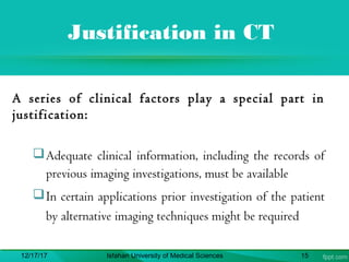
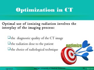
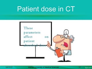
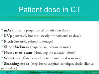
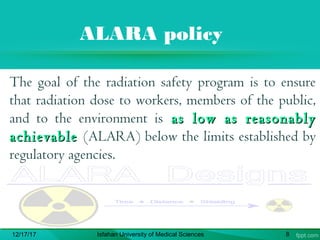
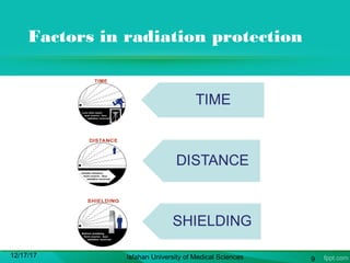
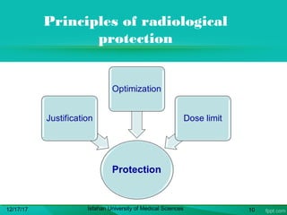
In the Name ofIn the Name of
GodGod
Radiation ProtectionRadiation Protection
in CTin CT
Seyedeh Shokoofeh Mousavi Gazafroudi
Seyedeh Shabnam Mousavi Gazafroudi
2
Table of Content
 Introduction to radiation
protection
 Review of CT
 Radiation protection in CT
 Equipment & layout of room
 Principles & guidelines
12/17/17 Isfahan University of Medical Sciences 3
Introduction to Radiation
Protection
Radiation risk
Riskis proportional to absorbed dose
Risk is quantified by determining the
“effective dose” and is expressed as
millisievert (mSv)
Atomic bomb survivors who experienced
doses that were slightly higherthan doses
encountered in CT demonstrated
increased cancers.
12/17/17 Isfahan University of Medical Sciences 5
12/17/17 Isfahan University of Medical
Sciences
6
12/17/17 7
ALARA policy
The goal of the radiation safety program is to ensure
that radiation dose to workers, members of the public,
and to the environment is as low as reasonablyas low as reasonably
achievableachievable (ALARA) below the limits established by
regulatory agencies.
12/17/17 Isfahan University of Medical Sciences 8
Factors in radiation protection
12/17/17 Isfahan University of Medical Sciences 9
Principles of radiological
protection
12/17/17 Isfahan University of Medical Sciences 10
Review of CT
CT examination
 "Computed tomography (CT)" means the production of a tomogram
by the acquisition and computer processing of x-ray transmission data.
Computed tomography includes the capability of producing axial
tomograms.
 CT examination is a “high dose” procedure and Justification in CT is of
particular importance for RP.
 The absorbed dose to tissues from CT can often approach or exceed the
levels known to increase the probability of cancer as shown in
epidemiological studies.
12/17/17 Isfahan University of Medical Sciences 12
Effective dose estimation
CTDI is equivalent to dose distribution integrated over the
z (longitudinal) patient axis divided by the slice thickness
12/17/17 Isfahan University of Medical Sciences 13
Radiation Protection in
CT
Justification in CT
A series of clinical factors play a special part in
justification:
Adequate clinical information, including the records of
previous imaging investigations, must be available
In certain applications prior investigation of the patient
by alternative imaging techniques might be required
12/17/17 Isfahan University of Medical Sciences 15
Optimization in CT
Optimal use of ionizing radiation involves the
interplay of the imaging process:
the diagnostic quality of the CT image
the radiation dose to the patient
the choice of radiological technique
12/17/17 Isfahan University of Medical Sciences 16
Patient dose in CT
12/17/17 Isfahan University of Medical Sciences 17
These
parameters
affect on
patient
absorbed dose :
Patient dose in CT
mAs ( directly proportional to radiation dose)
KVp ( inversely but not linearly proportional to dose)
Pitch (inversely related to dosage)
Slice thickness (requires an increase in mAs)
Number of scans (doubling the radiation dose)
Scan time (faster scans lead to an increased scan area)
Scanning mode (step-based vs.spiral technique, single-slice vs.
multi-slice)
12/17/17 Isfahan University of Medical Sciences 18
Equipment & layout of
room
Stationary CT rooms
 Computed
tomography rooms
typically have high
workloads and high
kilo voltage
technique settings.
gantry
coach
operator
Isodosecurves12/17/17 Isfahan University of Medical Sciences 20
Room layout
The official UK guidance on the design of
radiological facilities suggests that CT x-ray
rooms should be designed to a minimum
dimension of 38 m².
at least 1/16-Inch lead shielding or equivalent is
required for the walls, doors, floors, ceilings,
and operator's barrier. The concrete equivalence
would be about 4 to 6 inches of standard-density
concrete , depending on workload and distance
factors.12/17/17 Isfahan University of Medical Sciences 21
Equipment & facilities
12/17/17 Isfahan University of Medical Sciences 22
Equipment & facilities
Protective apron, glasses, gloves, collar & gonad
shields and of course film badge & other dosimeter
should be used.
All of devices should be certificated and
calibrated.
 Radiation safety committee and specially medical
physicist have important role in performing
radiation protection programs.
12/17/17 Isfahan University of Medical Sciences 23
Medical physicist’s tasks
Facilities shall be established and implemented under
the supervision of the medical physicist (MP)
MP must perform an initial or acceptance test of
each CT system prior to use on patients
MP must perform an annual evaluation of the CT
system and quality control program
Facility conducts a continuous quality control
program designed/overseen by the MP
12/17/17 Isfahan University of Medical Sciences 24
Principles &
Guidelines
ICRP recommendations
to reduce personnel dose
 Personal dosimeter shall be monitored (worn
properly, read at an appropriate frequency)
 Personal protective equipment to all staff should be
available
 Protective clothing should be worn by staff
 Duration of exposures, dose-rate and X-ray field
size should be minimized
 Positioning of personnel during examinations
should be assessed
12/17/17 Isfahan University of Medical Sciences 26
ICRP recommendations
to reduce patient dose
Actions for 3 groups are defined:
 Operator
 Manufacture
 Physicians
12/17/17 Isfahan University of Medical Sciences 27
What can operators do?
Limit the scanned volume
Reduce mAs values
Use automatic exposure control
Shielding of superficial organs
Use of spiral CT with pitch factor>1
Separate factors for children
Use of partial rotation
Adequate selection of image reconstruction
parameters
Record of dose, exposure factors12/17/17 Isfahan University of Medical Sciences 28
Actions for
manufacturers
 Introduce automatic exposure control
 Be conscious of high doses in CT
 Include safety features to avoid unnecessary dose
 Display of dose
 Convenience in using low dose protocols
 Draw attention of users to selecting separate
protocols for paediatric patients
12/17/17 Isfahan University of Medical Sciences 29
Actions for physician
 Ensure that patients are not irradiated unjustifiably
 Clinical guidelines advising which examinations are
appropriate and acceptable should be available
 Consider whether the required information be obtained by
MRI, ultrasonography
 Consider value of contrast medium enhancement
 Clinician has the responsibility to communicate to the
radiologist about previous CT examination
 CT examination of chest in young girls and young females
needs to be justified in view of high breast dose
12/17/17 Isfahan University of Medical Sciences 30
General rules in x-ray
departments
Using film badge & regular periodic dosimetry
Using standard devices , testing them periodically,
keeping according to manufacture order.
Applying protective equipment for all of patient     
 Training technologists in order to proper usage of
imaging machines & protective equipments
12/17/17 Isfahan University of Medical Sciences 31
Special rules in CT departments
Use of low dose scanning protocol
Warm up , calibration & check up of CT machines result
in patient dose reduction
CT technologist should assess patient information and
then choose best technique according to indication, size
and location of lesions
Accurate selection of slice tilt specially in brain scan
12/17/17 Isfahan University of Medical Sciences 32
AEOI recommendations
 Atomic Energy Organization of Iran (AEOI)
publishes guidelines and reports periodically
 latest guideline consists of 6 chapter about rules for
radiation activities in diagnostic departments
 objectives, applications, key words definitions, and
responsibilities are mentioned in introduction of
named report.
12/17/17 Isfahan University of Medical Sciences 33
AEOI report INRARP6CP02
( Chapter 3 )
Choose technical factors to acquire acceptable
quality images and low dose to patient
Use minimum slice number and minimum mAs
according to clinical goals
Don’t perform pre contrast scan routinely
Take care of lenses radiation by tilting the gantry
12/17/17 Isfahan University of Medical Sciences 34
AEOI report INRARP6CP02
( Chapter 3 ) cont.
Mount danger sign and warning light
Pregnant women should informed technician
 Occupational exposure should not exceed over
permitted dose limit
Fixed , portable and adjustable protective devices
should be available
12/17/17 Isfahan University of Medical Sciences 35
Thanks
36

radiation protection in ct

  • 1.
    In the NameofIn the Name of GodGod
  • 2.
    Radiation ProtectionRadiation Protection inCTin CT Seyedeh Shokoofeh Mousavi Gazafroudi Seyedeh Shabnam Mousavi Gazafroudi 2
  • 3.
    Table of Content Introduction to radiation protection  Review of CT  Radiation protection in CT  Equipment & layout of room  Principles & guidelines 12/17/17 Isfahan University of Medical Sciences 3
  • 4.
  • 5.
    Radiation risk Riskis proportionalto absorbed dose Risk is quantified by determining the “effective dose” and is expressed as millisievert (mSv) Atomic bomb survivors who experienced doses that were slightly higherthan doses encountered in CT demonstrated increased cancers. 12/17/17 Isfahan University of Medical Sciences 5
  • 6.
    12/17/17 Isfahan Universityof Medical Sciences 6
  • 7.
  • 8.
    ALARA policy The goalof the radiation safety program is to ensure that radiation dose to workers, members of the public, and to the environment is as low as reasonablyas low as reasonably achievableachievable (ALARA) below the limits established by regulatory agencies. 12/17/17 Isfahan University of Medical Sciences 8
  • 9.
    Factors in radiationprotection 12/17/17 Isfahan University of Medical Sciences 9
  • 10.
    Principles of radiological protection 12/17/17Isfahan University of Medical Sciences 10
  • 11.
  • 12.
    CT examination  "Computedtomography (CT)" means the production of a tomogram by the acquisition and computer processing of x-ray transmission data. Computed tomography includes the capability of producing axial tomograms.  CT examination is a “high dose” procedure and Justification in CT is of particular importance for RP.  The absorbed dose to tissues from CT can often approach or exceed the levels known to increase the probability of cancer as shown in epidemiological studies. 12/17/17 Isfahan University of Medical Sciences 12
  • 13.
    Effective dose estimation CTDIis equivalent to dose distribution integrated over the z (longitudinal) patient axis divided by the slice thickness 12/17/17 Isfahan University of Medical Sciences 13
  • 14.
  • 15.
    Justification in CT Aseries of clinical factors play a special part in justification: Adequate clinical information, including the records of previous imaging investigations, must be available In certain applications prior investigation of the patient by alternative imaging techniques might be required 12/17/17 Isfahan University of Medical Sciences 15
  • 16.
    Optimization in CT Optimaluse of ionizing radiation involves the interplay of the imaging process: the diagnostic quality of the CT image the radiation dose to the patient the choice of radiological technique 12/17/17 Isfahan University of Medical Sciences 16
  • 17.
    Patient dose inCT 12/17/17 Isfahan University of Medical Sciences 17 These parameters affect on patient absorbed dose :
  • 18.
    Patient dose inCT mAs ( directly proportional to radiation dose) KVp ( inversely but not linearly proportional to dose) Pitch (inversely related to dosage) Slice thickness (requires an increase in mAs) Number of scans (doubling the radiation dose) Scan time (faster scans lead to an increased scan area) Scanning mode (step-based vs.spiral technique, single-slice vs. multi-slice) 12/17/17 Isfahan University of Medical Sciences 18
  • 19.
  • 20.
    Stationary CT rooms Computed tomography rooms typically have high workloads and high kilo voltage technique settings. gantry coach operator Isodosecurves12/17/17 Isfahan University of Medical Sciences 20
  • 21.
    Room layout The officialUK guidance on the design of radiological facilities suggests that CT x-ray rooms should be designed to a minimum dimension of 38 m². at least 1/16-Inch lead shielding or equivalent is required for the walls, doors, floors, ceilings, and operator's barrier. The concrete equivalence would be about 4 to 6 inches of standard-density concrete , depending on workload and distance factors.12/17/17 Isfahan University of Medical Sciences 21
  • 22.
    Equipment & facilities 12/17/17Isfahan University of Medical Sciences 22
  • 23.
    Equipment & facilities Protectiveapron, glasses, gloves, collar & gonad shields and of course film badge & other dosimeter should be used. All of devices should be certificated and calibrated.  Radiation safety committee and specially medical physicist have important role in performing radiation protection programs. 12/17/17 Isfahan University of Medical Sciences 23
  • 24.
    Medical physicist’s tasks Facilitiesshall be established and implemented under the supervision of the medical physicist (MP) MP must perform an initial or acceptance test of each CT system prior to use on patients MP must perform an annual evaluation of the CT system and quality control program Facility conducts a continuous quality control program designed/overseen by the MP 12/17/17 Isfahan University of Medical Sciences 24
  • 25.
  • 26.
    ICRP recommendations to reducepersonnel dose  Personal dosimeter shall be monitored (worn properly, read at an appropriate frequency)  Personal protective equipment to all staff should be available  Protective clothing should be worn by staff  Duration of exposures, dose-rate and X-ray field size should be minimized  Positioning of personnel during examinations should be assessed 12/17/17 Isfahan University of Medical Sciences 26
  • 27.
    ICRP recommendations to reducepatient dose Actions for 3 groups are defined:  Operator  Manufacture  Physicians 12/17/17 Isfahan University of Medical Sciences 27
  • 28.
    What can operatorsdo? Limit the scanned volume Reduce mAs values Use automatic exposure control Shielding of superficial organs Use of spiral CT with pitch factor>1 Separate factors for children Use of partial rotation Adequate selection of image reconstruction parameters Record of dose, exposure factors12/17/17 Isfahan University of Medical Sciences 28
  • 29.
    Actions for manufacturers  Introduceautomatic exposure control  Be conscious of high doses in CT  Include safety features to avoid unnecessary dose  Display of dose  Convenience in using low dose protocols  Draw attention of users to selecting separate protocols for paediatric patients 12/17/17 Isfahan University of Medical Sciences 29
  • 30.
    Actions for physician Ensure that patients are not irradiated unjustifiably  Clinical guidelines advising which examinations are appropriate and acceptable should be available  Consider whether the required information be obtained by MRI, ultrasonography  Consider value of contrast medium enhancement  Clinician has the responsibility to communicate to the radiologist about previous CT examination  CT examination of chest in young girls and young females needs to be justified in view of high breast dose 12/17/17 Isfahan University of Medical Sciences 30
  • 31.
    General rules inx-ray departments Using film badge & regular periodic dosimetry Using standard devices , testing them periodically, keeping according to manufacture order. Applying protective equipment for all of patient       Training technologists in order to proper usage of imaging machines & protective equipments 12/17/17 Isfahan University of Medical Sciences 31
  • 32.
    Special rules inCT departments Use of low dose scanning protocol Warm up , calibration & check up of CT machines result in patient dose reduction CT technologist should assess patient information and then choose best technique according to indication, size and location of lesions Accurate selection of slice tilt specially in brain scan 12/17/17 Isfahan University of Medical Sciences 32
  • 33.
    AEOI recommendations  AtomicEnergy Organization of Iran (AEOI) publishes guidelines and reports periodically  latest guideline consists of 6 chapter about rules for radiation activities in diagnostic departments  objectives, applications, key words definitions, and responsibilities are mentioned in introduction of named report. 12/17/17 Isfahan University of Medical Sciences 33
  • 34.
    AEOI report INRARP6CP02 (Chapter 3 ) Choose technical factors to acquire acceptable quality images and low dose to patient Use minimum slice number and minimum mAs according to clinical goals Don’t perform pre contrast scan routinely Take care of lenses radiation by tilting the gantry 12/17/17 Isfahan University of Medical Sciences 34
  • 35.
    AEOI report INRARP6CP02 (Chapter 3 ) cont. Mount danger sign and warning light Pregnant women should informed technician  Occupational exposure should not exceed over permitted dose limit Fixed , portable and adjustable protective devices should be available 12/17/17 Isfahan University of Medical Sciences 35
  • 36.

Editor's Notes

  • #13 The effective dose in chest CT is in the order of 8 mSv (around 400 times more than chest radiograph dose) and in some CT examinations like that of pelvic region, it may be around 20 mSv
  • #22 Protection needs to extend from the floor to a height of not less than 2 m and be continuous.