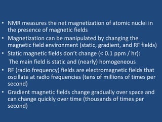
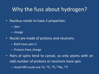
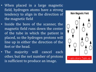
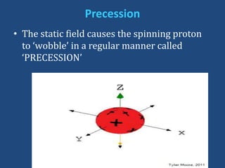
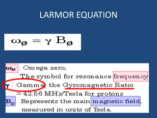
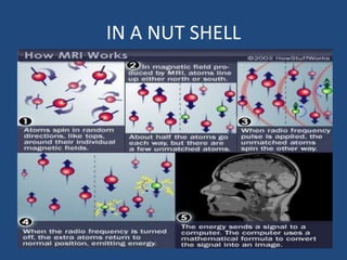
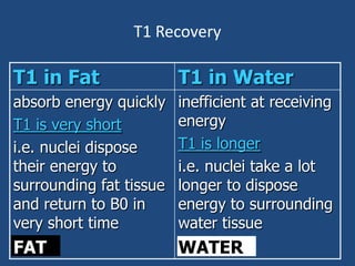
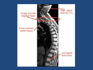
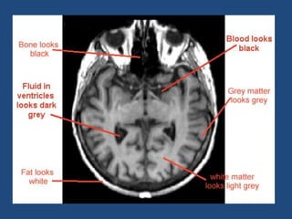
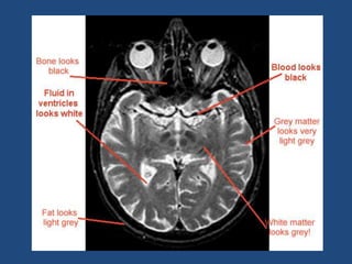
MRI PHYSICS
PEHCHAN KAUN
NOBEL PRIZE 2003
PETERMANSFIELD

Paul lauterbur
MRI: What is it?
Magnetic Resonance Imaging
Magnet
Radio Frequency = Resonance
Imaging
• NMR measures the net magnetization of atomic nuclei in
the presence of magnetic fields
• Magnetization can be manipulated by changing the
magnetic field environment (static, gradient, and RF fields)
• Static magnetic fields don’t change (< 0.1 ppm / hr):
The main field is static and (nearly) homogeneous
• RF (radio frequency) fields are electromagnetic fields that
oscillate at radio frequencies (tens of millions of times per
second)
• Gradient magnetic fields change gradually over space and
can change quickly over time (thousands of times per
second)
WE ARE MAGNETS!
Really?
• The adult human body is ~53%
water, and water is ~11% hydrogen by
mass but ~67% hydrogen by atomic
percent.
• Thus, most of the mass of the human
body is oxygen, but most of the atoms
in the human body are hydrogen
atoms.
• The average 70 kg adult human body
contains approximately 3 x 1027 atoms
of which 67% are hydrogen atoms!!!!
Why the fuss about hydrogen?
• Nucleus needs to have 2 properties:
– Spin
– charge

• Nuclei are made of protons and neutrons
– Both have spin ½
– Protons have charge

• Pairs of spins tend to cancel, so only atoms with an
odd number of protons or neutrons have spin
– Good MR nuclei are 1H, 13C, 19F, 23Na, 31P
Y HYDROGEN?? Cont…
• Simplest element with atomic number of 1
and atomic weight of 1
• When in ionic state (H+), it is nothing but a
proton.
• Proton is not only positively charged, but
also has magnetic spin (wobble)!
• MRI utilizes this magnetic spin property of
protons of hydrogen to elicit images!!
• We are magnets!
A Single Proton

There is electric charge
on the surface of the proton,
thus creating a small current
loop and generating
magnetic moment .

J

+

+

The proton also has
mass which generates
an
angular momentum
J when it is spinning.

+

Thus proton “magnet” differs from the magnetic bar in that it
also possesses angular momentum caused by spinning.
SPIN!!
• Protons and neutron spins are known as
nuclear spins.
• An unpaired component has a spin of ½
and two particles with opposite spins
cancel one another.

• In NMR it is the unpaired nuclear spins
that produce a signal in a magnetic field
But why we can’t act like magnets?
• The protons (i.e.
Hydrogen ions) in body
are spinning in a
haphazard fashion, and
cancel all the
magnetism. That is our
natural state!
• We need to discipline
them first, how?
• When placed in a large magnetic
field, hydrogen atoms have a strong
tendency to align in the direction of
the magnetic field
• Inside the bore of the scanner, the
magnetic field runs down the center
of the tube in which the patient is
placed, so the hydrogen protons will
line up in either the direction of the
feet or the head.
• The majority will cancel each
other, but the net number of protons
is sufficient to produce an image.

5-spin down 7spin up
Precession
• The static field causes the spinning proton
to ‘wobble’ in a regular manner called
‘PRECESSION’
LARMOR EQUATION
TIME TO WAKE UP…
REGARDING MRI SAY TRUE/FALSE
• The gyro magnetic ratio of a hydrogen atom is
42.57MHz/T
• The strength of magnetic field is measured in
gauss
• The Larmor frequency is defined as SUM of the
magnetic field and gyromagnetic ratio.
• An MR active nucleus spins at its own
precessional frequency.
• ONLY hydrogen nuclei are said to be MR active
SACH KA SAMNA….
REGARDING MRI SAY TRUE/FALSE
• The gyro magnetic ratio of a hydrogen atom is
42.57MHz/T T
• The strength of magnetic field is measured in
gauss T
• The Larmor frequency is defined as SUM of the
magnetic field and gyromagnetic ratio. F
• An MR active nucleus spins at its own precessional
frequency. T
• ONLY hydrogen nuclei are said to be MR active  F
IN A NUT SHELL
Now, its time to listen to radio in
RESONANCE.
►Pushing a swing in time with natural
interval of the swing will make the swing
higher and higher.
►Similarly, radio frequency pulses in
resonance push the aligned protons (H+) to
a higher energy level.
WATS RESONANCE????
Def:

Energy transition that occur when
object is subjected to frequency the
same as its own.
Here;

Radio Frequency "RF"
Same Frequency of H nuclei
At 90 degree to B0
2 things happen at Resonance:
1- Energy Absorption
Increase number of High energy Spin Up
nuclei
2- Phase Coherence
NMV precesses in transverse plane at Larmor
Frequency
• Energy Absorption:
–The MRI machine applies
radio frequency (RF)
pulse that is specific to
hydrogen.
–The RF pulses are applied
through a coil that is
specific to the part of the
body being scanned.
MR Signal
• NMV rotates around transverse plane.
It passes across Receiver Coil inducing
voltage in it.
• RF Removed  Signal decreased  Amplitude
of MR Signal decreased

• Free Induction Decay "FID":
– Free (No RF Pulse)
– ID (because of Decay of Induced signal in Receiver
Coil)
SUMMARY TILL NOW…..ENJOYY…
Measuring the MR Signal:
– the moving proton vector
induces a signal in the RF
antenna

Measuring the MR Signal
z

– The signal is picked up by a
coil and sent to the computer
system.
the received signal is
sinusoidal in nature
– The
computer
receives
mathematical data, which is
converted through the use of a
Fourier transform into an
image.

RF signal from
precessing protons

y

x

RF antenna
Now, we re-transmit the energy for
image processing
►The emitted energy is too small (despite
2500 times the magnetic field with
resonance RF pulse) to convert them into
images.
►Hence, repeated “ON-OFF” of RF pulses are
required.
►The emitted energy is stored (Kspace), analysed and converted into images.
• Imaging:
– When the RF pulse is turned off the
hydrogen protons slowly return to their
natural alignment within the magnetic field
and release their excess stored energy. This
is known as relaxation.
• What happens to the released energy?
– Released as heat
OR
– Exchanged and absorbed by other protons
OR
– Released as Radio Waves.
The Image
Relaxation Process
1-NMV recovers and
realign to B0
this process called "T1
Recovery"

2-Nuclei loose
Precessional
coherence or dephase
and NMV decay in the
transverse plane
this process called "T2
Decay"
T1 time & T2 Decay are an intrinsic contrast
parameter that are inherent to tissue being
imaged.

T1WI
Definitions
Echo Time "TE"
Time between RF excitation pulse and
collection of signal
(it affects the length of relaxation period
after removal of RF excitation pulse
and the peak of signal received in receiver
coil)
• Repetition Time "TR"
Time from application of one RF pulse
To the application of the next
(it affects the length of relaxation period
after application of one RF excitation pulse
to the beginning of the next).
Flip Angle
Angle throw which the NMV moved as
result of a RF excitation pulse
Parameters
Image contrast controlled by:
1- Extrinsic Contrast parameters:
TR, TE & Flip Angle
2- Intrinsic Contrast parameters:
T1 Recovery, T2 Decay, Proton Density, Flow &
Apparent Diffusion Coefficient
T1 Recovery
T1 Recovery
Caused by exchange of energy from
nuclei to their surrounding environment or lattice
"Spin Lattice Energy Transfer"
and realign in B0
this occur in exponential process
at different rates in different tissue
NB: Molecules are constantly in motion; Rotational and
Transitional
T1 Recovery

T1 in Fat

T1 in Water

absorb energy quickly
T1 is very short
i.e. nuclei dispose
their energy to
surrounding fat tissue
and return to B0 in
very short time

inefficient at receiving
energy
T1 is longer
i.e. nuclei take a lot
longer to dispose
energy to surrounding
water tissue

FAT

WATER
T1 Recovery
Short TR T1 contrast

(T1

• TR 300-600 ms
• TE 10-30 ms

Weighted)
T2 Decay
Fat much better at energy exchange than Water
Because T2 depends on:
1-How closely molecular motion of atoms matches
Larmor Frequency
2-Proximity of other spins
So;

Fat's T2 time is very short compared to water

FAT

WATER
T2 Decay
Long TE T2 contrast

(T2 Weighted)

• TR 2000 ms
• TE 70 ms
T2* DECAY
• T2* relaxation – Disturbances in magnetic
field (magnetic susceptibility) increase the
rate of spin coherence T2 relaxation
• The total NMR signal is a combination of the
total
number
of
nuclei
(proton
density), reduced by the T1, T2, and T2*
relaxation components
Proton Density
• Proton Density

Long TR Proton density

• TR 2000 ms
• TE 10-30 ms
Gradient Magnets
• Used to vary magnetic field in known
manner
• Each point has slightly different rate of
precession & Larmor Frequency.
• Variety of signal released by Protons
returning to z-plane can used to determine
the composition of exact location of each
point.
• function of "Gradient Magnets”
– Slice selection
– Frequency encoding
– Phase encoding
BRAIN STIMULATOR
REGARDING MRI……
• T1 Recovery is due to interactions of
intrinsic magnetic fields of adjacent nuclei.
• T2 Decay is due to dephasing caused by
inhomogenities of external magnetic field.
• Fat has short T1 Recovery time.
• Water has a short T1 Recovery time.
• On T1 fat appears as high signal.
ANSWERS
• T1 Recovery is due to interactions of
intrinsic magnetic fields of adjacent
nuclei F
• T2*Decay is due to dephasing caused by
inhomogenities of external magnetic
fieldT
• Fat has short T1 Recovery time T
• Water has a short T1 Recovery time F
• On T1 fat appears as high signal T
IMAGE QUALITY
• Factors that determine image quality
Signal: noise ratio ,
Contrast ,
Spatial Resolution,
Scan time.
SIGNAL : NOISE RATIO
• Noise is a random variation in the MR
signal, occurring at all frequencies and all
the time, due to -1. PATIENT(white noise)
2.ENVIRONMENT.
3. SCANNER.
• All noise reduces and obscures contrast
bbetween tissues and is worst in areas of
low PD and low signal.
IMPROVE SNR…
• INCREASING THE
SIGNAL BY
• INCREASING THE
VOXEL SIZE
• DECREASING TE AND
INCREASING TR/FLIP
ANGLE.
• SE SEQUENCES > GRE
• HIGH TESLA MACHINE

• REDUCING NOISE
BY
• INCREASING THE
NUMBNER OF
EXCITATIONS
• REDUCING
BANDWIDTH
• REDUCING CROSS
TALK
The great headache—ARTIFACTS!!
• Image Artifact is something observed in a scientific
investigation that is not naturally present but occurs
as a result of the investigative procedure.

• A structure not normally present, but visible as a
result of malfunction in the hardware or software of
the device, or a consequence of environmental
influences as heat or humidity or can be caused by
the human body itself.
WHICH ARTIFACT????
Motion artifacts( ghost)
SPECIAL THANKS TO…….

Mri physics

  • 1.
  • 2.
  • 3.
  • 4.
    MRI: What isit? Magnetic Resonance Imaging Magnet Radio Frequency = Resonance Imaging
  • 5.
    • NMR measuresthe net magnetization of atomic nuclei in the presence of magnetic fields • Magnetization can be manipulated by changing the magnetic field environment (static, gradient, and RF fields) • Static magnetic fields don’t change (< 0.1 ppm / hr): The main field is static and (nearly) homogeneous • RF (radio frequency) fields are electromagnetic fields that oscillate at radio frequencies (tens of millions of times per second) • Gradient magnetic fields change gradually over space and can change quickly over time (thousands of times per second)
  • 6.
  • 7.
    • The adulthuman body is ~53% water, and water is ~11% hydrogen by mass but ~67% hydrogen by atomic percent. • Thus, most of the mass of the human body is oxygen, but most of the atoms in the human body are hydrogen atoms. • The average 70 kg adult human body contains approximately 3 x 1027 atoms of which 67% are hydrogen atoms!!!!
  • 8.
    Why the fussabout hydrogen? • Nucleus needs to have 2 properties: – Spin – charge • Nuclei are made of protons and neutrons – Both have spin ½ – Protons have charge • Pairs of spins tend to cancel, so only atoms with an odd number of protons or neutrons have spin – Good MR nuclei are 1H, 13C, 19F, 23Na, 31P
  • 9.
    Y HYDROGEN?? Cont… •Simplest element with atomic number of 1 and atomic weight of 1 • When in ionic state (H+), it is nothing but a proton. • Proton is not only positively charged, but also has magnetic spin (wobble)! • MRI utilizes this magnetic spin property of protons of hydrogen to elicit images!! • We are magnets!
  • 10.
    A Single Proton Thereis electric charge on the surface of the proton, thus creating a small current loop and generating magnetic moment . J + + The proton also has mass which generates an angular momentum J when it is spinning. + Thus proton “magnet” differs from the magnetic bar in that it also possesses angular momentum caused by spinning.
  • 11.
    SPIN!! • Protons andneutron spins are known as nuclear spins. • An unpaired component has a spin of ½ and two particles with opposite spins cancel one another. • In NMR it is the unpaired nuclear spins that produce a signal in a magnetic field
  • 13.
    But why wecan’t act like magnets? • The protons (i.e. Hydrogen ions) in body are spinning in a haphazard fashion, and cancel all the magnetism. That is our natural state! • We need to discipline them first, how?
  • 14.
    • When placedin a large magnetic field, hydrogen atoms have a strong tendency to align in the direction of the magnetic field • Inside the bore of the scanner, the magnetic field runs down the center of the tube in which the patient is placed, so the hydrogen protons will line up in either the direction of the feet or the head. • The majority will cancel each other, but the net number of protons is sufficient to produce an image. 5-spin down 7spin up
  • 15.
    Precession • The staticfield causes the spinning proton to ‘wobble’ in a regular manner called ‘PRECESSION’
  • 17.
  • 18.
    TIME TO WAKEUP… REGARDING MRI SAY TRUE/FALSE • The gyro magnetic ratio of a hydrogen atom is 42.57MHz/T • The strength of magnetic field is measured in gauss • The Larmor frequency is defined as SUM of the magnetic field and gyromagnetic ratio. • An MR active nucleus spins at its own precessional frequency. • ONLY hydrogen nuclei are said to be MR active
  • 19.
    SACH KA SAMNA…. REGARDINGMRI SAY TRUE/FALSE • The gyro magnetic ratio of a hydrogen atom is 42.57MHz/T T • The strength of magnetic field is measured in gauss T • The Larmor frequency is defined as SUM of the magnetic field and gyromagnetic ratio. F • An MR active nucleus spins at its own precessional frequency. T • ONLY hydrogen nuclei are said to be MR active  F
  • 20.
    IN A NUTSHELL
  • 21.
    Now, its timeto listen to radio in RESONANCE. ►Pushing a swing in time with natural interval of the swing will make the swing higher and higher. ►Similarly, radio frequency pulses in resonance push the aligned protons (H+) to a higher energy level.
  • 22.
    WATS RESONANCE???? Def: Energy transitionthat occur when object is subjected to frequency the same as its own. Here; Radio Frequency "RF" Same Frequency of H nuclei At 90 degree to B0
  • 23.
    2 things happenat Resonance: 1- Energy Absorption Increase number of High energy Spin Up nuclei 2- Phase Coherence NMV precesses in transverse plane at Larmor Frequency
  • 24.
    • Energy Absorption: –TheMRI machine applies radio frequency (RF) pulse that is specific to hydrogen. –The RF pulses are applied through a coil that is specific to the part of the body being scanned.
  • 26.
    MR Signal • NMVrotates around transverse plane. It passes across Receiver Coil inducing voltage in it. • RF Removed  Signal decreased  Amplitude of MR Signal decreased • Free Induction Decay "FID": – Free (No RF Pulse) – ID (because of Decay of Induced signal in Receiver Coil)
  • 27.
  • 28.
    Measuring the MRSignal: – the moving proton vector induces a signal in the RF antenna Measuring the MR Signal z – The signal is picked up by a coil and sent to the computer system. the received signal is sinusoidal in nature – The computer receives mathematical data, which is converted through the use of a Fourier transform into an image. RF signal from precessing protons y x RF antenna
  • 29.
    Now, we re-transmitthe energy for image processing ►The emitted energy is too small (despite 2500 times the magnetic field with resonance RF pulse) to convert them into images. ►Hence, repeated “ON-OFF” of RF pulses are required. ►The emitted energy is stored (Kspace), analysed and converted into images.
  • 31.
    • Imaging: – Whenthe RF pulse is turned off the hydrogen protons slowly return to their natural alignment within the magnetic field and release their excess stored energy. This is known as relaxation. • What happens to the released energy? – Released as heat OR – Exchanged and absorbed by other protons OR – Released as Radio Waves.
  • 32.
  • 33.
    Relaxation Process 1-NMV recoversand realign to B0 this process called "T1 Recovery" 2-Nuclei loose Precessional coherence or dephase and NMV decay in the transverse plane this process called "T2 Decay"
  • 34.
    T1 time &T2 Decay are an intrinsic contrast parameter that are inherent to tissue being imaged. T1WI
  • 35.
    Definitions Echo Time "TE" Timebetween RF excitation pulse and collection of signal (it affects the length of relaxation period after removal of RF excitation pulse and the peak of signal received in receiver coil)
  • 36.
    • Repetition Time"TR" Time from application of one RF pulse To the application of the next (it affects the length of relaxation period after application of one RF excitation pulse to the beginning of the next).
  • 38.
    Flip Angle Angle throwwhich the NMV moved as result of a RF excitation pulse
  • 39.
    Parameters Image contrast controlledby: 1- Extrinsic Contrast parameters: TR, TE & Flip Angle 2- Intrinsic Contrast parameters: T1 Recovery, T2 Decay, Proton Density, Flow & Apparent Diffusion Coefficient
  • 40.
    T1 Recovery T1 Recovery Causedby exchange of energy from nuclei to their surrounding environment or lattice "Spin Lattice Energy Transfer" and realign in B0 this occur in exponential process at different rates in different tissue NB: Molecules are constantly in motion; Rotational and Transitional
  • 41.
    T1 Recovery T1 inFat T1 in Water absorb energy quickly T1 is very short i.e. nuclei dispose their energy to surrounding fat tissue and return to B0 in very short time inefficient at receiving energy T1 is longer i.e. nuclei take a lot longer to dispose energy to surrounding water tissue FAT WATER
  • 44.
    T1 Recovery Short TRT1 contrast (T1 • TR 300-600 ms • TE 10-30 ms Weighted)
  • 45.
    T2 Decay Fat muchbetter at energy exchange than Water Because T2 depends on: 1-How closely molecular motion of atoms matches Larmor Frequency 2-Proximity of other spins So; Fat's T2 time is very short compared to water FAT WATER
  • 47.
    T2 Decay Long TET2 contrast (T2 Weighted) • TR 2000 ms • TE 70 ms
  • 49.
    T2* DECAY • T2*relaxation – Disturbances in magnetic field (magnetic susceptibility) increase the rate of spin coherence T2 relaxation • The total NMR signal is a combination of the total number of nuclei (proton density), reduced by the T1, T2, and T2* relaxation components
  • 50.
    Proton Density • ProtonDensity Long TR Proton density • TR 2000 ms • TE 10-30 ms
  • 52.
    Gradient Magnets • Usedto vary magnetic field in known manner • Each point has slightly different rate of precession & Larmor Frequency. • Variety of signal released by Protons returning to z-plane can used to determine the composition of exact location of each point. • function of "Gradient Magnets” – Slice selection – Frequency encoding – Phase encoding
  • 53.
    BRAIN STIMULATOR REGARDING MRI…… •T1 Recovery is due to interactions of intrinsic magnetic fields of adjacent nuclei. • T2 Decay is due to dephasing caused by inhomogenities of external magnetic field. • Fat has short T1 Recovery time. • Water has a short T1 Recovery time. • On T1 fat appears as high signal.
  • 54.
    ANSWERS • T1 Recoveryis due to interactions of intrinsic magnetic fields of adjacent nuclei F • T2*Decay is due to dephasing caused by inhomogenities of external magnetic fieldT • Fat has short T1 Recovery time T • Water has a short T1 Recovery time F • On T1 fat appears as high signal T
  • 55.
    IMAGE QUALITY • Factorsthat determine image quality Signal: noise ratio , Contrast , Spatial Resolution, Scan time.
  • 56.
    SIGNAL : NOISERATIO • Noise is a random variation in the MR signal, occurring at all frequencies and all the time, due to -1. PATIENT(white noise) 2.ENVIRONMENT. 3. SCANNER. • All noise reduces and obscures contrast bbetween tissues and is worst in areas of low PD and low signal.
  • 57.
    IMPROVE SNR… • INCREASINGTHE SIGNAL BY • INCREASING THE VOXEL SIZE • DECREASING TE AND INCREASING TR/FLIP ANGLE. • SE SEQUENCES > GRE • HIGH TESLA MACHINE • REDUCING NOISE BY • INCREASING THE NUMBNER OF EXCITATIONS • REDUCING BANDWIDTH • REDUCING CROSS TALK
  • 59.
    The great headache—ARTIFACTS!! •Image Artifact is something observed in a scientific investigation that is not naturally present but occurs as a result of the investigative procedure. • A structure not normally present, but visible as a result of malfunction in the hardware or software of the device, or a consequence of environmental influences as heat or humidity or can be caused by the human body itself.
  • 74.
  • 75.
  • 79.