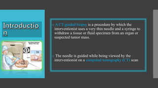
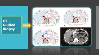
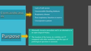
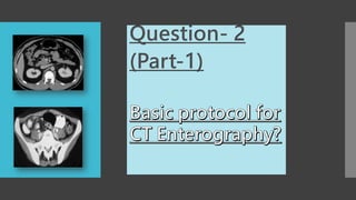
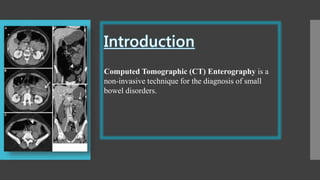
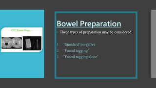
Question-1
o A CT-guided biopsy is a procedure by which the
interventionist uses a very thin needle and a syringe to
withdraw a tissue or fluid specimen from an organ or
suspected tumor mass.
o The needle is guided while being viewed by the
interventionist on a computed tomography (CT) scan
CT
Guided
Biopsy
1. Biopsy ore-biopsy of malignancy for targeted therapy
2. Enlarging solitary nodule
3. Suspicious lung, pleural or mediastinal lesions
4. Suspicious lesion located deep inside the body and
sample can’t be taken on Ultrasound
5. Pulmonary lesion inaccessible to bronchoscopy
6. Mediastinal or pleural mass
7. Focal Parenchymal infiltrates
 Lack of safe access
 Uncorrectable bleeding diathesis
 Respiratory disease
 Poor respiratory function or reserve
 Uncooperative patient
 Minimally invasive procedure and is an alternative to
an open surgical biopsy.
 The location of the lesion, its visibility on CT
compared with other modalities, and the type of
pathological specimen is assessed.
 Consent form
 If Allergic to contrast , must informed to the doctor
 Don’t eat or drink before the procedure
 coagulation profile:
• International normalized ratio (INR) ≤ 1.5
• Normal prothrombin time (PT)
• Partial thromboplastin time (PTT)
 Patient lies supine, Prone or on either side, depending on
where needle is inserted
 To locate site for needle insertion, first CT Scan is done.
 Needle is inserted through skin
 Another CT Scan is performed to confirm the tip of
needle lies at desired location
 Then biopsy specimen is withdrawn through needle
Techniqu
e
1-Bleeding
2-Infection
3-Pneumothorax
4-Allergic
Reaction
1- Not indicated for patients with bleeding disorder
2- Procedure is performed on almost all organs of body except
3- Spleen because there is high risk of severe post-biopsy
hemorrhage.
 Differentiation of necrotic vs. visible portions of tumor
 higher image resolution
 Ability to visualize the abdominal organs and viscera
 Clear depiction of all anatomical structures
 Better characterization of retroperitoneal structures
 Readily recognized any complications
 Exposure to radiations
 Expensive
 Time consuming
 Patients must be brought to CT room
Question- 2
(Part-1)
Computed Tomographic (CT) Enterography is a
non-invasive technique for the diagnosis of small
bowel disorders.
• Evaluation of Crohn disease or ulcerative colitis (location,
severity and complications
• Bowel obstruction, tumors
• Gastrointestinal tract bleeding
• Localizing sites of GI tract bleeding
• Detecting small bowel neoplasms
1- Contrast media allergy
2- Chronic kidney disease
3- Patients in post-operative period in whom abscess or anastomotic
leak is considered
4- Patients who have had multiple CT examination in their lifetime
5- Pregnancy
1. Abstain from all food and drink 4-6 hours before the exam.
2. Patients drinks about 1.5 L of oral contrast over 30-60 min.
3. Adequate luminal distension is necessary as it allows better
mucosal enhancement and is important in evaluation of Crohn
disease.
4. CT enterography utilizes negative or neutral contrast e.g. water,
mannitol etc
 Patients are asked to withhold all oral intake, starting 4
hours before the examination.
 To improve visualization of the mucosa and achieve
better bowel distention, a negative oral contrast agent is
administered.
 After the oral contrast agent is ingested, a bolus of
intravenous contrast material (125 ml ) followed by 50
mL of saline solution is administered
 Allergic reaction to contrast media
 Pregnancy
 High radiation Exposure
The main limitation of CT enterography is secondary to the
use of water as the single neutral contrast.
1-CT entero-graphy
visualizes the entire
thickness of the bowel
wall.
2-It eliminates the need
for video capsule
endoscopy (VCE) and
its complications.
The main disadvantages of the technique are
the requisite exposure of patients to ionizing radiation
the frequent need for repeated imaging
the necessity of using intravenous contrast material
 CT colonography also known as ‘’Virtual Colonoscopy’’
 Radiological examination of choice for detection of colonic
neoplasia of large bowel
CT
Colon
o-
graph
y
 Screening for colorectal cancer (CRC)
 Incomplete colonoscopy
 Patients in high-risk group or who have aversion to colonoscopy.
 Obstructing lesion preventing full colonoscopy
 Three types of preparation may be considered:
1. ’Standard’ purgative
2. ’Faecal tagging’
3. ’Faecal tagging alone’
1. Patient is instructed to go to the toilet before procedure.
2. 20mg Buscopan is given.
3. Air or carbon dioxide is insufflated in patients colon.
4. CT scout is performed to check gaseous large bowel distention.
5. Scan both in supine and prone position.
 Patient should be advised that cramping sensations are
normal for next 24 hours
 Patient should be advised to consume additional fluids
for 24 hours
 Contact the department in severe pain
 Perforations are rare
 Discomfort
 Adverse reaction to contrast
CT Guided Biopsy.pptx

CT Guided Biopsy.pptx

  • 3.
  • 4.
    o A CT-guidedbiopsy is a procedure by which the interventionist uses a very thin needle and a syringe to withdraw a tissue or fluid specimen from an organ or suspected tumor mass. o The needle is guided while being viewed by the interventionist on a computed tomography (CT) scan
  • 5.
  • 6.
    1. Biopsy ore-biopsyof malignancy for targeted therapy 2. Enlarging solitary nodule 3. Suspicious lung, pleural or mediastinal lesions 4. Suspicious lesion located deep inside the body and sample can’t be taken on Ultrasound 5. Pulmonary lesion inaccessible to bronchoscopy 6. Mediastinal or pleural mass 7. Focal Parenchymal infiltrates
  • 7.
     Lack ofsafe access  Uncorrectable bleeding diathesis  Respiratory disease  Poor respiratory function or reserve  Uncooperative patient  Minimally invasive procedure and is an alternative to an open surgical biopsy.  The location of the lesion, its visibility on CT compared with other modalities, and the type of pathological specimen is assessed.
  • 8.
     Consent form If Allergic to contrast , must informed to the doctor  Don’t eat or drink before the procedure  coagulation profile: • International normalized ratio (INR) ≤ 1.5 • Normal prothrombin time (PT) • Partial thromboplastin time (PTT)
  • 9.
     Patient liessupine, Prone or on either side, depending on where needle is inserted  To locate site for needle insertion, first CT Scan is done.  Needle is inserted through skin  Another CT Scan is performed to confirm the tip of needle lies at desired location  Then biopsy specimen is withdrawn through needle
  • 10.
  • 11.
    1-Bleeding 2-Infection 3-Pneumothorax 4-Allergic Reaction 1- Not indicatedfor patients with bleeding disorder 2- Procedure is performed on almost all organs of body except 3- Spleen because there is high risk of severe post-biopsy hemorrhage.
  • 12.
     Differentiation ofnecrotic vs. visible portions of tumor  higher image resolution  Ability to visualize the abdominal organs and viscera  Clear depiction of all anatomical structures  Better characterization of retroperitoneal structures  Readily recognized any complications  Exposure to radiations  Expensive  Time consuming  Patients must be brought to CT room
  • 13.
  • 14.
    Computed Tomographic (CT)Enterography is a non-invasive technique for the diagnosis of small bowel disorders.
  • 15.
    • Evaluation ofCrohn disease or ulcerative colitis (location, severity and complications • Bowel obstruction, tumors • Gastrointestinal tract bleeding • Localizing sites of GI tract bleeding • Detecting small bowel neoplasms
  • 16.
    1- Contrast mediaallergy 2- Chronic kidney disease 3- Patients in post-operative period in whom abscess or anastomotic leak is considered 4- Patients who have had multiple CT examination in their lifetime 5- Pregnancy
  • 17.
    1. Abstain fromall food and drink 4-6 hours before the exam. 2. Patients drinks about 1.5 L of oral contrast over 30-60 min. 3. Adequate luminal distension is necessary as it allows better mucosal enhancement and is important in evaluation of Crohn disease. 4. CT enterography utilizes negative or neutral contrast e.g. water, mannitol etc
  • 18.
     Patients areasked to withhold all oral intake, starting 4 hours before the examination.  To improve visualization of the mucosa and achieve better bowel distention, a negative oral contrast agent is administered.  After the oral contrast agent is ingested, a bolus of intravenous contrast material (125 ml ) followed by 50 mL of saline solution is administered
  • 19.
     Allergic reactionto contrast media  Pregnancy  High radiation Exposure The main limitation of CT enterography is secondary to the use of water as the single neutral contrast.
  • 20.
    1-CT entero-graphy visualizes theentire thickness of the bowel wall. 2-It eliminates the need for video capsule endoscopy (VCE) and its complications. The main disadvantages of the technique are the requisite exposure of patients to ionizing radiation the frequent need for repeated imaging the necessity of using intravenous contrast material
  • 22.
     CT colonographyalso known as ‘’Virtual Colonoscopy’’  Radiological examination of choice for detection of colonic neoplasia of large bowel CT Colon o- graph y
  • 23.
     Screening forcolorectal cancer (CRC)  Incomplete colonoscopy  Patients in high-risk group or who have aversion to colonoscopy.  Obstructing lesion preventing full colonoscopy
  • 24.
     Three typesof preparation may be considered: 1. ’Standard’ purgative 2. ’Faecal tagging’ 3. ’Faecal tagging alone’
  • 25.
    1. Patient isinstructed to go to the toilet before procedure. 2. 20mg Buscopan is given. 3. Air or carbon dioxide is insufflated in patients colon. 4. CT scout is performed to check gaseous large bowel distention. 5. Scan both in supine and prone position.
  • 26.
     Patient shouldbe advised that cramping sensations are normal for next 24 hours  Patient should be advised to consume additional fluids for 24 hours  Contact the department in severe pain
  • 27.
     Perforations arerare  Discomfort  Adverse reaction to contrast