Difference Between Single Slice and Multi Slice CT Scanner
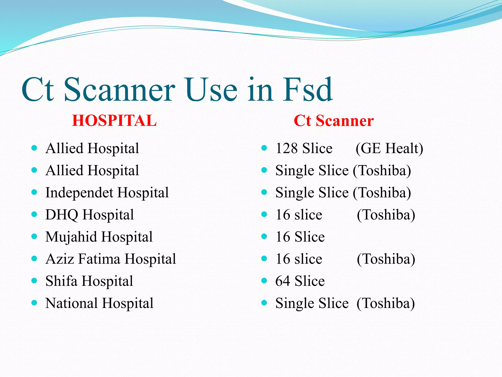
A single slice CT scanner uses a single x-ray source and detector that rotate around the patient to acquire a series of transmission measurements from different angles. A multi-slice CT scanner uses multiple detectors arranged in a row, allowing it to acquire multiple slices of data simultaneously with each rotation. This provides advantages like faster scanning, reduced motion artifacts, and the ability to perform 3D imaging. Common hospitals in the area have CT scanners ranging from single slice to 128 slice models.
Difference Between Single Slice and Multi Slice CT Scanner
1.
Difference Between SingleSlice
and Multi Slice CT Scanner
Presented By.
Talha Saleeme 21305
Asad Rasool 21307
Ali Arslan 21309
Zartash Gul 21310
2.
CT Scan
Themeaning of the word “tomography.” This Greek word
comes from two distinct words “tomos” and “graphe.”
“Tomos” means “section or slice” while “graphe” means
“drawing.”
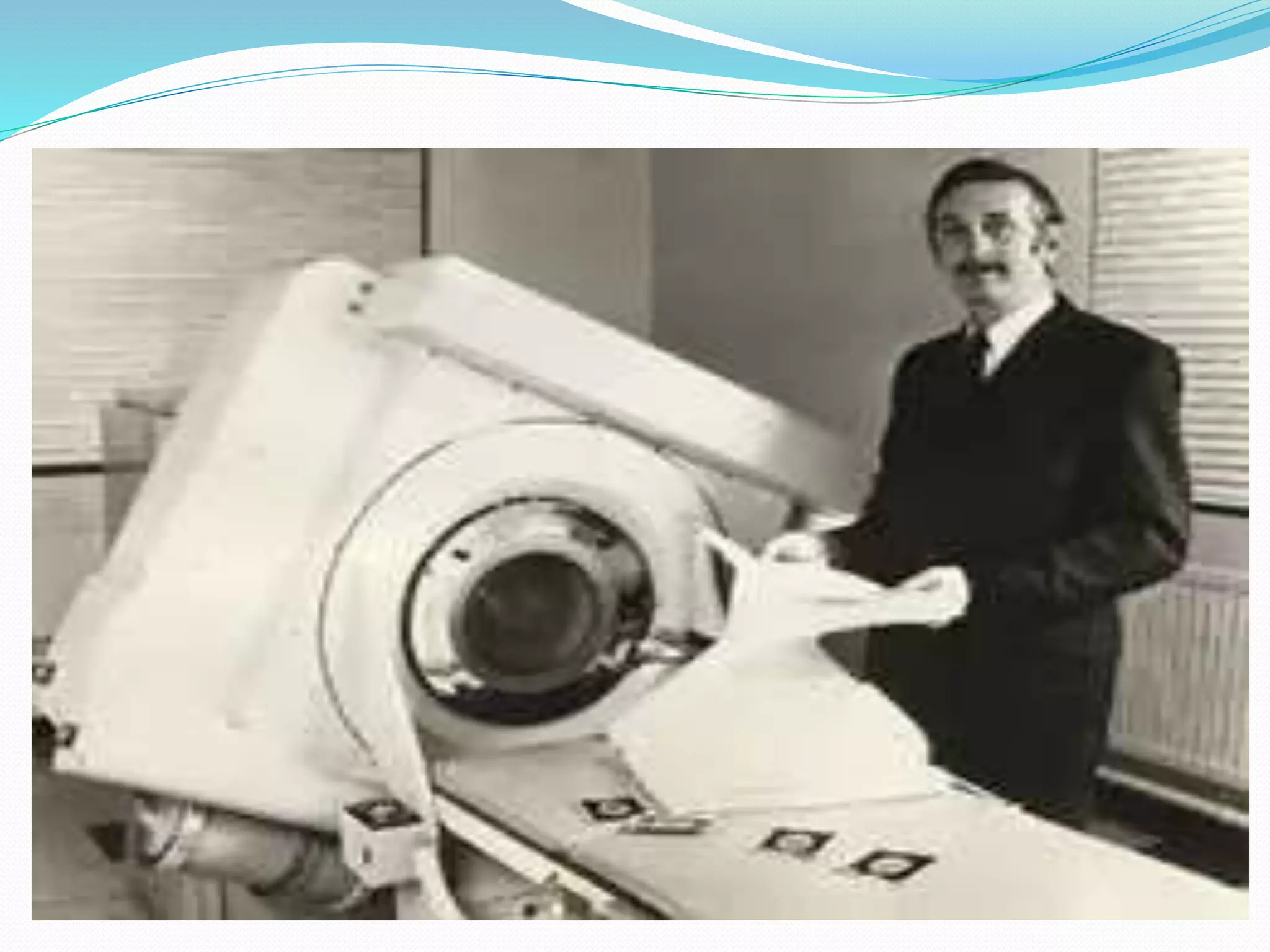
CT was invented in 1972 by British engineer Godfrey
Hounsfield of EMI Laboratories, England
5.
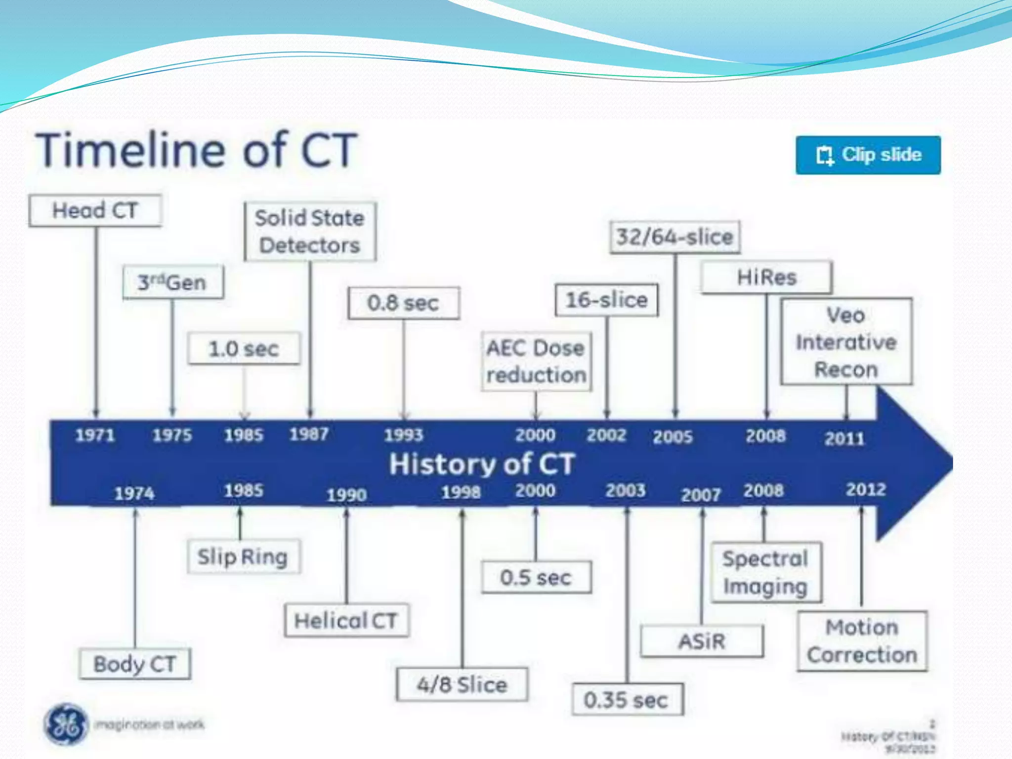
During its25-year history, CT has made great
improvements in speed, patient comfort, and resolution.
As CT scan times have gotten faster, more anatomy can be
scanned in less time. Faster scanning helps to eliminate
artifacts from patient motion such as breathing or
peristalsis
6.
How Single Slicework?
1. The single slice Ct scan had an x-ray source and a single
detector.
2. Data acquisition involved moving both the tube and
detector across the scanning plane to acquire a serious of
transmission measurements.
3. All data collected through a 180 degree rotation.
7.
Parts of singleslice CT scan
1. Gantry
X-ray tube
High voltage Generator
Detector
Pre patient collimator
Post patient collimator
2. Table
3. Ups
4. Control panel
Multi Slice CTScan
The 1980s saw incremental development of CT scanner
technology: shorter scan times and increased matrix sizes,
until by the late 1980s scan times were down to only 3
seconds and matrix sizes were up to 1024 x 1024.
Development continued through the 1990s, with the
introduction of spiral (continuous) scanning in the early
1990s and the development of multi-slice scanners, with
4-slice scanners and 0.5 second scan times being 'state-of-
the-art' by the end of the century.
10.
Current Use ofCT Scan
Development of CT scanner technology continued through
the early years of the 21st century, particularly with multi-
slice scanners. At the time of writing, high-end scanners
were offering up to 320 slices, dual-source and dual-
energy x-ray sources
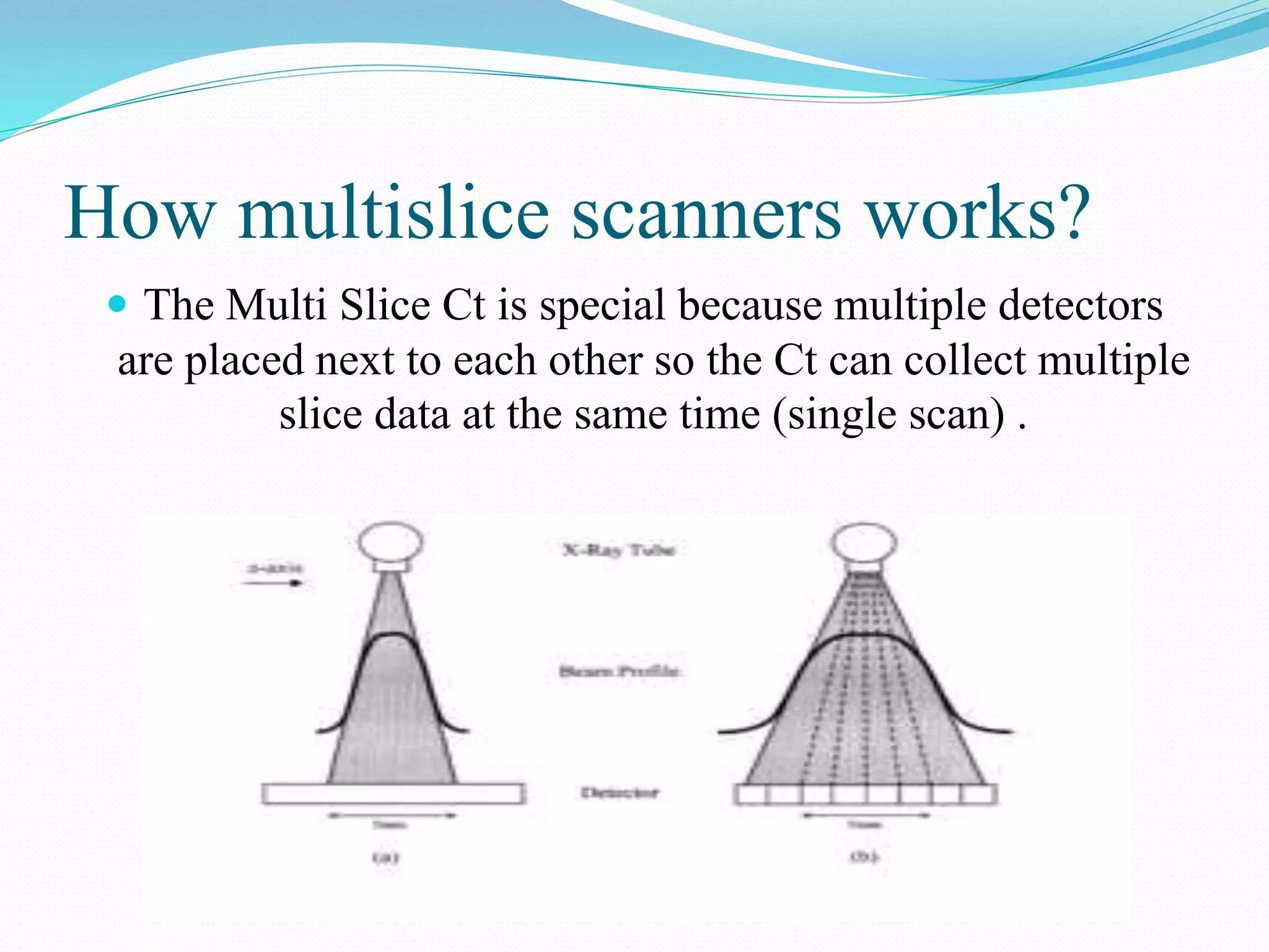
How multislice scannersworks?
The Multi Slice Ct is special because multiple detectors
are placed next to each other so the Ct can collect multiple
slice data at the same time (single scan) .
13.
The Multi slicecan work sequential and spiral mode also.
In the simplest Multi slice Ct there are rows of detectors.
In these, the radiographer/ assistant can set the slice
thickness with the help of the collimator just as the
conventional Ct.
An important thing is that, usually the number of
measurable slices is differ from the number of detectors.
14.
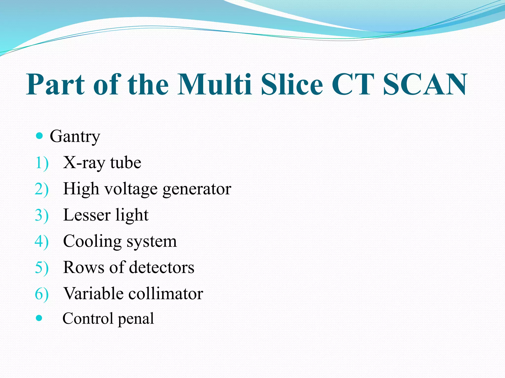
Part of theMulti Slice CT SCAN
Gantry
1) X-ray tube
2) High voltage generator
3) Lesser light
4) Cooling system
5) Rows of detectors
6) Variable collimator
Control penal
15.
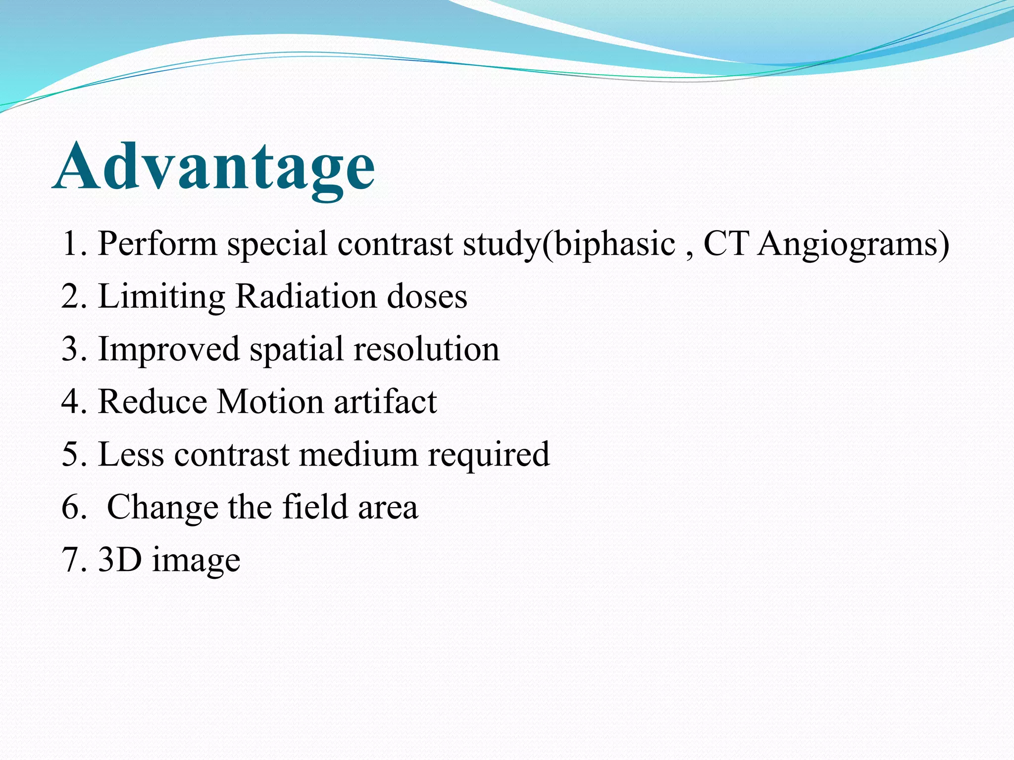
Advantage
1. Perform specialcontrast study(biphasic , CT Angiograms)
2. Limiting Radiation doses
3. Improved spatial resolution
4. Reduce Motion artifact
5. Less contrast medium required
6. Change the field area
7. 3D image
3rd Generation:
Singlex ray tube
Detector 400-1000
Beam fan shape
Rotate and rotate
Use slip ring technology
20.
4th Generation:
Rotateand stationary
X-ray tube Rotate at 360 degree
Detector stationary
Detectors in ring form
Imaging time in sub seconds
22.
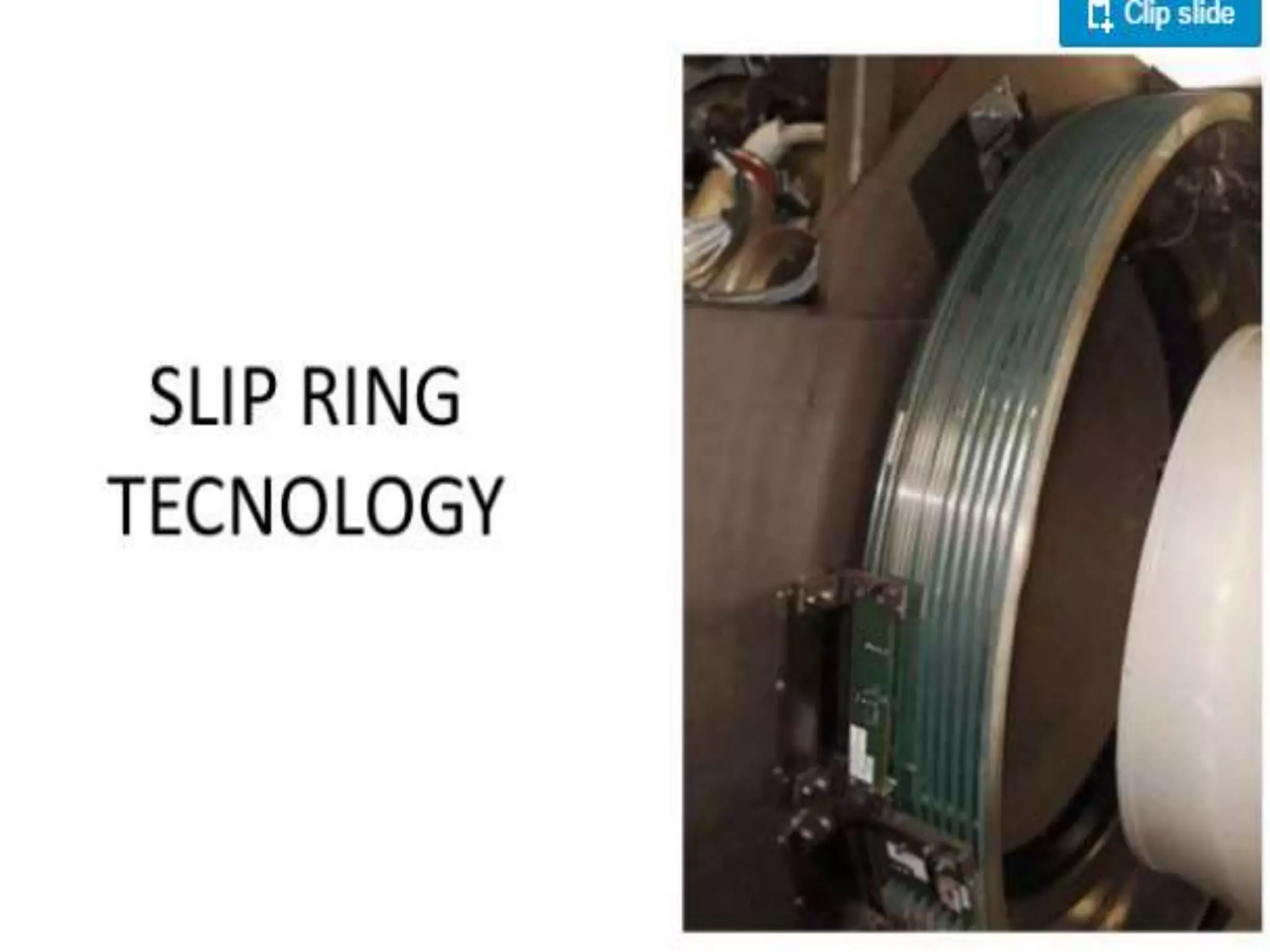
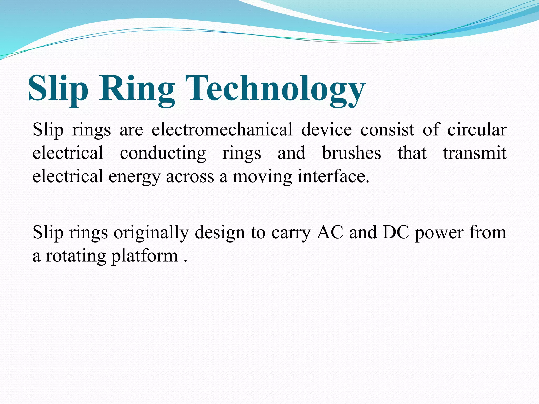
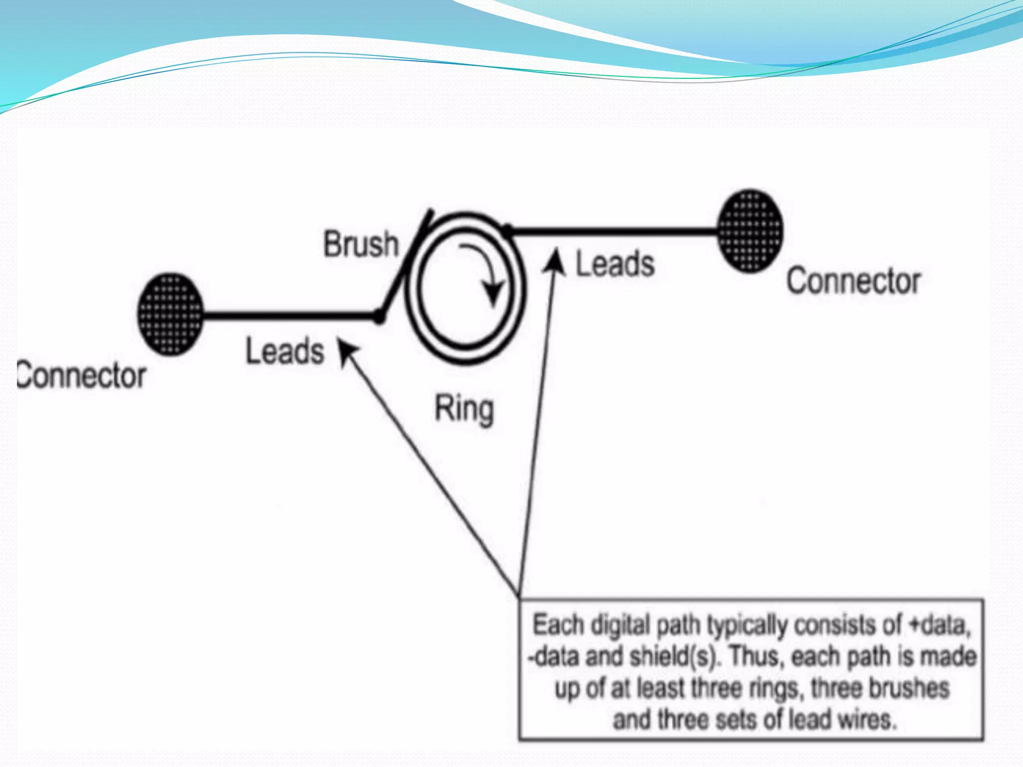
Slip Ring Technology
Sliprings are electromechanical device consist of circular
electrical conducting rings and brushes that transmit
electrical energy across a moving interface.
Slip rings originally design to carry AC and DC power from
a rotating platform .
24.
The fivepair of larger brushes provide the voltage required by
the x ray tube and the 3 pair of smaller one transfer signals
from gantry controller.
Brushes are used to transmit electrical power to the CT
scanner components.
There are two types of brushes that can be used wire and
composite.