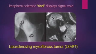
MSK
SIGNS
DR. DALIA EL SAIED MD
GENERAL ORGANIZATION FOR TEACHING
HOSPITALS AND INSTITUTES.
Adhesive Capsulitis
Thickened coraco-humeral ligament (long arrow) obliterating the rotator cuff interval ….
Long head of biceps (short arrow).
Thickened IGHL obliterating the axillary recess.
Mouzopoulos sign (blue M)
Light bulb sign
Posterior shoulder
dislocation
Ivory vertebra
Metastases
Paget’s
Mushroom sign
CHPS Rectosigmoid
Endometriosis
Facet
arthropathy
Geyser sign
ACJ cyst
in case of chronic rotator cuff
tendon tear.
Split Fat sign
 Thin peripheral rim of fat best seen
on planes along long axis of the
lesion in non fat suppressed
sequences
Peripheral nerve
sheath tumors.
Fascicular sign
 Multiple small ring-like structures
Peripheral nerve sheath
tumors.
Target sign
 Peripheral high T2 signal
 Central low signal
Peripheral nerve sheath tumors.
Dot in circle sign
Madura foot
Bow tie sign
Unilateral facet dislocation
Hamato-lunate impingement
syndrome
Occurs with type II lunate morphology
(existence of a medial facet on the distal lunate
for articulation with the hamate)
Scaphoid nonunion advanced collapse
(SNAC).
Occur with Non-
union of scaphoid
fractures.
SCAL / SNAC
(Advanced collapse)
 = Collapse of the carpal arches.
 Complications of scaphoid fractures:-
 AVN (edema =viability)
 Non union
Scapholunate advanced collapse (SLAC)
Stage I (SLAC)
Stage II (SLAC)
 Following untreated
scaphoLunate dissociation
(arrow).
 Advanced osteoarthritis.
SLAC
 - Initially affect radial styloid and scaphoid
articulation.
 - Later ---- whole radioscaphoid articulation, then
proximal capitate migration.
SLAC
* Clenched fist view
* Stress view for
scapholunate ligament
dissociation
#Differentials
SNAC
Complication of Non united
scaphoid fracture
Radioscaphoid osteoarthritis.
Then proximal capitate migration.
SLAC
Complication of
scaphoLunate dissociation
Radioscaphoid osteoarthritis.
Then proximal capitate migration.
SLAC associations
- Cortical ring sign (caused by scaphoid
malalignment/ rotatory subluxation).
- DISI
SL & CL angles.
Arcs of Gilula
Advanced collapse = Disturbance any of the 3 carpal arches.
Treatment of SNAC / SLAC
Four-corner fusion arthrodesis.
- Partial fusion technique.
- Used in advanced degenerative changes
- To reduce pain.
Scaphoidectomy and four corner fusion.
#Differentials
Ulnar impaction
Positive ulnar variance
Ulnar side marrow edema.
Kienböck disease
Negative ulnar variance
Diffuse marrow edema.
#Differentials (Pain and soft tissue edema)
CRPS
Diffuse patchy
("bone marrow edema") on STIR images.
- BME fluctuate or migrate to other areas.
- Soft tissue edema.
Acute Charcot
Focal distribution
bone marrow edema of the mid foot.
- BME Regress steadily.
- Soft tissue edema.
Charcot/ Neuropathic arthropathy
Acute
- Mid foot-
marrow edema.
Chronic
- No bone marrow
edema.
- Rocker bottom
deformity.
Chronic with
superimposed
OM
- Ulcer (arrow).
- Edema in cuboid/
near the ulcer.
Chronic Charcot
 Malalignment of the mid foot intertarsal
articulations with consequent collapse of
the navicular bone.
 Obliteration mounting to Reversal of the
longitudinal plantar arch creating
pressure point at the mid foot level.
Chronic Charcot with superimposed
osteomyelitis.
Focal skin interruption/ ulcer
(arrow) along the level of the
bony protuberance of the
cuboid.
Femoroacetabular impingement (FAI)
Dysplastic osseous bump at the femoral
head-neck junction
Labral ossification leads over-coverage of
the femoral head by the acetabulum
Leads to premature labral tear and premature osteoarthritis.
Femoroacetabular impingement (FAI)
#Differentials
AVN
Marrow edema of the
femoral head.
TOH
Marrow edema of the
head and neck.
Arthritis
Marrow edema of both
articular surfaces.
Classification of AVN
Stage I :-
Negative
Stage II :-
Mixed subchondral
sclerosis and cysts. Stage III :-
Femoral head
collapse.
Stage IV :-
Collapse and OA.
Classification of AVN
Metatropic dysplasia
Severe platyspondyly.
Narrow chest
Short diaphysis.
Mushroom shaped metaphysis.
Onion peel periosteal reaction
Ewing’s sarcoma
Periosteal reaction
More malignantLess malignant
Osteofibrous
dysplasia
- Young age (10 y)
- Size: 7 cm
- Bubbly
appearance.
- Ground glass on
CT.
Adamantinoma
- Older age/ 2nd -
3rd decade.
- Size: 10-17 cm
- Lytic areas
interspersed with
areas of sclerosis.
- Destruction/
locally malignant
# Differentials
Both has eccentric epicenter
Osteofibrous
dysplasia
- Young age (10 y)
- Lesion begins in
anterior cortex.
- Anterior bowing of
tibia.
Fibrous dysplasia
- Older age/ 2nd -
3rd decade.
- No osteoblastic
rim.
- Endosteal
scalloping.
# Differentials
FCD NOF
< 3 cm > 3 cm
Rind sign
Fibrous dysplasia
[Layer of thick, sclerotic reactive bone (rind)]
Peripheral sclerotic “rind” displays signal void.
Liposclerosing myxofibrous tumor (LSMFT)
Tibialization of the fibula
Weismann netter syndrome.
(Posterior cortical thickening as well as anterior bowing
of tibia and fibula)
Normal
Peri-lunate dislocation
Line of ligamentous injury. Line of Osseous injury.
Peri lunate dislocation
*** Fractures will be within
zone of vulnerability:-
o Radial styloid,
o Waist of the scaphoid or
capitate,
o Proximal pole of the
hamate, triquetrum or ulnar
styloid.
Line of Osseous injury
Transradial scaphoid peri lunate fracture dislocation
Complications
• Median nerve dysfunction.
• Post-traumatic arthrosis.
• Complex regional pain $.
• Tendon problems.
• Carpal instability
lunate dislocation
Transradial styloid lunate fracture dislocation
Epimysial injury (peripheral injury)
The epimysium is the fibrous tissue/
fascia that lies at the edge of the muscle.
With epimysial injury, fluid tends to leak
out and collect in perifacial space-------
 Epimysial pattern of edema.
Wrisberg rib
- Characteristic longitudinal
tear of the posterior horn of
the lateral meniscus.
- The most common mensical
injury with ACL tear.
- May be used as a secondary
sign of ACL disruption.
Wrisberg ligament
Mechanism of injury
- Ligament of Wrisberg (small arrow
heads) coursing from the medial
femoral condyle to the posterior horn
laterally.
- So anterior tibial translation creates
meniscal injury passing through root
into the PHLL (Red arrow) at the site of
the Wrisberg attachment.
Deep notch sign
ACL tear
Posterolateral corner
Fibular collateral ligament
Biceps
Popliteus
BicepsPopliteus
FCL
Fibular collateral ligament
Biceps
FCL
FCL
Popliteus tendon
Popliteus
FCL
Popliteofibular ligament
Popliteus FCL
PFL
PFLPFL
Fabellofibular ligament
Arcuate ligament
PLC injury
Avulsion of distal biceps
femoris tendon from
fibular head
Proximal tear of fibular
collateral ligament
Popliteus injury Torn
popliteofibular
ligament
Torn arcuate ligament
Arcuate fracture
Avulsion fracture of fibular styloid process
Talonavicular coverage angle
With pes cavus , there is medial peritalar subluxation
of the navicular
Normally navicular bone ends with the same edge of
talus.
Hallux valgus
assessment
Tibial tubercle to trochlear groove
distance (TT-TG distance).
- Used to assess patellar translation /
patellar instability .
- Performed by superimposing axial
images of the femoral condyles
and tibial tuberosity.
Draw a line along the posterior femoral condyles, and then draw the following lines
perpendicular to this line:
Bisecting the tibial tuberosity (TT)
—Bisecting the trochlear groove sulcus (TG)
žMeasure the distance between TT and TG = TT-TG distance.
Normal distance <15 mm .
Assessment of hindfoot abnormality
on plain X-ray
Angle between the tibial shaft axis and the
calcaneal axis (adapted line to the medial
and lateral surfaces of the calcaneus).
Clubfoot
 Clubfoot, or talipes equinovarus, is a congenital deformity consisting of
hindfoot equinus, hindfoot varus, and forefoot varus.
Hindfoot deformities
Talocalcaneal angle. (AP view)
Formed between the long axis of the talus and a line
drawn along the lateral surface of the calcaneus,
Note that the long axis of talus normally extends along
the first metatarsal, and the central long axis of
calcaneus normally extends along the fourth metatarsal.
Talocalcaneal angle (AP view)
- If < 20 degrees then varus deformity
- If > 40 degrees then valgus deformity
Talocalcaneal angle (Lateral view)
angle formed between the longitudinal axes of the
talus and calcaneus
if < 35 degrees then varus deformity >>>>
clubfoot, pes cavus foot.
if > 50 degrees then valgus deformity congenital
vertical talus, flatfoot
- The midline of the calcaneal
tuberosity (arrow) normally lies
slightly lateral to the mid-
diaphyseal axis of the tibia,
giving a normal hindfoot angle
of 0 to 5 degrees valgus.
Hindfoot valgus, pes planus
Increased Talo calcaneal angle.
Calcaneal pitch
Angle formed by a line from the base of heel &
inferior cortex of calcaneus.
if < 20 degrees then pes planus/ flat foot.
if > 25 degrees then pes alta
Normal talar - 1st metatarsal (Meary's
angle).
Standing lateral X ray.
Normal -0, straight line
Sinus tarsi
Middle subtalar joint
Mortise view
Achilles tendon tear
 Insertional
 Non insertional 2-6 cm away from insertion (hypovascular area).
 Complete/ full thickness.
 Partial thickness.
 Torn fibers; overlapped, gab, retraction.
Tibialis posterior dysfunction
 Torn tibialis posterior tendon-----------> flat foot.
 As tendon has slips going to all tarsal bones.
Tibialis posterior dysfunction
It occurs when the posterior tibial tendon becomes
inflamed or torn
There is Significant link between PTTD and the
stabilization of plantar arch, like the spring ligament
plantar fascia and tarsal sinus ligaments injuries.
Hindfoot valgus
Spring ligament
Chronic tear spring ligament
Thickening of the supero-
medial component
Cervical ligament injury
Irregularity of the fibers of the cervical
ligament with edema in the tarsal sinus.
Flat foot
 The red lines show the
impaired longitudinal axes
of the talus and the first
metatarsal bone.
FHL tenosynovitis
(sites) behind talus between medial and
lateral tubercle,
behind sustantaculum tali,
and at the tendon intersection (MKH)
between the sesamoid bone of the big
toe,
 Between the two sesamoid bone of
the big toe.
Master knot of Henry
- The crossing of the flexor
digitorum longus tendon
obliquely over the flexor hallucis
longus tendon in the midfoot, at
the level of the navicular bone
- One of the locations of flexor
hallucis longus tenosynovitis
"Boomerang" appearance
Torn peroneus brevis tendon
 Precedes split tear of the peroneus brevis muscle
ATFL...attach to neck of talus at lateral talar process
Calcaneofibular ligament
Peronei tendons are lateral to calcaneofibular ligament.
Peroneus longus tendon
Radiopaedia (calcaneonavicular lig)
Spring ligament (blue)
Tibiospring(superficial deltoid) arrow
Spring ligament
Spring ligament/ superomedial
portion (comes off
sustentaculum tali to hug the
head of talus)
Medioplantar oblique (Heads) …. MPO
Inferoplantar longitudinal (arrow)… IPL
Middle subtalar joint= middle facet
Sinus tarsi (cervical ligament blue)
interosseous ligament (Red)
Talocalcaneal coalition…failure of
segmentation
Middle subtalar joint coalition/
at middle facet.
Change in the orientation of the middle facet (shd be transverse) if sloping
downward laterally = coalition… DUE TO change in orientation of bone with body
weight.
Subacute osteomyelitis/ Brodie’s
Key word:--
- Alternating bands of low
and high signal.
- Extensive reactive
edema.
"Penumbra Sign“
Osteomyelitis
‘’ Rim of granulation tissue eliciting high signal in T1 relative to the cavity and isointense signal relative to the
muscles’’
Body of calcaneus;
the most common
site for fracture due
to fall from height
Subtalar joint; anterior, middle, posterior
Peronei related to
peroneal tubercle of
calcaneus.
peroneal tubercle
Thickened Joint capsule…. Humeral
ligaments
Rotator interval
Rotator interval
SGHL (T sign)
Biceps pulley, ‘’Red arrows’’
with the coracohumeral
ligament. ‘’Arrow heads’’
Lesser tuberosity… ‘’Blue line’’
Biceps sling…if disrupted…dislocation tendon
Saddlebag appearance over the
subscapularis muscle (arrow).
Communicates with the
glenohumeral joint
Superior subscapularis recess
Subcoracoid bursa
Located anterior to the
subscapularis muscle.
Does not communicate with
the glenohumeral joint.
- The foot can be divided into
three anatomical sections called
the hindfoot, midfoot, and
forefoot.
- The hindfoot consists of the
talus and the calcaneus.
Lateral hindfoot impingement
( Extraarticular lateral ankle impingement).
 It can be divided into:-
o Talocalcaneal impingement.
o Subfibular impingement.
o Combined talocalcaneal-
subfibular impingements.
Lateral hindfoot impingement
Causes
 Posterior tibial tendon insufficiency.
 Congenital flat foot.
 Healed intraarticular calcaneal fracture.
 Neuropathic arthropathy (Diabetes).
 Inflammatory arthritis.
Chronic hindfoot valgus malalignment
which is often due to
posterior tibial tendon
insufficiency
‘’ tendon crucial in
maintaining the
longitudinal arch of the
foot’’
Measured by angle
between the medial
calcaneal cortex and the
long axis of the tibia just
posterior to the
sustentaculum tali.
Hindfoot valgus measurement
Normal angle < 6 degrees.
Hindfoot valgus measurement
Distance between the mid tibial
axis and mid calcaneal axis
should be less than 10 mm.
Here it is 17.6 mm.
MRI features
Subcortical cystic
changes and bone
marrow edema at
talocalcaneal
(arrowheads), and the
calcaneofibular region
Talocalcaneal impingement
Thickened calcaneofibular
ligament.
(Red arrow)
PT: peroneal tendons
Subfibular impingement
Differential diagnosis
 Sinus tarsi syndrome.
 Inflammatory arthropathies (such as rheumatoid
arthritis)
.
 Calcaneal osteotomy is often necessary to correct hindfoot
valgus.
Treatment
Mandibular lesions
• Odontogenic
• Non odontogenicCysts
• Odontogenic
• Non odontogenic
Benign
tumors
• Primary/ odontogenic.
• Secondary/ metastatic.
Malignant
tumors
Odontogenic benign tumors
Radiolucent
• Ameloblastoma
• Odontogenic myxoma.
Radio-
opaque
• Odontoma.
• Cementoma.
Mixed • Calcifying epithelial odontogenic tumor.
Soap bubble appearance
Ameloblastoma
Tennis racket appearance
Odontogenic myxoma
Mandible
Benign
tumors
#Differentials
CPPD arthropathy
- Bilateral
- Arthropathic changes mainly at the radiocarpal
joint.
- Scapholunate separation.
- Calcifications at TFCC. (chondrocalcinosis)
SLAC
- Unilateral/ post traumatic.
- Arthropathic changes mainly at the
radiocarpal joint.
- Scapholunate separation.
- No calcifications at TFCC.
#Differentials
(Arthropathy at DIP)
Hyperpara-
thyroidism
- Whiskering &
- Erosions at radial side
of middle phalanx.
Psoriasis
-Ill defined
osteophytes/
Whiskering.
- Central collapse
Erosive OA
- Sharp osteophytes.
- Central joint
collapse.
Gout
- Eccentric erosions,
Overhanging edge.
- Juxta articular Soft
tissue mass/ tophus.
Carpal boss
Degenerative
•Degenerative osteophyte formation
Os styloideum
Accessory ossicle of the wrist.
Hypertrophied bony protuberance at the base of the second or third metacarpals on the dorsal surface.
Pronator fat pad sign.
Ventral bulging of the fat pad overlying the
pronator quadratus muscle (PQM).
= Radial fracture
Tear drop sign
Ankle joint effusion.
Teardrop shaped opacity at the inferior part of the
anterior compartment of the ankle.
Knee joint effusion
Well-defined homogeneous soft tissue density
within the suprapatellar recess and Hoffa’s fat pad
on a lateral radiograph.
Fat pads in the knee:-
Suprapatellar and prefemoral fat pads.
Soft tissue density in-between the two
fat pads, indicates effusion.
- Simple effusion (haemarthrosis):- think
of sever ligamentous, mensical or intra-
articular bone injury.
- Fat fluid level (lipohaemarthrosis):-
think of intra-articular fracture.
Lipohaemarthrosis
= intraarticular fracture.
Fat/ fluid leveling ‘’ fat, serum, cells’’
Gout, erosions and chalky material deposition at TMT
Soft tissue lesions with fluid/ fluid levels.
Cavernous
haemangioma
(>2/3rd of the lesion)
Synovial
sarcoma MPNST.
(of the right intercostal
space)
(Fl/Fl level <
2/3rd of the
lesion due to
necrosis) =
high grade
sarcomas
Bone lesions with fluid/ fluid levels.
ABC Telangiectatic
osteosarcoma GCT with
secondary ABC
Benign soft tissue tumors
Upper limb
Child :-
Haemangioma.
Old :- GCT
tendon sheath.
Lower limb
Child:-
Granuloma
annulare.
Old:- Lipoma.
Malignant soft tissue tumors
Upper limb
Child :-
Fibrosarcoma.
Angiosarcoma.
Old :- MFH
Leiomyosarcoma.
Lower limb
Child:- Fibrosarcoma
rhabdomyosarcoma.
Old:- MFH
Liposarcoma.
( of the iliopsoas muscle)
Cerebrotendinous xanthomatosis
Achilles tendon xanthomahoroid Plexus Xanthogranuloma
Juxtacortical ossifications
Myositis
ossificans
(Egg shell ca+ &
central lucency)
Tumoral
calcinosis
- Cloud-like.
- Periarticular
distribution.
Calcific
myonecrosis
-Rare pos-traumatic
- Linear sheet.
-In lower limb
Subungual
exostosis
Nora
lesion
Juxtacortical ossifications
Extra-skeletal
osteosarcoma
(Codman triangle)
Paraosteal
osteosarcoma
Juxtacortical
chondroma/ sarcoma
(buttress periosteal reaction)
Soft tissue lesions
Group I
High SI T1 and intermediate T2 (relative to bone marrow) Lipoma, melanoma
(rare).
Group II
Intermediate SI T1 and high T2 haematoma, haemangioma, synovial
sarcoma.
Group III
Low SI T1 and high T2 ganglion, myxoma, myoid liposarcoma.
myxofibrosarcoma.
Group IV
Low to Intermediate SI T1 and low T2 Desmoid, PVNS.
- Fat signal with
fine internal
septations.
Lipoma
• Old
• - Intralesional
dark signal soft
tissue masses in
T1 WIs. (Asterix).
Liposarcoma
Children.
- Intralesional
dark signal soft
tissue masses in T1
WIs.
Lipoblastoma
Group I
Group II
Subacute
haematoma
Low signal areas of
liquefaction
Haemangioma
Bag of worm appearance in
STIR
Synovial sarcoma
Periarticular
Group III
Ganglion
cyst
Myxoid
liposarcoma
Fat- T1
Myxoid –
high T2,
Intense
Myxoma
Low T1
Very high T2
Little
enhancement
Myxofibrosarcoma
Areas of necrosis
Very high T2 signal
Group IV
Musculoaponeurotic
fibromatosis/ desmoid
Collagen areas --low T1/T2 (Arrows)
Cellular part – High T2, enhance
(circles)
Pigmented villonodular synovitis
(PVNS)
Low T1/T2/ T2*
( of the iliopsoas muscle) .
Broccoli sign
Lipoma arborescens
Lasagna sign
Fibroelastoma dorsi
Fat component:- high T1/T2, Fibrous component :- Low T1/T2.
Fascicular sign
Fibrolipomatous
hamartoma of the nerve
Fascicular sign
Malignant peripheral
nerve sheath tumor
Multiple hypointense ring-like structures that correspond to fascicular bundles
Split fat sign
Peripheral nerve sheath
tumor
Fatty rim around the lesion
Target sign
Malignant peripheral nerve
sheath tumor
Low signal intensity centrally and high signal intensity peripherally on T2 weighted sequences
Bone marrow and soft tissue edema
= Cortical lesion.
Focal cortical thickening
Osteoid
osteoma
(lucent nidus)
Stress
fracture
(endosteal and
periosteal
callus)
Infection
/Osteomyelitis
Athletic pubalgia
Superior cleft sign
Hyperintense area along
the superior pubic ramus
= Rectus
abdominus/adductor
longus attachment
microtear.
Secondary cleft
sign
Hyperintense area along
the inferior pubic ramus
= Short adductor
attachment microtear.
Osteitis
pubis
SA short adductor RA rectus abdominus
AL adductor longus
Shoulder labrum variations
(a) Normal anatomy; (b) Sublabral recess (sublabral sulcus); (c) Sublabral foramen (sublabral
hole); (d) Buford complex. LHBT: long head of biceps tendon, SGHL: superior glenohumeral
ligament, MGHL: middle glenohumeral ligament, IGHL: inferior glenohumeral ligament.
Sublabral recess
an increased linear signal undercutting the contour of the superior glenoid labrum (arrows,A)
following the contour of the glenoid cartilage without extension posterior to the biceps
anchor.
Sublabral Foramen
It is located between the one o’clock and three o’clock position and provides a communication between the glenohumeral
joint and the subscapularis recess (white arrows).
Normal appearing anterior superior labrum (arrowheads), and middle glenohumeral ligament (black arrows)
Buford complex
A cord-like middle glenohumeral ligament (white arrow) associated with an absent anterior superior labrum (black arrow)
mimicking a labral tear with normal posterior labrum. Subscapularis tendon (arrowhead).
#Differentials Elbow Joint
Osteochondral injury
Following repeated stress injury.
Lies along the anterior aspect of
capitellum.
Posterior capitellar impaction
Following posterior elbow dislocation.
Lies along the posterior aspect.
Muscle edema
Strain/contusion
Following trauma
Muscle edema (e.g:-
feathery appearance).
Myositis
Non traumatic
changes
Muscle and fascial
edema.
Denervation.
(Early phase)
Nerve affection
e.g:- Enlarged cervical
roots --->
Diffuse
arthrofibrosis
Focal arthrofibrosis
Focal nodular scar formation anterior
to the graft
Cyclops lesion
(Eye sign)
ACL graft
complications
Thank you

Msk signs edited