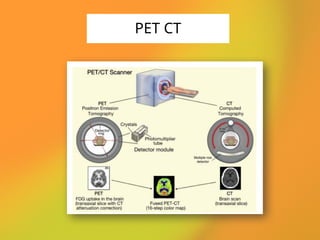
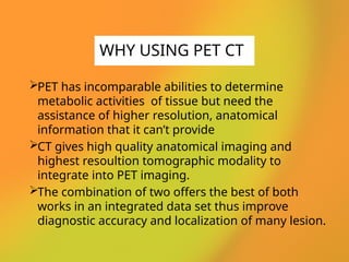
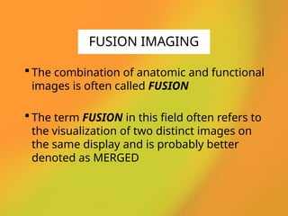
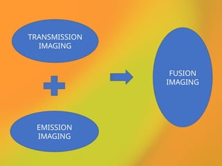
RADIOLOGY IN THE NUTSHELL
TOPIC – PET CT
LECTURE BY – NABANITA DAS
• PET CT stands for Positron Emission
Tomography Computed Tomography
• It is a medical imaging technique using a device
combines in a single gantry system both PET and
CT
• It gives high accuracy than PET and CT alone
• PET CT is the combination of functional and
anatomical imaging.
Note: It will decrease radiation dose and cost
effective.
INTRODUCTION
@radiology_in_thenutshell
PET CT DISCOVERY
@radiology_in_thenutshell
⚫The first PET-CT system constructed by David
Townsend(at the University of Geneva) and
Ronald Nutt (at CPS Innovations in Knoxville,
TN) with help from colleagues.
⚫The combinations of PET and CT scanners was
first suggested by R. Raylman in his 1991 Ph.D.
thesis.
PET stands for Positron Emission Tomography.
A nuclear medical imaging technique which
uses a small amount of radioactive compound
(tracers) to help in the diagnosis of disease.
CT stands for Computed Tomography.
It gives cross sectional image and accurate
diagnostic information about the distribution of
structure inside the body.
PET VS CT
PET CT
PET has incomparable abilities to determine
metabolic activities of tissue but need the
assistance of higher resolution, anatomical
information that it can’t provide
CT gives high quality anatomical imaging and
highest resoultion tomographic modality to
integrate into PET imaging.
The combination of two offers the best of both
works in an integrated data set thus improve
diagnostic accuracy and localization of many lesion.
WHY USING PET CT
 The combination of anatomic and functional
images is often called FUSION
 The term FUSION in this field often refers to
the visualization of two distinct images on
the same display and is probably better
denoted as MERGED
FUSION IMAGING
TRANSMISSION
IMAGING
EMISSION
IMAGING
FUSION
IMAGING
FUSION IMAGING
Consists of three main components
CT SCANNER
PET SCANNER
A COAXIAL BED
DESIGN OF PET CT
 The CT scan is in the first part of the scanner
The function and capability of the scanner are
similar to those of the stand-alone CT system
The only difference is that the gantry of the CT
scanner has no tilt capability.
Main application was to supplement the
functional information from PET with the
intrinsically associated anatomy as imaged by
CT
CT COMPONENT OF
PET CT
The PET scan is in the second part of the
scanner
PET subsystems offer detector rings
completely surrounding the patient.
The individual detector elements are
composed of scintillation crystals
connected to photomultiplier tubes
PET COMPONENT
OF PET CT
A single common table is for both the
scanner
60-70 cm distance between the two machine
PET/CT scanners feature a port diameter
of 70 cm & axial length of 100 cm, the
larger aperture also increases patient
comfort
A shared console for viewing the fused
image
COAXIAL TABLE
CROSS SECTION OF
PET CT
Fasting
• Overnight fasting is mandatory
Restrictions
• Avoid strenous activity
• Avoid intake flourine contain meal
• Avoid caffeine intake
Blood Glucose Level
• Should be 150mg/dl
PATIENT PREPARATION
The typical dose of FDG is 10 mCi injected
intravenously
Imaging is initiated approximately 60
minutes following the injection The patient
lie supine on the couch with arms above or
side of the body
A whole-body PET study (neck through
pelvis) follows an enhanced whole-body CT
study
CT scan takes 60 – 70 sec
PET scan takes 30 – 45 min depend on the
study
PROCEDURE
At first the CT scanner takes a topogram
 The topogram used to outline the region of the body
to be scanned
The CT generates 100s of trans axial image through
the body
The blue-purple rectangle represents CT coverage
during the study, and each overlapping green
rectangle represents PET coverage
Now the table moves to the PET scanner part of the
machine which began the detecting the radiation
emitted from patients performing the emission part
The patient is stepped through the scanner images a
swath of about 14-15 cm through the patient, and
generates about 30-40 contiguous trans axial image
CONTD
The enormous data is generated from the
scan
100s of trans axial PET & CT images are
firstly reconstructed
 The image is often reformatted into coronal
and saggital images
Finally a fusion image of PET and CT
combines and image is generated
IMAGE GENERATION
FLOW CHART OF
TYPICAL PET CT
OPERATION
THANK YOU
@radiology_in_thenutshell

PET CT (positron emission tomography-computed tomography scan)

  • 1.
    RADIOLOGY IN THENUTSHELL TOPIC – PET CT LECTURE BY – NABANITA DAS
  • 2.
    • PET CTstands for Positron Emission Tomography Computed Tomography • It is a medical imaging technique using a device combines in a single gantry system both PET and CT • It gives high accuracy than PET and CT alone • PET CT is the combination of functional and anatomical imaging. Note: It will decrease radiation dose and cost effective. INTRODUCTION @radiology_in_thenutshell
  • 3.
    PET CT DISCOVERY @radiology_in_thenutshell ⚫Thefirst PET-CT system constructed by David Townsend(at the University of Geneva) and Ronald Nutt (at CPS Innovations in Knoxville, TN) with help from colleagues. ⚫The combinations of PET and CT scanners was first suggested by R. Raylman in his 1991 Ph.D. thesis.
  • 4.
    PET stands forPositron Emission Tomography. A nuclear medical imaging technique which uses a small amount of radioactive compound (tracers) to help in the diagnosis of disease. CT stands for Computed Tomography. It gives cross sectional image and accurate diagnostic information about the distribution of structure inside the body. PET VS CT
  • 5.
  • 6.
    PET has incomparableabilities to determine metabolic activities of tissue but need the assistance of higher resolution, anatomical information that it can’t provide CT gives high quality anatomical imaging and highest resoultion tomographic modality to integrate into PET imaging. The combination of two offers the best of both works in an integrated data set thus improve diagnostic accuracy and localization of many lesion. WHY USING PET CT
  • 7.
     The combinationof anatomic and functional images is often called FUSION  The term FUSION in this field often refers to the visualization of two distinct images on the same display and is probably better denoted as MERGED FUSION IMAGING
  • 8.
  • 9.
  • 10.
    Consists of threemain components CT SCANNER PET SCANNER A COAXIAL BED DESIGN OF PET CT
  • 11.
     The CTscan is in the first part of the scanner The function and capability of the scanner are similar to those of the stand-alone CT system The only difference is that the gantry of the CT scanner has no tilt capability. Main application was to supplement the functional information from PET with the intrinsically associated anatomy as imaged by CT CT COMPONENT OF PET CT
  • 12.
    The PET scanis in the second part of the scanner PET subsystems offer detector rings completely surrounding the patient. The individual detector elements are composed of scintillation crystals connected to photomultiplier tubes PET COMPONENT OF PET CT
  • 13.
    A single commontable is for both the scanner 60-70 cm distance between the two machine PET/CT scanners feature a port diameter of 70 cm & axial length of 100 cm, the larger aperture also increases patient comfort A shared console for viewing the fused image COAXIAL TABLE
  • 14.
  • 15.
    Fasting • Overnight fastingis mandatory Restrictions • Avoid strenous activity • Avoid intake flourine contain meal • Avoid caffeine intake Blood Glucose Level • Should be 150mg/dl PATIENT PREPARATION
  • 16.
    The typical doseof FDG is 10 mCi injected intravenously Imaging is initiated approximately 60 minutes following the injection The patient lie supine on the couch with arms above or side of the body A whole-body PET study (neck through pelvis) follows an enhanced whole-body CT study CT scan takes 60 – 70 sec PET scan takes 30 – 45 min depend on the study PROCEDURE
  • 17.
    At first theCT scanner takes a topogram  The topogram used to outline the region of the body to be scanned The CT generates 100s of trans axial image through the body The blue-purple rectangle represents CT coverage during the study, and each overlapping green rectangle represents PET coverage Now the table moves to the PET scanner part of the machine which began the detecting the radiation emitted from patients performing the emission part The patient is stepped through the scanner images a swath of about 14-15 cm through the patient, and generates about 30-40 contiguous trans axial image CONTD
  • 18.
    The enormous datais generated from the scan 100s of trans axial PET & CT images are firstly reconstructed  The image is often reformatted into coronal and saggital images Finally a fusion image of PET and CT combines and image is generated IMAGE GENERATION
  • 19.
    FLOW CHART OF TYPICALPET CT OPERATION
  • 20.