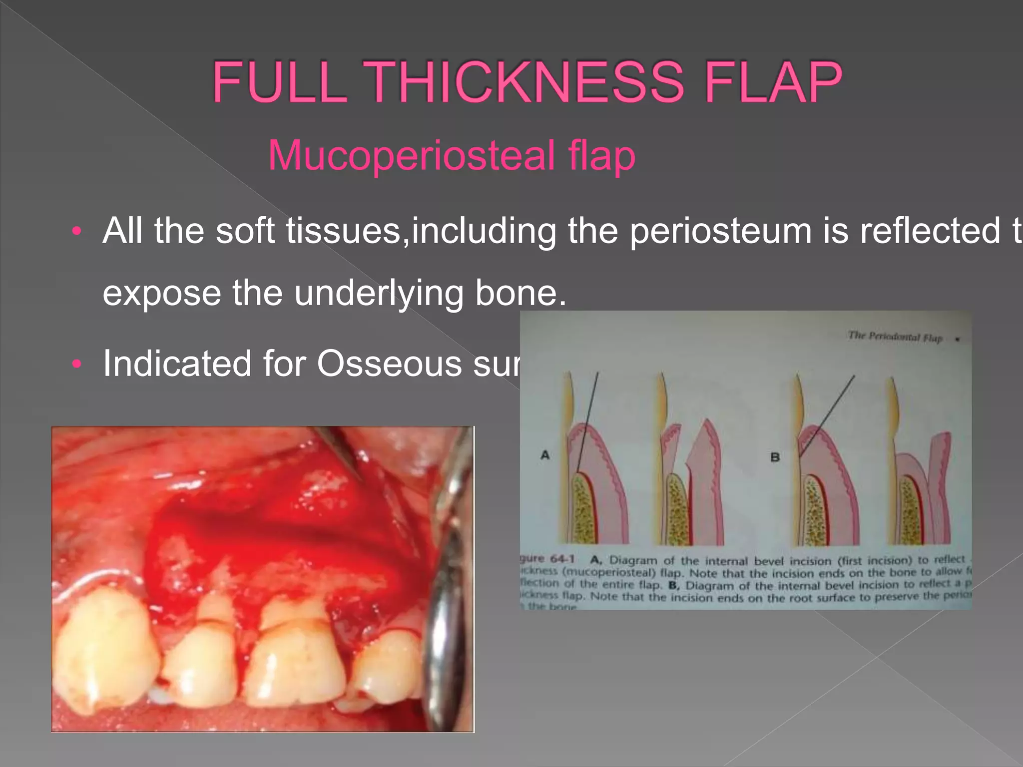
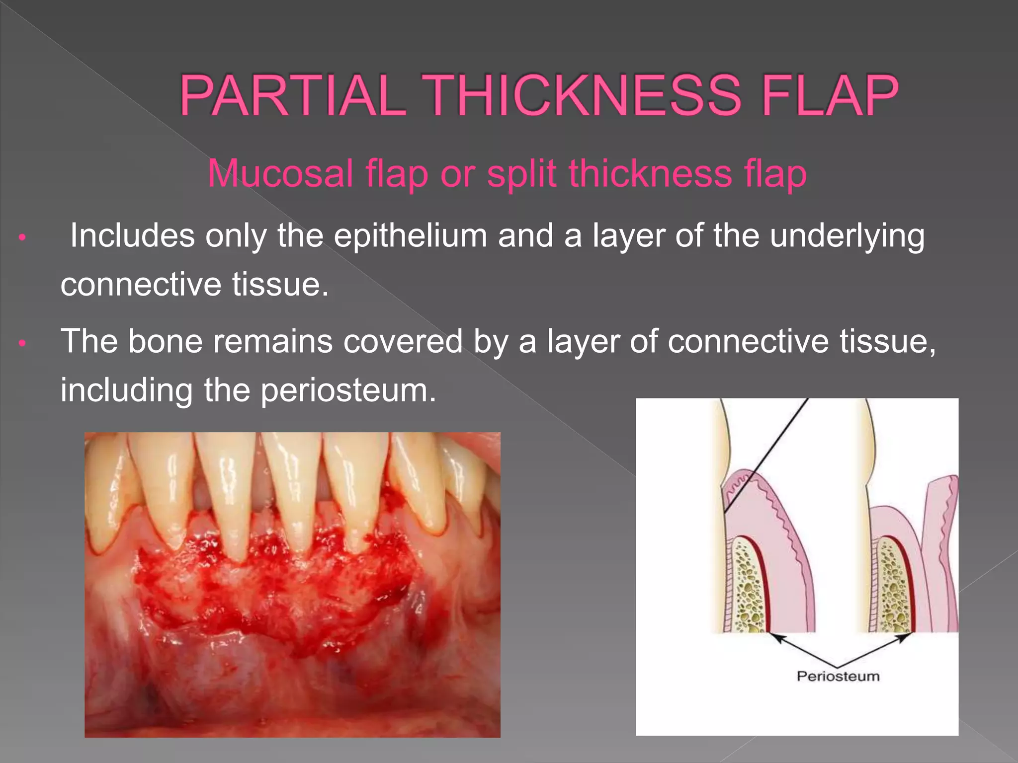
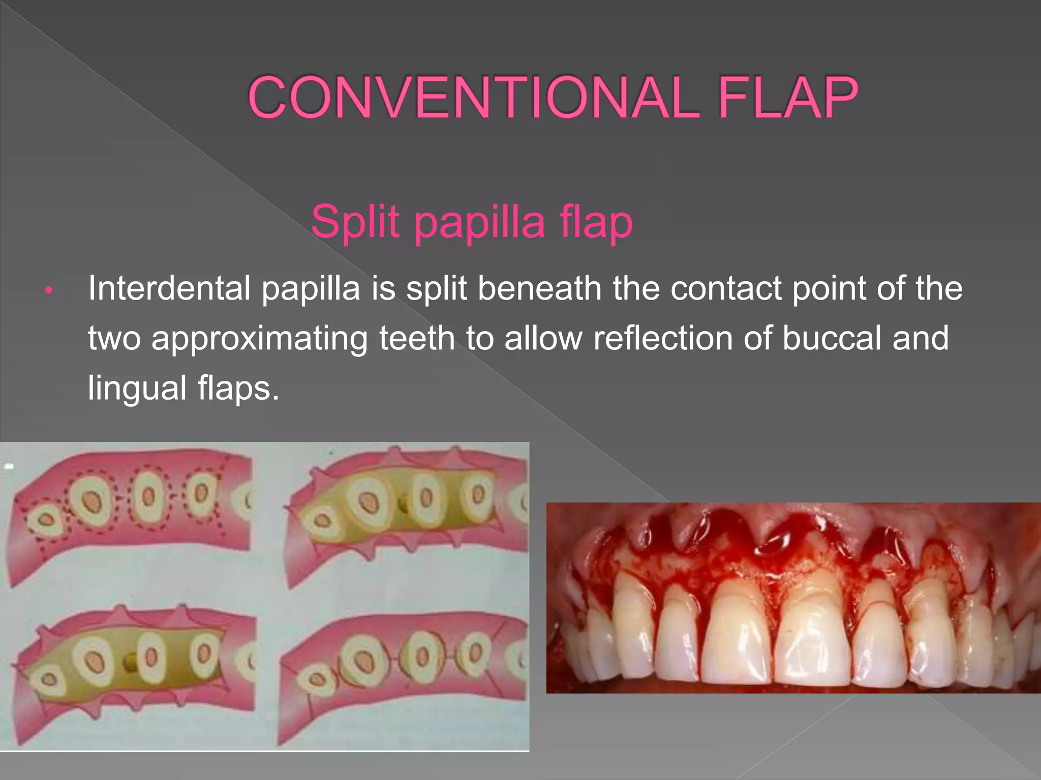
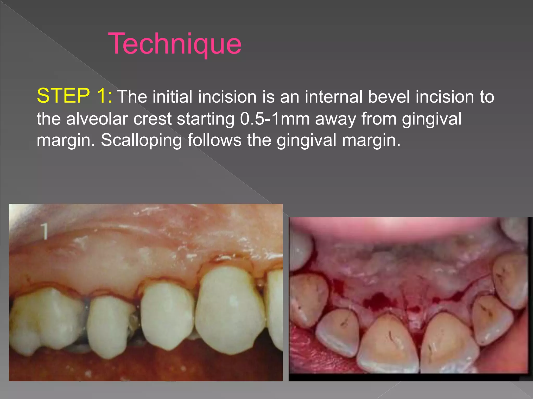
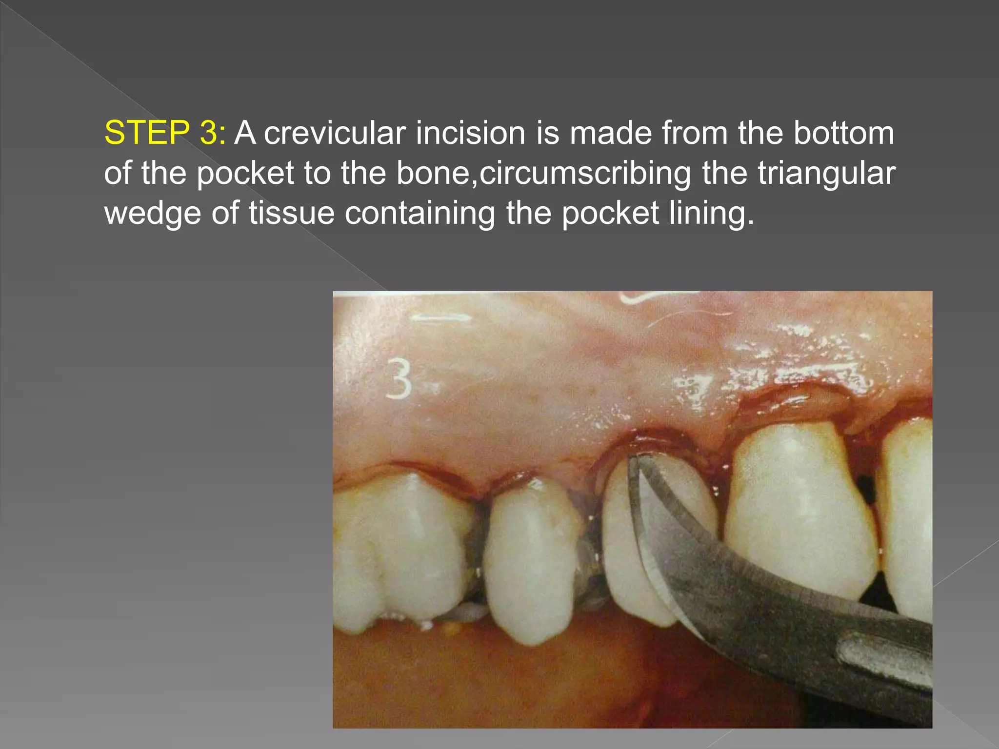
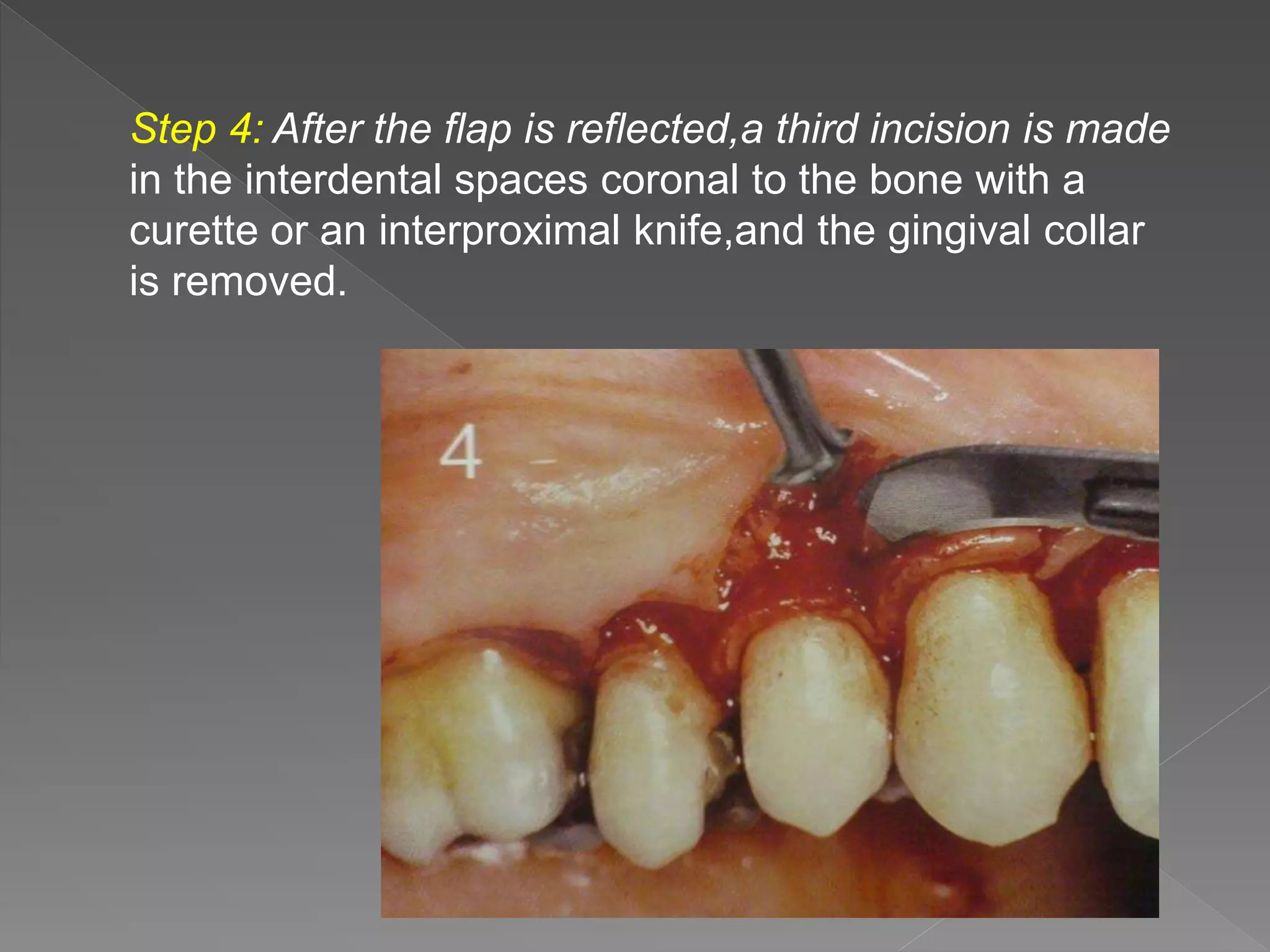
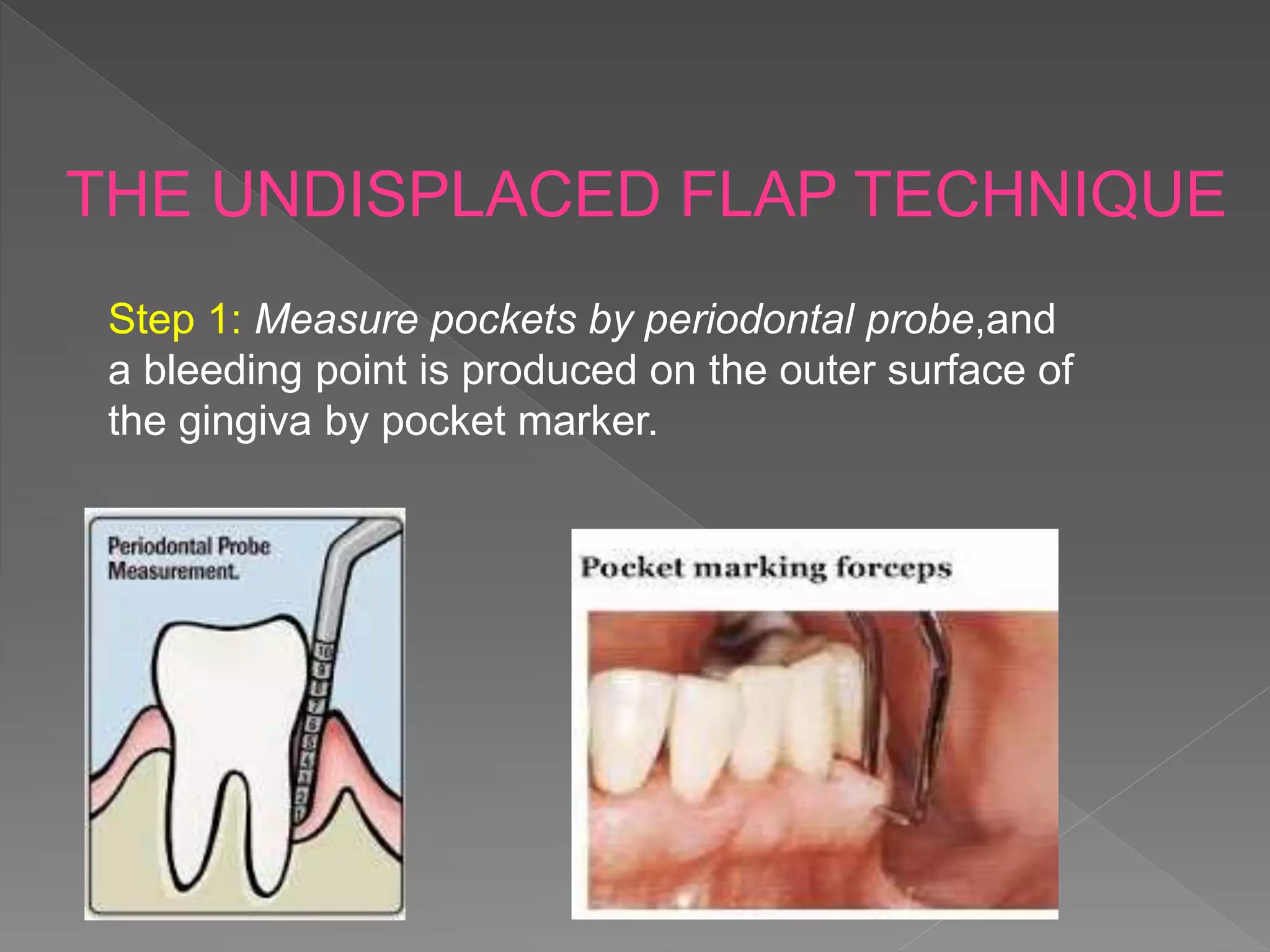
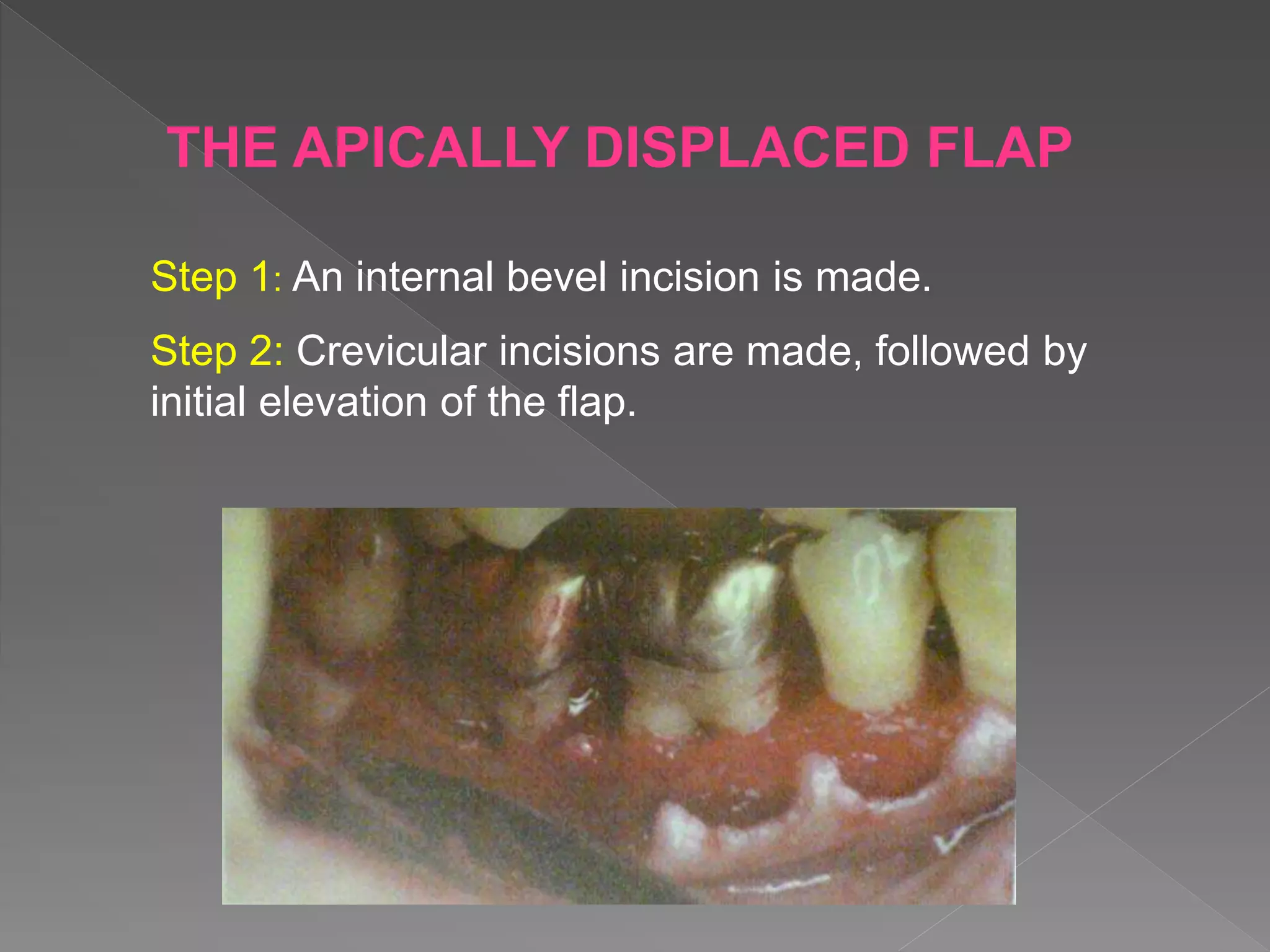
DEFINATION
INDICATIONS AND CONTRAINDICATIONS
CLASSIFICATION
PLANNING FOR FLAP SURGERY
INCISIONS IN FLAP SURGERY
ELEVATION OF FLAP SURGERY
HEALING AFTER FLAP SURGERY
FLAP TECHNIQUES
 Definition
It is a section of gingiva and/or
mucosa surgically separated from the
underlying tissues to provide visibility of
and access to the bone and root surface.
INDICATIONS
• Irregular bony contours.
• Pockets on teeth in which a complete removal
of root irritants is not clinically possible.
• Grade II or III furcation involvement.
• Root resection / hemisection.
• Infrabony pockets on distal areas of
second molars.
CONTRAINDICATIONS
• Poor plaque control
• High caries rate
• Unrealistic patient expectations or desires
• Uncontrolled medical conditions such as
‐unstable angina
‐uncontrolled diabetes
‐uncontrolled hypertension
‐myocardial infarction / stroke within 6 months.
• Based on bone exposure after reflection
1. Full thickness(mucoperiosteal)flap
2. Partial thickness(split thickness)flap
• Placement of flap after surgery
1. Non-displaced flap
2. Displaced flap
a)Apical displaced flaps
b)Coronal displaced flaps
c)Lateral displaced flaps
• Management of papilla
1. Conventional flap
2. Papilla preservation flap
Mucoperiosteal flap
• All the soft tissues,including the periosteum is reflected to
expose the underlying bone.
• Indicated for Osseous surgery.
Mucosal flap or split thickness flap
• Includes only the epithelium and a layer of the underlying
connective tissue.
• The bone remains covered by a layer of connective tissue,
including the periosteum.
Flap is returned and Sutured in its original
position
• Currently,It is the most commonly performed type of
periodontal surgery.
• Soft tissue pocket wall is removed with the initial
incision.
• Thus,it may be considered an internal bevel
Gingivectomy.
They are placed apically, coronally,or laterally to
their original position
• Both full thickness and partial thickness flaps
can be displaced
• The attached gingiva has to be totally separated from the
underlying bone,thereby enabling the unattached portion of
the gingiva to be movable.
• Dictated by the surgical judgement of the
operator and may depend on the objectives
of the operation.
• Two basic flap designs are used.
 Conventional flap
 Papilla preservation flap
Split papilla flap
• Interdental papilla is split beneath the contact point of the
two approximating teeth to allow reflection of buccal and
lingual flaps.
• Incorporates the entire papilla in one of the flaps by means of
crevicular interdental incisions to serve the connective tissue
attachment.
• Horizontal incision at the base of the papilla.
 Should be done in detail before procedure
› Based on clinical & radiographic findings of case
› It should include the following
 Type of flap
 Location and type of incisions
 Management of underlying bone
 Final placement of flap
 Sutures used
› Plan may be modified in case of any variations
 Horizontal incisions
Types of incisions
 Internal bevel incision
 Crevicular incision
 Interdental incision
 Vertical incision
Initial or first incision
Reverse bevel incision.
 Direction of incision
• 2-3mm from gingival margin
• Aimed at Crest of alveolar
bone and Apical to crest of
bone
• BP blade #11 or #15 used
• Basic to flap surgery
Exposure of root and
underlying bone
• Removes pocket lining
• Conserves uninvolved
outer gingiva
• Produces a sharp, thin
flap margin
• Places the connective
tissue close to the root.
Second incision
Made from base of the sulcus
to the crest of bone
• Forms “V” shaped
wedge of tissue,contains
Infected granulation
tissue,Junctional
epithelium &
Supracrestal fibers.
• BP blade #12 used.
Third incision
• A periosteal elevator is inserted into
the initial internal bevel incision, and
the flap is separated from the bone.
• It separates the collar of gingiva that is
left around the tooth.
• The Orban knife is usually used for
this incision.
Oblique or releasing incision
• Depend upon flap design and purpose.
• Can be used on one or both sides of the flap.
• Flap without vertical incision is called as
“Envelop Flap”.
• Areas of incision
At line angles of papilla
 Full thickness flap
Also called as blunt
dissection.
• With periosteal elevator
moved in mesial, distal
and apical direction
• Optimum bone exposure
achieved.
Types
Full thickness flap
Partial thickness flap
 0-24 hours
› Clot connects flap and tooth/bone surface
› Contains fibrin network of
 PMNLs and erythrocytes
 Cell debris
 Capillaries at wound edge
› Exudate results
 1-3 days
› Space between bone/tooth and flap is thinner
› Epithelial cells migrate over wound edge
› Minimal inflammatory response seen
 1 week
› Epithelial attachment is established by
 Hemidesmosomes and basal lamina
› Blood clot replaced by granulation tissue from
 Gingival connective tissue; bone marrow and PDL
 2 weeks
› Collagen fibers parallel to tooth surface
› Clinically normal but immature junction
 4 weeks
› Fully epithelized sulcus; well defined attachment
› Functional orientation of supra-crestal fibers
• The Modified Widman flap
• The Undisplaced flap
• The Apically displaced flap
• Flaps for regenerative surgery
The Papilla preservation flap
Conventional flap
• Distal molar surgery
FLAP TECHNIQUES
THE MODIFIED WIDMAN FLAP
Ramfjord and Nissle(1974):
They presented Modified Widman Flap.
Technique
STEP 1: The initial incision is an internal bevel incision to
the alveolar crest starting 0.5-1mm away from gingival
margin. Scalloping follows the gingival margin.
STEP 2: The gingival is reflected with a periosteal
elevator.
STEP 3: A crevicular incision is made from the bottom
of the pocket to the bone,circumscribing the triangular
wedge of tissue containing the pocket lining.
Step 4: After the flap is reflected,a third incision is made
in the interdental spaces coronal to the bone with a
curette or an interproximal knife,and the gingival collar
is removed.
Step 5: Tissue tags and granulation tissue are
removed with a curette.
Step 6: Bone architecture is not corrected except if it
prevents good tissue adaptation to the necks of the
teeth.
Adapt the facial and lingual interproximal
tissue adjacent to each other for no interproximal bone
remains exposed at the time of suturing.
Step 7: Interrupted direct sutures are placed in
each interdental space and covered with
tetracycline ointment and with a periodontal
surgical pack.
THE UNDISPLACED FLAP TECHNIQUE
Step 1: Measure pockets by periodontal probe,and
a bleeding point is produced on the outer surface of
the gingiva by pocket marker.
Step 2: The initial, internal bevel incision is made after
the scalloping of the bleeding marks on
the gingiva.
Step 3: Crevicular incision is made from the bottom of the
pocket to the bone to detach the connective tissue from
the bone.
Step 4: The flap is reflected with a periosteal elevator (blunt
dissection) from the internal bevel incision.
Step 5: The interdental incision is made with an
interdental knife.
Step 6: The triangular wedge of tissue is removed with
curette.
Step 7: The area is debrided,removing all tissue tags
and granulation tissue using sharp curette.
Step 8: After the necessary scaling and root planing.
Step 9: Flaps are placed in their original site and sutured.
THE APICALLY DISPLACED FLAP
Step 1: An internal bevel incision is made.
Step 2: Crevicular incisions are made, followed by
initial elevation of the flap.
Step 3: Vertical incisions are made extending beyond
the mucogingival junction.
Step 4: After removal of all granulation tissue, scaling
and root planing, and osseous surgery if needed, the
flap is displaced apically.
Step 5: Suture around the tooth prevents the flap from
sliding to a position more apical.
After 1 week, dressings and sutures are removed. The
area is usually repacked for another week.
FLAPS FOR REGENERATIVE SURGERY
1)The Papilla Preservation Flap
Step 1: A crevicular incision is made with no
incisions across the interdental papilla.
Step 2: The preserved papilla can be incorporated into
the facial or lingual/palatal flap.
Step 3: An Orban knife is used in interdental papilla.
Step 4: The flap is reflected.
2)Conventional Flap
Step 1: Using a #12 blade,incise the tissue at the
bottom of the pocket and to the crest of the bone.
Step 2: Reflect the flap maintaining it as thick as
possible.
Complicated by the presence of bulbous fibrous tissue
over the maxillary tuberosity or prominent retromolar
pads in the mandible.
This surgery was described by “Robinson and Braden”.
DISTAL MOLAR SURGERY
Maxillary Molars
• The treatment of distal pockets on the maxillary arch is
usually more simple than the treatment of a similar lesion
on the mandibular arch.
• Tuberosity presents a greater amount of fibrous
attached gingiva.
Technique: Two parallel incisions.
Mandibular Molars
• Does not usually present as much fibrous attached
gingiva.
• The two incisions distal to the molar area with the
greatest amount of attached gingiva.
• Incisions directed distolingually or distofacially
• Incision designs for surgical procedures distal to the
mandibular second molar.
Periodontal flap

Periodontal flap

  • 2.
    DEFINATION INDICATIONS AND CONTRAINDICATIONS CLASSIFICATION PLANNINGFOR FLAP SURGERY INCISIONS IN FLAP SURGERY ELEVATION OF FLAP SURGERY HEALING AFTER FLAP SURGERY FLAP TECHNIQUES
  • 3.
     Definition It isa section of gingiva and/or mucosa surgically separated from the underlying tissues to provide visibility of and access to the bone and root surface.
  • 4.
    INDICATIONS • Irregular bonycontours. • Pockets on teeth in which a complete removal of root irritants is not clinically possible. • Grade II or III furcation involvement. • Root resection / hemisection. • Infrabony pockets on distal areas of second molars.
  • 5.
    CONTRAINDICATIONS • Poor plaquecontrol • High caries rate • Unrealistic patient expectations or desires • Uncontrolled medical conditions such as ‐unstable angina ‐uncontrolled diabetes ‐uncontrolled hypertension ‐myocardial infarction / stroke within 6 months.
  • 6.
    • Based onbone exposure after reflection 1. Full thickness(mucoperiosteal)flap 2. Partial thickness(split thickness)flap • Placement of flap after surgery 1. Non-displaced flap 2. Displaced flap a)Apical displaced flaps b)Coronal displaced flaps c)Lateral displaced flaps • Management of papilla 1. Conventional flap 2. Papilla preservation flap
  • 7.
    Mucoperiosteal flap • Allthe soft tissues,including the periosteum is reflected to expose the underlying bone. • Indicated for Osseous surgery.
  • 8.
    Mucosal flap orsplit thickness flap • Includes only the epithelium and a layer of the underlying connective tissue. • The bone remains covered by a layer of connective tissue, including the periosteum.
  • 9.
    Flap is returnedand Sutured in its original position • Currently,It is the most commonly performed type of periodontal surgery. • Soft tissue pocket wall is removed with the initial incision. • Thus,it may be considered an internal bevel Gingivectomy.
  • 10.
    They are placedapically, coronally,or laterally to their original position • Both full thickness and partial thickness flaps can be displaced • The attached gingiva has to be totally separated from the underlying bone,thereby enabling the unattached portion of the gingiva to be movable.
  • 11.
    • Dictated bythe surgical judgement of the operator and may depend on the objectives of the operation. • Two basic flap designs are used.  Conventional flap  Papilla preservation flap
  • 12.
    Split papilla flap •Interdental papilla is split beneath the contact point of the two approximating teeth to allow reflection of buccal and lingual flaps.
  • 13.
    • Incorporates theentire papilla in one of the flaps by means of crevicular interdental incisions to serve the connective tissue attachment. • Horizontal incision at the base of the papilla.
  • 14.
     Should bedone in detail before procedure › Based on clinical & radiographic findings of case › It should include the following  Type of flap  Location and type of incisions  Management of underlying bone  Final placement of flap  Sutures used › Plan may be modified in case of any variations
  • 15.
     Horizontal incisions Typesof incisions  Internal bevel incision  Crevicular incision  Interdental incision  Vertical incision
  • 16.
    Initial or firstincision Reverse bevel incision.  Direction of incision • 2-3mm from gingival margin • Aimed at Crest of alveolar bone and Apical to crest of bone • BP blade #11 or #15 used
  • 17.
    • Basic toflap surgery Exposure of root and underlying bone • Removes pocket lining • Conserves uninvolved outer gingiva • Produces a sharp, thin flap margin • Places the connective tissue close to the root.
  • 18.
    Second incision Made frombase of the sulcus to the crest of bone • Forms “V” shaped wedge of tissue,contains Infected granulation tissue,Junctional epithelium & Supracrestal fibers. • BP blade #12 used.
  • 19.
    Third incision • Aperiosteal elevator is inserted into the initial internal bevel incision, and the flap is separated from the bone. • It separates the collar of gingiva that is left around the tooth. • The Orban knife is usually used for this incision.
  • 20.
    Oblique or releasingincision • Depend upon flap design and purpose. • Can be used on one or both sides of the flap. • Flap without vertical incision is called as “Envelop Flap”.
  • 21.
    • Areas ofincision At line angles of papilla
  • 22.
     Full thicknessflap Also called as blunt dissection. • With periosteal elevator moved in mesial, distal and apical direction • Optimum bone exposure achieved. Types Full thickness flap Partial thickness flap
  • 23.
     0-24 hours ›Clot connects flap and tooth/bone surface › Contains fibrin network of  PMNLs and erythrocytes  Cell debris  Capillaries at wound edge › Exudate results  1-3 days › Space between bone/tooth and flap is thinner › Epithelial cells migrate over wound edge › Minimal inflammatory response seen
  • 24.
     1 week ›Epithelial attachment is established by  Hemidesmosomes and basal lamina › Blood clot replaced by granulation tissue from  Gingival connective tissue; bone marrow and PDL  2 weeks › Collagen fibers parallel to tooth surface › Clinically normal but immature junction  4 weeks › Fully epithelized sulcus; well defined attachment › Functional orientation of supra-crestal fibers
  • 25.
    • The ModifiedWidman flap • The Undisplaced flap • The Apically displaced flap • Flaps for regenerative surgery The Papilla preservation flap Conventional flap • Distal molar surgery FLAP TECHNIQUES
  • 26.
    THE MODIFIED WIDMANFLAP Ramfjord and Nissle(1974): They presented Modified Widman Flap.
  • 27.
    Technique STEP 1: Theinitial incision is an internal bevel incision to the alveolar crest starting 0.5-1mm away from gingival margin. Scalloping follows the gingival margin.
  • 28.
    STEP 2: Thegingival is reflected with a periosteal elevator.
  • 29.
    STEP 3: Acrevicular incision is made from the bottom of the pocket to the bone,circumscribing the triangular wedge of tissue containing the pocket lining.
  • 30.
    Step 4: Afterthe flap is reflected,a third incision is made in the interdental spaces coronal to the bone with a curette or an interproximal knife,and the gingival collar is removed.
  • 31.
    Step 5: Tissuetags and granulation tissue are removed with a curette.
  • 32.
    Step 6: Bonearchitecture is not corrected except if it prevents good tissue adaptation to the necks of the teeth. Adapt the facial and lingual interproximal tissue adjacent to each other for no interproximal bone remains exposed at the time of suturing.
  • 33.
    Step 7: Interrupteddirect sutures are placed in each interdental space and covered with tetracycline ointment and with a periodontal surgical pack.
  • 34.
    THE UNDISPLACED FLAPTECHNIQUE Step 1: Measure pockets by periodontal probe,and a bleeding point is produced on the outer surface of the gingiva by pocket marker.
  • 35.
    Step 2: Theinitial, internal bevel incision is made after the scalloping of the bleeding marks on the gingiva.
  • 36.
    Step 3: Crevicularincision is made from the bottom of the pocket to the bone to detach the connective tissue from the bone.
  • 37.
    Step 4: Theflap is reflected with a periosteal elevator (blunt dissection) from the internal bevel incision. Step 5: The interdental incision is made with an interdental knife.
  • 38.
    Step 6: Thetriangular wedge of tissue is removed with curette. Step 7: The area is debrided,removing all tissue tags and granulation tissue using sharp curette.
  • 39.
    Step 8: Afterthe necessary scaling and root planing. Step 9: Flaps are placed in their original site and sutured.
  • 40.
    THE APICALLY DISPLACEDFLAP Step 1: An internal bevel incision is made. Step 2: Crevicular incisions are made, followed by initial elevation of the flap.
  • 41.
    Step 3: Verticalincisions are made extending beyond the mucogingival junction. Step 4: After removal of all granulation tissue, scaling and root planing, and osseous surgery if needed, the flap is displaced apically.
  • 42.
    Step 5: Suturearound the tooth prevents the flap from sliding to a position more apical. After 1 week, dressings and sutures are removed. The area is usually repacked for another week.
  • 43.
    FLAPS FOR REGENERATIVESURGERY 1)The Papilla Preservation Flap Step 1: A crevicular incision is made with no incisions across the interdental papilla.
  • 44.
    Step 2: Thepreserved papilla can be incorporated into the facial or lingual/palatal flap. Step 3: An Orban knife is used in interdental papilla. Step 4: The flap is reflected.
  • 45.
    2)Conventional Flap Step 1:Using a #12 blade,incise the tissue at the bottom of the pocket and to the crest of the bone. Step 2: Reflect the flap maintaining it as thick as possible.
  • 46.
    Complicated by thepresence of bulbous fibrous tissue over the maxillary tuberosity or prominent retromolar pads in the mandible. This surgery was described by “Robinson and Braden”. DISTAL MOLAR SURGERY
  • 47.
    Maxillary Molars • Thetreatment of distal pockets on the maxillary arch is usually more simple than the treatment of a similar lesion on the mandibular arch. • Tuberosity presents a greater amount of fibrous attached gingiva. Technique: Two parallel incisions.
  • 48.
    Mandibular Molars • Doesnot usually present as much fibrous attached gingiva. • The two incisions distal to the molar area with the greatest amount of attached gingiva. • Incisions directed distolingually or distofacially
  • 49.
    • Incision designsfor surgical procedures distal to the mandibular second molar.