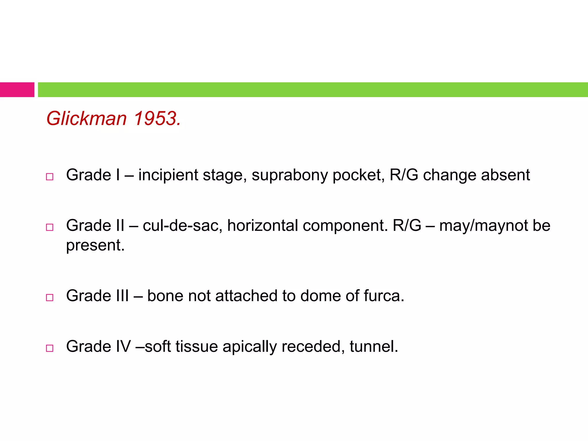
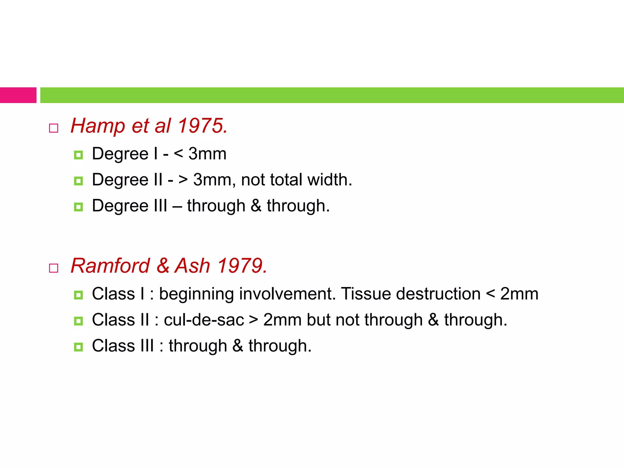
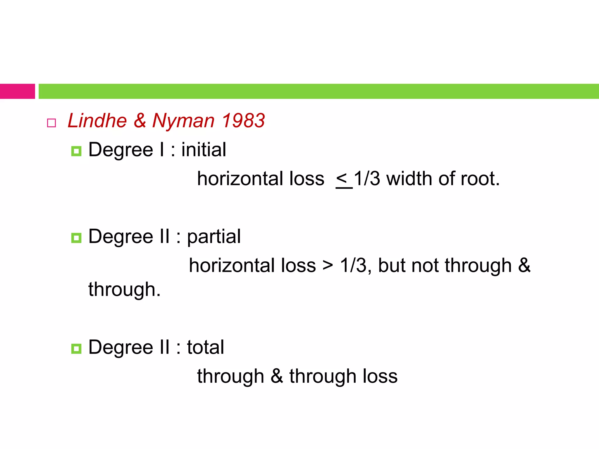
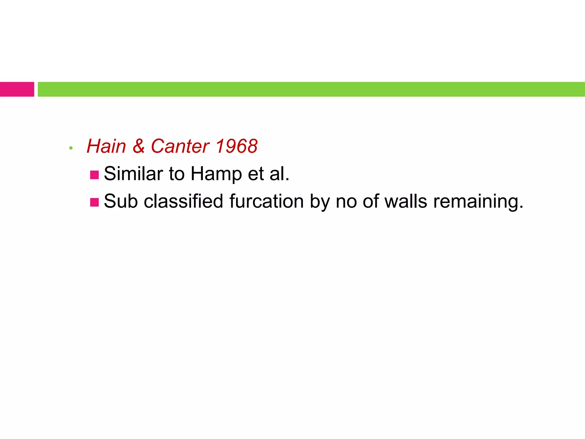
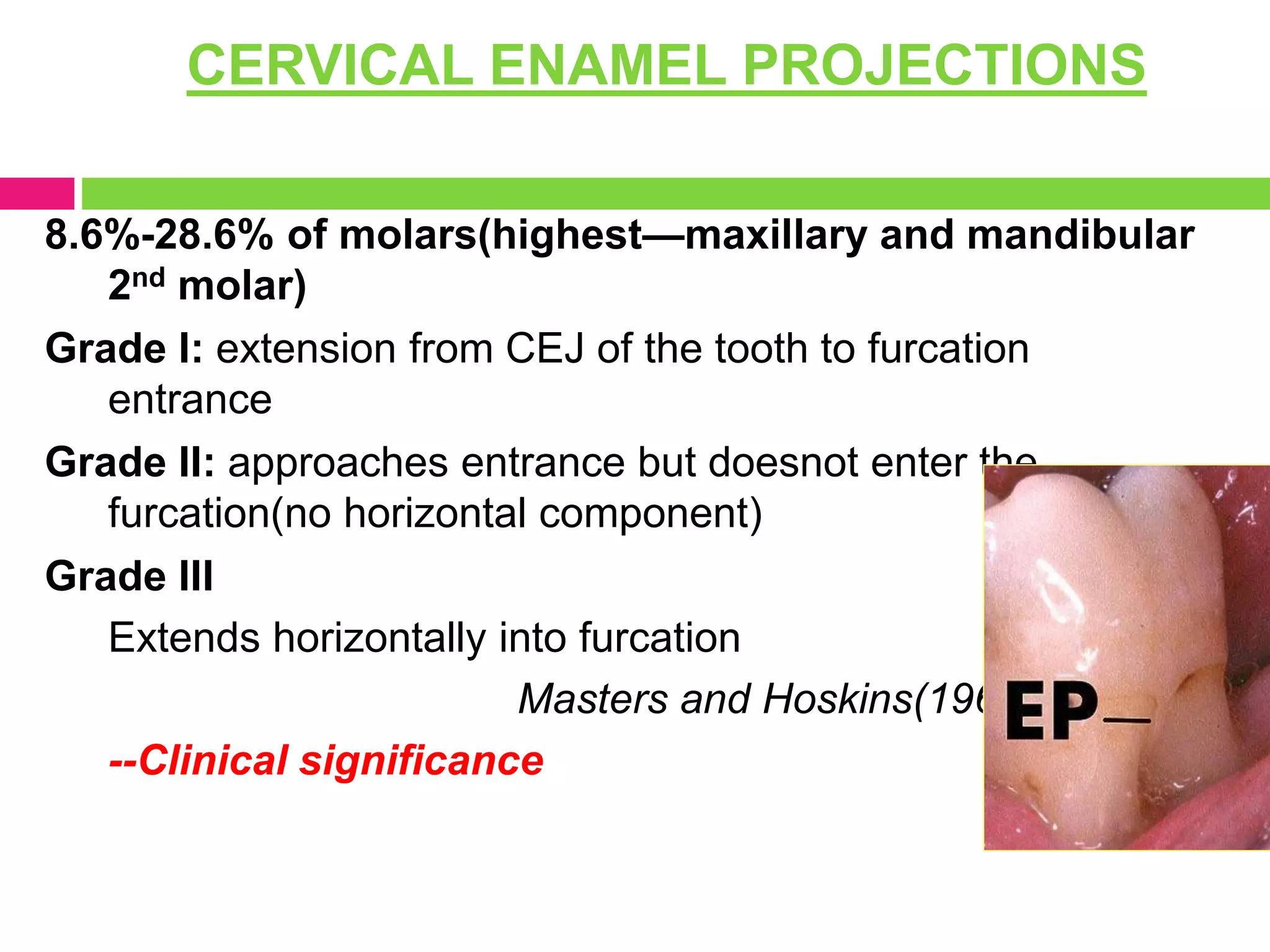
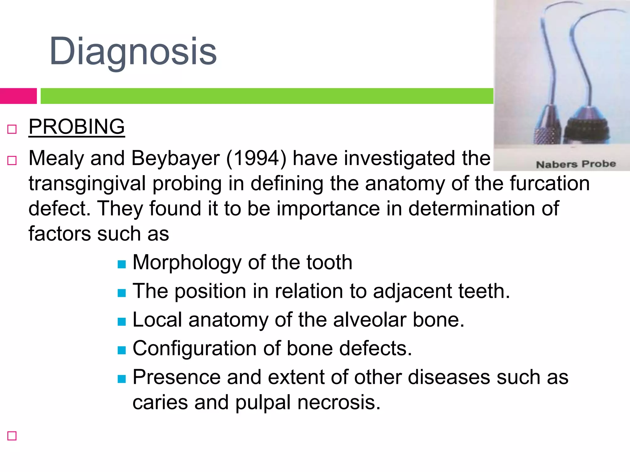
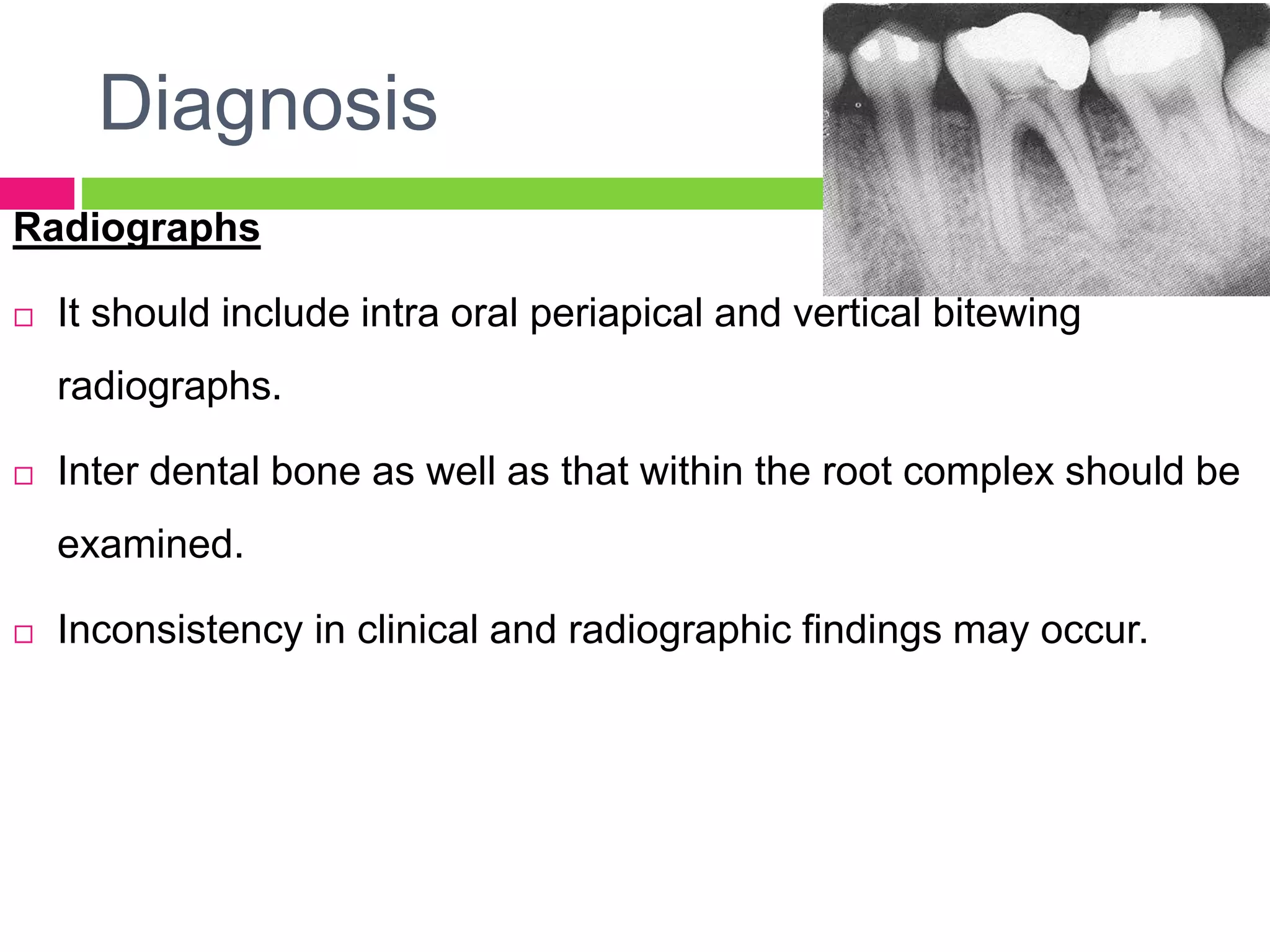
3. Introduction
Furcation is an area of complex anatomic morphology,
that once involved is difficult to debride by routine
periodontal instrumentation.
4. Furcation – anatomic area of a multirooted tooth where the
roots diverge.
AAP
1992
Furcation invasion – pathologic resorption of bone within
the furcation.
AAP 1992
Furcation involvement – invasion of the bifurcations/
trifurcations of multirooted tooth by periodontal disease.
8. Glickman 1953.
Grade I – incipient stage, suprabony pocket, R/G change absent
Grade II – cul-de-sac, horizontal component. R/G – may/maynot be
present.
Grade III – bone not attached to dome of furca.
Grade IV –soft tissue apically receded, tunnel.
Classification
10. Title
Goldman 1958
Grade I : incipient.
Grade II : cul-de-sac.
Grade III : through & through.
11. Hamp et al 1975.
Degree I - < 3mm
Degree II - > 3mm, not total width.
Degree III – through & through.
Ramford & Ash 1979.
Class I : beginning involvement. Tissue destruction < 2mm
Class II : cul-de-sac > 2mm but not through & through.
Class III : through & through.
Classification
12. Title
Lindhe & Nyman 1983
Degree I : initial
horizontal loss < 1/3 width of root.
Degree II : partial
horizontal loss > 1/3, but not through &
through.
Degree II : total
through & through loss
13. Title
Riochetti 1982
Class I : 1mm of horizontal measurement.
Class I a : 1-2 mm of horizontal invasion, earliest
damage.
Class II : 2-4 mm
Class II a : 4-6 mm
Class III : > 6mm
14. Title
Basaraba 1990
Class I : initial/incipient furcal invasion
Class II : partial/ patent furcal invasion
Class III : communicating furcal invasion.
15. Title
• Hain & Canter 1968
Similar to Hamp et al.
Sub classified furcation by no of walls remaining.
16. Title
Fedi 1985
Combined Glickman & Hamp classification.
Grade I to IV.
Grade II furcations subdivided
degree I (< 3mm) degree II (> 3mm)
17. Title
Vertical component
Tarnow & Fletcher 1984.
Subclass A – denotes furcation
involvements with vertical
bone loss of 3mm or less.
Subclass B – vertical bone
loss of 4-6 mm
Subclass C – bone loss from the
fornix of 7mm or more
18. Title
Eskow & Kapin 1984
Same classification as Tarnow & Fletcher, but thirds instead of
3mm units are used.
19. Title
Easley & Drenman1969
Based on the ledges & morphology of remaining
bone.
Class I : fluting coronal to furcation affected. no definite
horizontal component.
Class II : Type I – essentially horizontal, no
buccal/lingual ledge.
Type II - buccal/lingual ledge, definite vertical
component.
Class III : through & through.
20. Epidemiology
Incidence and Distribution
Lorato (1981) found that:
Average no. of furcation involvement increased with
age.
Furcation involvement most common in 1st permanent
molars
In maxillary molars, buccal aspects more often
invaded.
Maxillary premolars-Lower incidence of furcation.
22. Plaque associated Inflammation
Extension of inflammatory periodontal disease in
furcation area leads to inter-radicular bone resorption—
reduction of bone height and formation of furcation
defect.
Furcation involvement is a phase in root ward extension
of periodontal pocket(Glickman, 1950)
Cellular and fluid inflammatory exudation---epithelial
proliferation
24
23. Trauma from Occlusion
• Suspected etiologic/contributing factor in isolated
furcation defects—crater like or angular deformities in
bone---bone destruction is localized to one of the
roots--controversial
• Glickman(1950)
• Waerhaug (1980)
inflammation +oedema=extrude tooth
Traumatized and sensitive
25
25. 27
CERVICAL ENAMEL PROJECTIONS
8.6%-28.6% of molars(highest—maxillary and mandibular
2nd molar)
Grade I: extension from CEJ of the tooth to furcation
entrance
Grade II: approaches entrance but doesnot enter the
furcation(no horizontal component)
Grade III
Extends horizontally into furcation
Masters and Hoskins(1964)
--Clinical significance
26. 2. Root Trunk length:
•Teeth with shorter trunks are more prone for development of
furcation defects compared to ones with longer root trunk length.
3. Root length
•Determines the amount of attachment or support that a tooth
will have.
•Teeth with long root trunks and short roots would have lost
significant amount of support by the time the furcation is
affected.
27. 3. Root form:
Flutings on the root surface coupled with developmental
grooves and concavities-- plaque retentive areas hastening
the process of periodontal breakdown leading to early
furcation involvement.
4. Anatomy of the furcation
•Bifurcational ridges, concavity in the root and accessory
canals may jeopardize plaque control as well as treatment
outcome.
29. Enamel Pearls
Incidence: 1.1% - 9.7%
(moskow & canut et
al(1990)
In ,Maxillary 2nd molar -
found near the CEJ
extending into molar
bifurcations
Prevent connective
tissue attachment.
30. Diagnosis
PROBING
Mealy and Beybayer (1994) have investigated the role of
transgingival probing in defining the anatomy of the furcation
defect. They found it to be importance in determination of
factors such as
Morphology of the tooth
The position in relation to adjacent teeth.
Local anatomy of the alveolar bone.
Configuration of bone defects.
Presence and extent of other diseases such as
caries and pulpal necrosis.
31. Diagnosis
Probing of the furcation.
Instruments. : Nabers probe.
Buccal and lingual furcation can be easily
probed.
Proximal furcations are difficult for probing
particularly when broad contacts are
present n adjacent teeth.
In maxillary molars mesial furcation is
located more palatally than to the buccal
tooth surface. It should therefore be probed
from the palatal aspect.
32. Diagnosis
In contrast the distal furcation is located
midway bucco-lingually and can therefore
be probed either from the buccal or palatal
aspect
Furcation probing in maxillary premolar is
very difficult due to the presence of
anatomic variations such as longitudinal
furrows, invaginations opening at varying
distances from the CEJ.
33. Diagnosis
Radiographs
It should include intra oral periapical and vertical bitewing
radiographs.
Inter dental bone as well as that within the root complex should be
examined.
Inconsistency in clinical and radiographic findings may occur.
35. Differential Diagnosis
2. Trauma from occlusion
Increase occlusal forces may cause tissue destruction or adaptation
within the inter radicular area of multirooted tooth.
In such cases a radiolucency may be evident in the root complex and
tooth may exhibit increase mobility.
Probing however fails to detect any furcations involvement.
In such situations occlusal adjustment should precede periodontal
therapy.
If the defect is of occlusal origin the tooth becomes stabilized and the
defect disappears within weeks following occlusal correction.
36. Management
Objectives of furcation therapy
To facilitate maintenance.
To prevent further attachment loss.
Obliterate furcation defects as a periodontal
maintenance problem.
37. Factors to be considered when deciding for mode of
therapy
Degree of involvement
Crown-root ratio
Length of root
Degree of root separation
Strategic value of tooth
Root anatomy
Residual tooth mobility
Ability to eliminate the defect
Endodontic therapy & complications
Prosthetic requirements
Periodontal condition of adjacent teeth.
38. Can be broadly classified as put forth by Kalkwarf and
Reinhardt as follows. (1988)
Maintain the furcation
Increase the access to furcation
Removal of furcation
Closure of furcation with new
attachment.
39. Maintain the furcation
Hirschfeld and Wasserman evaluated 600 patients who
had been treated for periodontal disease and followed with
maintenance appointments every 4-6 months for at least
15 years.
Many furcations were primarily treated by sub gingival
scaling.
During the 22 year span of the study of the 1464 molars
involved 460 were lost
40. Maintain the furcation
Waerhaug has noted a close association of 0.91 mm
between the sub gingival plaque front and attachment
fibers in areas of attachment loss in furcations.
Some amount of plaque was always left behind when it
was present in the central area prior to treatment.
He concluded that subgingival plaque removal in the
furcation was invariably incomplete.
41. Maintain the furcation
Obliteration of the furcation: Baer et al (1983) proposed
the elimination of anatomic niches by filling advanced
furcation defects with biocompatible material.
Furcation areas of 50 maxillary teeth and 20 mandibular
molars were surgically exposed and 2 weeks later packed
with Intermediate Restorative Material (IRM).
Clinical success was reported for up to 5 years.
They concluded that IRM was physiologically acceptable to
gingival tissues, prevented caries and simplified plaque
control.
They also advised IRM placement in advancement
42. Increasing access to the furcation
Gingivectomy / Apically positioned flap.
Increases access for plaque
control and allows
resolution of
periodontal inflammation.
43. Odontoplasty
It is the reshaping of the tooth coronal to the furcation to
improve access for plaque control.
It increases entrance to the furca and reduces its horizontal
depth.
Mainly advised for Grade I and Grade II furcation defects.
Caution should be exercised with regard to
Hypersensitivity
Pulpal irritation leading to permanent damage
Pulp exposure
Increase risk of root caries.
44. b. Osteoplasty and Ostectomy:
Osteoplasty: Reshaping surfaces of bone without removing
tooth supporting bone
Ostectomy: Reshaping and removal of tooth supporting
bone.
Improved plaque control through osteoplasty is reported to
be accomplished by--
--Creating bony ramps into the furcation area allowing the
gingival to tuck into tooth concavities
--Removing lip of the bony defect to decrease horizontal depth
of the involvement
--Reducing pocket depth by allowing apical adaptation of the
flap.
45. Recommended for Grade I and II furcation involvements.
In advanced cases of Grade II and Grade III furcations
ostectomy may be extended into create a tunnel to expose
the entire furcation area.
47. Such treatment should be restricted to
Cases where other surgical procedures are contra indicated.
Roots are divergent to allow adequate postoperative plaque
control with inter proximal brushes
Patient has demonstrated a high level of plaque control in the
past.
Hamp et al 1975—5 year clinical trial(7 teeth)—4/7 caries
Helden et al 1989– 149 teeth(mean of 37.5 months)---75%
functional
48. Grant and Stern advocate that such procedures often
result in a reverse architerature, which may encourage
further plaque accumulation.
The main advantage of this technique
-- avoidance of prosthetic reconstruction and endodontic
therapy
Anatomical consideration--maxillary
49. III. Elimination of the furcation:
Terminology
Tooth sectioning
Root resection
Hemisection /Root
separation/bicuspidization
Root amputation
50. III. Elimination of the furcation:
Root resection / Hemisection
Objectives
To resect the open root furcation area and make possible
debridement of the residual root.
To eliminate the periodontal pocket by removal of the
furcation.
To improve the furcation form for dental hygiene.
To preserve maximum periodontal tissue to the residual root.
To control inter dental sparse (embrasure) in the area
adjacent to the root.
To treat teeth with severe caries.
51. Indications for root resection and hemisection.
--Advanced furcation involvement Grade II and Grade III is
an absolute indication for root resection or hemisection.
52. Contra indications
Insufficient periodontal support to save the roots
Fused roots
Extremely long root trunks.
In conditions where endodontic treatment of the
resected root is impossible.
In patients who are unable to establish appropriate
hygiene or where the strategic value of maintaining
dental arch continuity is lacking.
53. From a prognostic point of view the following factors should be
considered.
Periodontal considerations.
A long and wide root with a large crown is the ideal form
After root resection / hemisection the crater like osseous defect
around the residual root is removed by osseous resection and
the periodontal pocket eliminated.
A form that facilitates plaque control must be achieved.
Patient must be capable of following through oral hygiene and
keeping up the professional schedule.
58. Guided tissue regeneration in the treatment of degree III
furcation defects in maxillary molars
Pontoriero R, Lindhe T, JCP, 1995; 22:810-812.
11 subjects with generalized periodontitis and advanced lesions
in the maxillary molar regions, bilateral mesial distal, but not
buccal degree III furcation defects in 1st and 2nd molars
--some reduction in PD and some gain in CAL had occurred at
both test and control sites
--none of the furcation defects had closed, but retained the
characteristics of degree III furcation defects.
59. Guided Tissue Regeneration in the treatment of
degree II furcations in maxillary molars
Pontoriero R, Lindhe J JCP 1995;
22:756-763.
18 inter proximal (10M, 8 D) and 10 buccal pairs
Re-entry after 6 months.
The addition of GTR enhanced the result by promoting
probing attachment and bone gain and decreases soft
tissue recession on buccal furcation only
None seen on mesial / distal furcation.
60. GTR in degree II furcation involved mandibular molars.
Pontoriero R, Lindhe J JCP 1988: 15:247-254.
21 subjects
--more than 90% of the sites treated with GTR complete
resolution of the defect occurred.
--Conventional therapy attained the same result in < 20%
cases.
61.
62. EXTRACTION OF FURCATION
INVOLVED TEETH
when attachment loss is so extensive that no root can
be maintained or when treatment would result in a tooth
form where plaque control by the patient is difficult to
achieve.
when no improvement of the overall treatment plan is
expected or carious lesions or endodontic problems may
cause the preserved tooth to be a future risk with regard
to long-term prognosis.
63. I. Patient factors
Adequate plaque control is a must for successful outcome.
Cigarette smoking
Rosenberg (1994), Tonetti et al (1995) showed less attachment
gains in intra bony defects following GTR in smokers than non
smokers.
FACTORS AFFECTING CLINICAL
OUTCOME
64. II. Defect factors
Degree 1 and 2 buccal and lingual furcations of 1st and 2nd
mandibular molars respond well to GTR therapy.
However in maxillary molars location of the defect plays a critical
role as no improvement beyond that of debridement as obtained
in interproximal degree II defects compared to buccal defect
(Ponteriero et al 1995)
65. Direct correlation between initial depth of the defect and
gain in attachment of GTR is observed.
Based on present evidence successful outcome of GTR
treatment can be expected only in mandibular and maxillary
buccal degree II furcations.
66. III. Technical factors
A flap management technique that places wound margins
away from the entrance to the healing defects is essential for
+ve outcome of regenerative therapy of furcation defects.
Sander and Karring (1995) studied significance of bacterial
contamination in monkeys. The findings of this study showed
that new attachments and bone formation was favored
considerably if bacterial contamination of the membrane was
prevented during wound healing.
67. IV. Antibiotic therapy.
Nyland & Egelberg (1990) studied effect of tetracycline
irrigation of the site as compared to saline
One-year evaluation of attachmentlevels and pocket depths
showed similar clinically negligible (<1 mm) variation
68. Prognosis
In a 5-year study of Hamp et al (1975) 175 teeth with
various degree of furcation involvement in 100 patients
were noted.
Of the 175 teeth.
32 (18%) - SRP
49 (28%) - SRP + Furcationplasty
87 (50%) - Root resection.
7 (4%) – Tunnel preparation.
Patients were enrolled in a maintenance program of
periodic recall of 3-6 months.
Plaque and gingivitis scores measured ensured that oral
hygiene maintenance was of the highest quality.
None of the teeth were lost during 5 years of the study.
69. Prognosis
Key to long term success
--thorough diagnosis
--selection of patients with good oral hygiene
--careful surgical and restorative management
70. Conclusion
The presence of furcation involvement is one clinical
finding that can lead to a diagnosis of advanced
periodontitis and potentially to a less favorable prognosis
for the affected tooth or teeth.
Furcation involvement therefore presents both diagnostic
and therapeutic dilemmas.
71. References
Carranza’s clinical periodontology. 10th edition.
Clinical periodontology and implant dentistry. Jan
Lindhe. 5th edition.
Diagnosis and epidemiology of periodontal osseous
lesions. Periodontology 2000, vol 22, 2000.
The conservative approach in the treatment of furcation
lesions
Periodontology 2000, vol 22, 2000.
72. References
Management of furcation involvement
Periodontology 2000, vol. 9, 1995, 69-89
Current status for furcation involvements – dcna 35, no 3,
1991.
Molar root anatomy & management of furcation defects –
jcp 2001; 28;730.
Atlas ofcosmetic and reconstructive periodontal surgery
3rd edition, Edward Cohen
Root complex: Is the portion of a tooth that is located apical to the CEJ is the portion that is normally covered with a root cementum.
Root trunk: Represents the undivided region of the root. Height of the root trunk is defined as the distance between the CEJ and the separation line (furcation) between two root cones.
Root cone: it is included in the divided region of the root complex.
Degree of separation: it is the angle of separation between two root cones.
Divergence: it is the distance between two roots; this distance normally increases in apical direction.
Entrance: the transitional area between the undivided and the divided part of the root
Fornix: the roof of the furcation….81% orifice is less than 1mm or less and 58% 0.75mm or less bowers 1979
Most sensitive to injury from excessive occlusal forces
Affect plaque control removal,complicate srp,local factor for devt of gingivitis and pditis…shud b removed to facilitate maintenance
Closely approximated or fused roots…teeth wid widely separated roots..mor treatment options and readily treated
Careful probing is required to determine the presence and extent of furcation involvement, the position of the attachment relative to the furca and the extent and configuration of the furcation defect.
In some patients furcation involvement in these teeth may be first identified after the elevation of a soft tissue flap.
Eg. Buccal furcation involvement of a max. molar may be obscured on a radiograph by the large palatal root--Change of angulations of the central beam should be done to aid proper diagnosis.
In such cases vitality of the tooth should be evaluated.
If negative endodontic treatment should be done first. In many of the case the soft tissue and hard tissue of the furcation respond after endodontic treatment with obliterations of the defect.
If signs of healing fail to occur in 2 months of endodontic treatment the lesions should be given appropriate periodontal therapy.
Flutes above the furcation may also be incorporated in the restoration but flat and use of supra gingival margins is advocated whenever possible.
6 onths surgical re enty
In such cases strategic extraction of the involved tooth should be planned
Detailed knowledge of the morphology of the multirooted teeth and their position in the dental arch is a fundamental pre-requisite for a proper understanding of problems, which may occur when such teeth become involved with destructive periodontal disease. Routine home care methods may not keep the furcation area free of plaque.