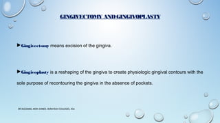
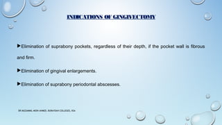
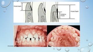
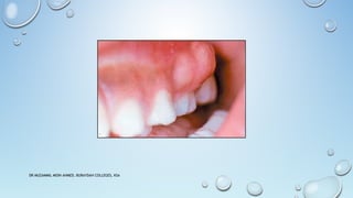
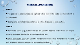
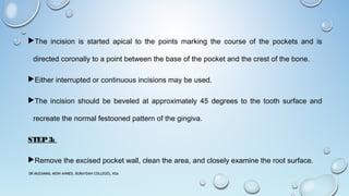
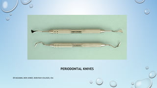
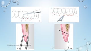
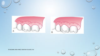
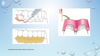
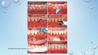
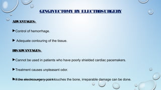
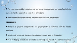
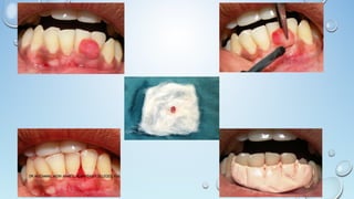
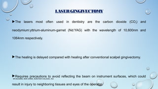
GINGIVECTOMY & GINGIVOPLASTY
DR. MUZAMMIL MOIN AHMED
ASSISTANT PROFESSOR
DIVISION OF PERIODONTICS
DEPARTMENT OF PREVENTIVE DENTAL SCIENCES
BURAYDAHCOLLEGE OF PHARMACY ANDDENTISTRY
BURAIDAH, AL-QASSIM, KSA.
DR MUZAMMIL MOIN AHMED, BURAYDAH COLLEGES, KSA
GINGIVECTOMY ANDGINGIVOPLASTY
Gingivectomy means excision of the gingiva.
Gingivoplasty is a reshaping of the gingiva to create physiologic gingival contours with the
sole purpose of recontouring the gingiva in the absence of pockets.
DR MUZAMMIL MOIN AHMED, BURAYDAH COLLEGES, KSA
INDICATIONS OF GINGIVECTOMY
Elimination of suprabony pockets, regardless of their depth, if the pocket wall is fibrous
and firm.
Elimination of gingival enlargements.
Elimination of suprabony periodontal abscesses.
DR MUZAMMIL MOIN AHMED, BURAYDAH COLLEGES, KSA
CONTRAINDICATIONS OF GINGIVECTOMY
The need for bone surgery or examination of the bone shape and morphology.
Situations in which the bottom of the pocket is apical to the mucogingival junction.
Esthetic considerations, particularly in the anterior maxilla.
DR MUZAMMIL MOIN AHMED, BURAYDAH COLLEGES, KSA
VARIOUS TECHNIQUES OF GINGIVECTOMY
Surgical gingivectomy
Gingivectomy by electrosurgery
Laser gingivectomy
Gingivectomy with chemosurgery
DR MUZAMMIL MOIN AHMED, BURAYDAH COLLEGES, KSA
SURGICAL GINGIVECTOMY
STEP1:
The pockets on each surface are explored with a periodontal probe and marked with a
pocket marker.
 Each pocket is marked in several areas to outline its course on each surface.
STEP2:
Periodontal knives (e.g., Kirkland knives) are used for incisions on the facial and lingual
surfaces and those distal to the terminal tooth in the arch.
Orban periodontal knives are used for interdental incisions. Bard-Parker blades #12 and
#15, as well as scissors, are used as auxiliary instruments.
DR MUZAMMIL MOIN AHMED, BURAYDAH COLLEGES, KSA
The incision is started apical to the points marking the course of the pockets and is
directed coronally to a point between the base of the pocket and the crest of the bone.
Either interrupted or continuous incisions may be used.
The incision should be beveled at approximately 45 degrees to the tooth surface and
recreate the normal festooned pattern of the gingiva.
STEP3:
Remove the excised pocket wall, clean the area, and closely examine the root surface.
DR MUZAMMIL MOIN AHMED, BURAYDAH COLLEGES, KSA
STEP4:
Carefully curette the granulation tissue and remove any remaining calculus and necrotic
cementum to leave a smooth and clean surface.
STEP5:
Cover the area with a surgical pack.
DR MUZAMMIL MOIN AHMED, BURAYDAH COLLEGES, KSA
PERIODONTAL KNIVES
DR MUZAMMIL MOIN AHMED, BURAYDAH COLLEGES, KSA
GINGIVECTOMY BY ELECTROSURGERY
ADVANTAGES:
Control of hemorrhage.
 Adequate contouring of the tissue.
DISADVANTAGES:
Cannot be used in patients who have poorly shielded cardiac pacemakers.
Treatment causes unpleasant odor.
If the electrosurgery point touches the bone, irreparable damage can be done.DR MUZAMMIL MOIN AHMED, BURAYDAH COLLEGES, KSA
The heat generated by injudicious use can cause tissue damage and loss of periodontal
support when the electrode is used close to the bone
 when electrode touches the root, areas of cementum burn are produced.
TECHNIQUE:
Removal of gingival enlargements and gingivoplasty is performed with the needle
electrode.
Small, ovoid loop or the diamond shaped electrodes are used for festooning.
In all reshaping procedures, electrode is activated and moved in a concise “shaving”
DR MUZAMMIL MOIN AHMED, BURAYDAH COLLEGES, KSA
LASERGINGIVECTOMY
The lasers most often used in dentistry are the carbon dioxide (CO2) and
neodymium:yttrium-aluminum-garnet (Nd:YAG) with the wavelength of 10,600nm and
1064nm respectively.
The healing is delayed compared with healing after conventional scalpel gingivectomy.
Requires precautions to avoid reflecting the beam on instrument surfaces, which could
result in injury to neighboring tissues and eyes of the operator.
DR MUZAMMIL MOIN AHMED, BURAYDAH COLLEGES, KSA
HEALING AFTERGINGIVECTOMY
Initially, formation of a protective surface clot.
Underlying tissue becomes acutely inflamed, with some necrosis.
The clot is then replaced by granulation tissue.
By 24 hours, there is an increase in new connective tissue cells mainly angioblasts, below
the surface of inflammation.
By the third day, numerous young fibroblasts are located in the area.
This highly vascularized connective tissue grows coronally, creating a new, free gingival
margin and sulcus.
DR MUZAMMIL MOIN AHMED, BURAYDAH COLLEGES, KSA
After 12 to 24 hours, epithelial cells at the margins of the wound start migrating over the
granulation tissue.
Epithelial activity reaches a peak in 24 to 36 hours.
After 5 to 14 days, surface epithelialization is generally complete.
Complete repair takes about 1 month.
DR MUZAMMIL MOIN AHMED, BURAYDAH COLLEGES, KSA
GINGIVOPLASTY
Gingivoplasty is a reshaping of the gingiva to create physiologic gingival contours with the
sole purpose of recontouring the gingiva in the absence of pockets.
Gingival and periodontal disease often produces deformities in the gingiva that is
conducive for plaque accumulation and food debris, which prolongs and aggravates the
disease process.
Such deformities include (1) gingival clefts and craters, (2) craterlike interdental papillae
DR MUZAMMIL MOIN AHMED, BURAYDAH COLLEGES, KSA
Gingivoplasty may be accomplished with a periodontal knife, a scalpel, rotary coarse
diamond stones, or electrodes.
The technique resembles that of festooning of a artificial denture, which consists of
tapering the gingival margin, creating a scalloped marginal outline, thinning the attached
gingiva , creating vertical interdental grooves, and shaping the interdental papillae.
DR MUZAMMIL MOIN AHMED, BURAYDAH COLLEGES, KSA
THANK YOU
DR MUZAMMIL MOIN AHMED, BURAYDAH COLLEGES, KSA

GINGIVECTOMY AND GINGIVOPLASTY

  • 1.
    GINGIVECTOMY & GINGIVOPLASTY DR.MUZAMMIL MOIN AHMED ASSISTANT PROFESSOR DIVISION OF PERIODONTICS DEPARTMENT OF PREVENTIVE DENTAL SCIENCES BURAYDAHCOLLEGE OF PHARMACY ANDDENTISTRY BURAIDAH, AL-QASSIM, KSA. DR MUZAMMIL MOIN AHMED, BURAYDAH COLLEGES, KSA
  • 2.
    DR MUZAMMIL MOINAHMED, BURAYDAH COLLEGES, KSA
  • 3.
    GINGIVECTOMY ANDGINGIVOPLASTY Gingivectomy meansexcision of the gingiva. Gingivoplasty is a reshaping of the gingiva to create physiologic gingival contours with the sole purpose of recontouring the gingiva in the absence of pockets. DR MUZAMMIL MOIN AHMED, BURAYDAH COLLEGES, KSA
  • 4.
    INDICATIONS OF GINGIVECTOMY Eliminationof suprabony pockets, regardless of their depth, if the pocket wall is fibrous and firm. Elimination of gingival enlargements. Elimination of suprabony periodontal abscesses. DR MUZAMMIL MOIN AHMED, BURAYDAH COLLEGES, KSA
  • 5.
    DR MUZAMMIL MOINAHMED, BURAYDAH COLLEGES, KSA
  • 6.
    DR MUZAMMIL MOINAHMED, BURAYDAH COLLEGES, KSA
  • 7.
    CONTRAINDICATIONS OF GINGIVECTOMY Theneed for bone surgery or examination of the bone shape and morphology. Situations in which the bottom of the pocket is apical to the mucogingival junction. Esthetic considerations, particularly in the anterior maxilla. DR MUZAMMIL MOIN AHMED, BURAYDAH COLLEGES, KSA
  • 8.
    VARIOUS TECHNIQUES OFGINGIVECTOMY Surgical gingivectomy Gingivectomy by electrosurgery Laser gingivectomy Gingivectomy with chemosurgery DR MUZAMMIL MOIN AHMED, BURAYDAH COLLEGES, KSA
  • 9.
    SURGICAL GINGIVECTOMY STEP1: The pocketson each surface are explored with a periodontal probe and marked with a pocket marker.  Each pocket is marked in several areas to outline its course on each surface. STEP2: Periodontal knives (e.g., Kirkland knives) are used for incisions on the facial and lingual surfaces and those distal to the terminal tooth in the arch. Orban periodontal knives are used for interdental incisions. Bard-Parker blades #12 and #15, as well as scissors, are used as auxiliary instruments. DR MUZAMMIL MOIN AHMED, BURAYDAH COLLEGES, KSA
  • 10.
    The incision isstarted apical to the points marking the course of the pockets and is directed coronally to a point between the base of the pocket and the crest of the bone. Either interrupted or continuous incisions may be used. The incision should be beveled at approximately 45 degrees to the tooth surface and recreate the normal festooned pattern of the gingiva. STEP3: Remove the excised pocket wall, clean the area, and closely examine the root surface. DR MUZAMMIL MOIN AHMED, BURAYDAH COLLEGES, KSA
  • 11.
    STEP4: Carefully curette thegranulation tissue and remove any remaining calculus and necrotic cementum to leave a smooth and clean surface. STEP5: Cover the area with a surgical pack. DR MUZAMMIL MOIN AHMED, BURAYDAH COLLEGES, KSA
  • 12.
    PERIODONTAL KNIVES DR MUZAMMILMOIN AHMED, BURAYDAH COLLEGES, KSA
  • 13.
    DR MUZAMMIL MOINAHMED, BURAYDAH COLLEGES, KSA
  • 14.
    DR MUZAMMIL MOINAHMED, BURAYDAH COLLEGES, KSA
  • 15.
    DR MUZAMMIL MOINAHMED, BURAYDAH COLLEGES, KSA
  • 16.
    DR MUZAMMIL MOINAHMED, BURAYDAH COLLEGES, KSA
  • 17.
    GINGIVECTOMY BY ELECTROSURGERY ADVANTAGES: Controlof hemorrhage.  Adequate contouring of the tissue. DISADVANTAGES: Cannot be used in patients who have poorly shielded cardiac pacemakers. Treatment causes unpleasant odor. If the electrosurgery point touches the bone, irreparable damage can be done.DR MUZAMMIL MOIN AHMED, BURAYDAH COLLEGES, KSA
  • 18.
    The heat generatedby injudicious use can cause tissue damage and loss of periodontal support when the electrode is used close to the bone  when electrode touches the root, areas of cementum burn are produced. TECHNIQUE: Removal of gingival enlargements and gingivoplasty is performed with the needle electrode. Small, ovoid loop or the diamond shaped electrodes are used for festooning. In all reshaping procedures, electrode is activated and moved in a concise “shaving” DR MUZAMMIL MOIN AHMED, BURAYDAH COLLEGES, KSA
  • 19.
    DR MUZAMMIL MOINAHMED, BURAYDAH COLLEGES, KSA
  • 20.
    LASERGINGIVECTOMY The lasers mostoften used in dentistry are the carbon dioxide (CO2) and neodymium:yttrium-aluminum-garnet (Nd:YAG) with the wavelength of 10,600nm and 1064nm respectively. The healing is delayed compared with healing after conventional scalpel gingivectomy. Requires precautions to avoid reflecting the beam on instrument surfaces, which could result in injury to neighboring tissues and eyes of the operator. DR MUZAMMIL MOIN AHMED, BURAYDAH COLLEGES, KSA
  • 21.
    LASERGINGIVECTOMY The lasers mostoften used in dentistry are the carbon dioxide (CO2) and neodymium:yttrium-aluminum-garnet (Nd:YAG) with the wavelength of 10,600nm and 1064nm respectively. The healing is delayed compared with healing after conventional scalpel gingivectomy. Requires precautions to avoid reflecting the beam on instrument surfaces, which could result in injury to neighboring tissues and eyes of the operator. DR MUZAMMIL MOIN AHMED, BURAYDAH COLLEGES, KSA
  • 22.
    HEALING AFTERGINGIVECTOMY Initially, formationof a protective surface clot. Underlying tissue becomes acutely inflamed, with some necrosis. The clot is then replaced by granulation tissue. By 24 hours, there is an increase in new connective tissue cells mainly angioblasts, below the surface of inflammation. By the third day, numerous young fibroblasts are located in the area. This highly vascularized connective tissue grows coronally, creating a new, free gingival margin and sulcus. DR MUZAMMIL MOIN AHMED, BURAYDAH COLLEGES, KSA
  • 23.
    After 12 to24 hours, epithelial cells at the margins of the wound start migrating over the granulation tissue. Epithelial activity reaches a peak in 24 to 36 hours. After 5 to 14 days, surface epithelialization is generally complete. Complete repair takes about 1 month. DR MUZAMMIL MOIN AHMED, BURAYDAH COLLEGES, KSA
  • 24.
    GINGIVOPLASTY Gingivoplasty is areshaping of the gingiva to create physiologic gingival contours with the sole purpose of recontouring the gingiva in the absence of pockets. Gingival and periodontal disease often produces deformities in the gingiva that is conducive for plaque accumulation and food debris, which prolongs and aggravates the disease process. Such deformities include (1) gingival clefts and craters, (2) craterlike interdental papillae DR MUZAMMIL MOIN AHMED, BURAYDAH COLLEGES, KSA
  • 25.
    Gingivoplasty may beaccomplished with a periodontal knife, a scalpel, rotary coarse diamond stones, or electrodes. The technique resembles that of festooning of a artificial denture, which consists of tapering the gingival margin, creating a scalloped marginal outline, thinning the attached gingiva , creating vertical interdental grooves, and shaping the interdental papillae. DR MUZAMMIL MOIN AHMED, BURAYDAH COLLEGES, KSA
  • 26.
    THANK YOU DR MUZAMMILMOIN AHMED, BURAYDAH COLLEGES, KSA