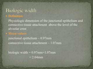
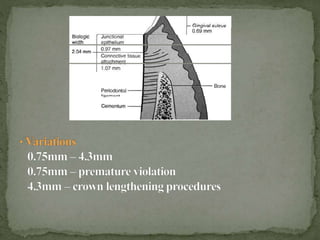
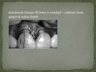
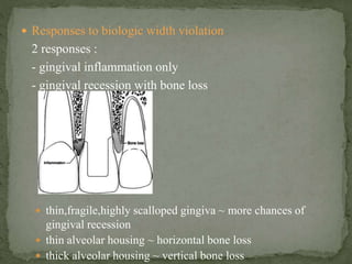
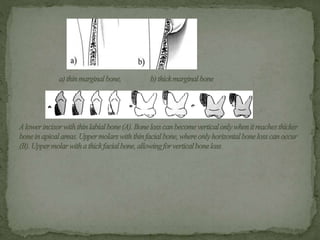
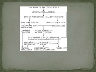
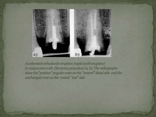
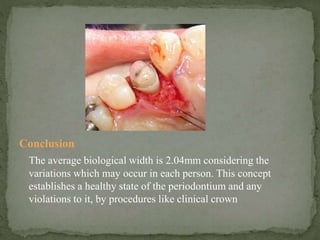
BIOLOGIC WIDTH & ITS IMPLICATIONS
 Definition
Physiologic dimension of the junctional epithelium and
connective tissue attachment above the level of the
alveolar crest
 Mean values
junctional epithelium – 0.97mm
connective tissue attachment – 1.07mm
biologic width = 0.97mm+1.07mm
= 2.04mm
 Margin placement & biologic width
- supra-, sub-, equi-gingival
- subgingival placement
 esthetic
 extension of caries
 retention & resistance form
 Margin within about 1mm of gingival sulcus ~ ideal
 Biologic width evaluation
- variations to be considered
- radiographic ~ superimposition
- discomfort on probing
- ‘sounding to bone’ ~ probing through anaesthetised
attachment tissues till bone is touched ~ subtract from
gingival sulcus depth
 Responses to biologic width violation
2 responses :
- gingival inflammation only
- gingival recession with bone loss
 thin,fragile,highly scalloped gingiva ~ more chances of
gingival recession
 thin alveolar housing ~ horizontal bone loss
 thick alveolar housing ~ vertical bone loss
 Correcting biologic width violation
- 2 options
 Surgery
 Orthodontic extrusion
- surgery ~ more rapid, bone removed away from margin
by measured distance of ideal biological width for that patient
+ additional 5mm for ‘ferrule’ effect
- interproximal violation ~ surgery contraindiated
- orthodontic extrusion ~ interproximal violations
2 types ;
- low orthodontic extrusion force ~ bone & gingiva follows
~osseous contouring
- rapid orthodontic extrusion force with supracrestal
fibrotomy~ gingival inflammaton ~ bone does not follow
 Crown lengthening procedures & biologic width
- short clinical forms
- retention & resistance form
- extention of caries
 Methods
- removal of soft tissue only ~ adequate attached gingiva &
> 3mm of tissue coronal to the bone crest or
- removal of both soft tissue and alveolar bone ~ inadequate
attached gingiva & < 3mm of tissue coronal to bone crest
- gingivectomy or flap technique
- subgingival extention of caries ~ additional 1mm of bone
removed to provide ‘ferrule’ effect
Conclusion
The average biological width is 2.04mm considering the
variations which may occur in each person. This concept
establishes a healthy state of the periodontium and any
violations to it, by procedures like clinical crown
lengthening, inadverent placement of margins of restoration
breaching the attachment apparatus, will hinder the healthy
state of the periodontium resulting in periodontal problems as
has been mentioned. Thus it is essential to properly evaluate
the biologic width of the particular case and preserve this entity
before and while performing such treatment modalities.
References
 Clinical Periodontology by Carranza
 www.wikipedia.com
 Stomatologija,Baltic Dental & Maxillofacial Journal,2006
 www.pubmed.com
Biologic width

Biologic width

  • 1.
    BIOLOGIC WIDTH &ITS IMPLICATIONS
  • 2.
     Definition Physiologic dimensionof the junctional epithelium and connective tissue attachment above the level of the alveolar crest  Mean values junctional epithelium – 0.97mm connective tissue attachment – 1.07mm biologic width = 0.97mm+1.07mm = 2.04mm
  • 4.
     Margin placement& biologic width - supra-, sub-, equi-gingival - subgingival placement  esthetic  extension of caries  retention & resistance form  Margin within about 1mm of gingival sulcus ~ ideal  Biologic width evaluation - variations to be considered - radiographic ~ superimposition - discomfort on probing - ‘sounding to bone’ ~ probing through anaesthetised
  • 5.
    attachment tissues tillbone is touched ~ subtract from gingival sulcus depth
  • 6.
     Responses tobiologic width violation 2 responses : - gingival inflammation only - gingival recession with bone loss  thin,fragile,highly scalloped gingiva ~ more chances of gingival recession  thin alveolar housing ~ horizontal bone loss  thick alveolar housing ~ vertical bone loss
  • 9.
     Correcting biologicwidth violation - 2 options  Surgery  Orthodontic extrusion - surgery ~ more rapid, bone removed away from margin by measured distance of ideal biological width for that patient + additional 5mm for ‘ferrule’ effect - interproximal violation ~ surgery contraindiated - orthodontic extrusion ~ interproximal violations 2 types ; - low orthodontic extrusion force ~ bone & gingiva follows ~osseous contouring - rapid orthodontic extrusion force with supracrestal fibrotomy~ gingival inflammaton ~ bone does not follow
  • 11.
     Crown lengtheningprocedures & biologic width - short clinical forms - retention & resistance form - extention of caries  Methods - removal of soft tissue only ~ adequate attached gingiva & > 3mm of tissue coronal to the bone crest or - removal of both soft tissue and alveolar bone ~ inadequate attached gingiva & < 3mm of tissue coronal to bone crest - gingivectomy or flap technique - subgingival extention of caries ~ additional 1mm of bone removed to provide ‘ferrule’ effect
  • 12.
    Conclusion The average biologicalwidth is 2.04mm considering the variations which may occur in each person. This concept establishes a healthy state of the periodontium and any violations to it, by procedures like clinical crown
  • 13.
    lengthening, inadverent placementof margins of restoration breaching the attachment apparatus, will hinder the healthy state of the periodontium resulting in periodontal problems as has been mentioned. Thus it is essential to properly evaluate the biologic width of the particular case and preserve this entity before and while performing such treatment modalities. References  Clinical Periodontology by Carranza  www.wikipedia.com  Stomatologija,Baltic Dental & Maxillofacial Journal,2006  www.pubmed.com