Downloaded 249 times
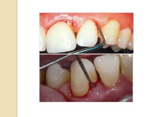
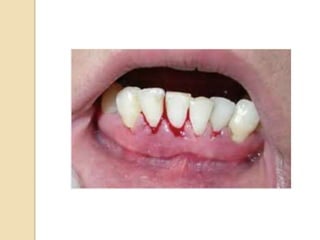
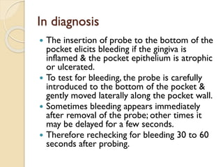
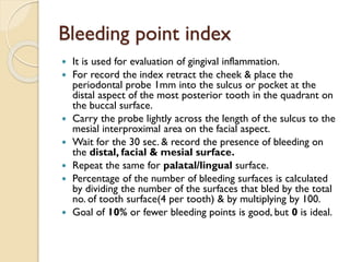


Bleeding on probing is an early sign of gingival inflammation and is commonly used to assess periodontal disease status. It occurs when increased crevicular fluid and breakdown of gingival tissues due to inflammation allows blood vessels to rupture upon gentle probing. Local factors like poor oral hygiene and systemic conditions like vitamin deficiencies or coagulation disorders can contribute to abnormal gingival bleeding. The bleeding point index is used to evaluate gingival inflammation by recording the number of bleeding sites after probing specific areas in the mouth.


