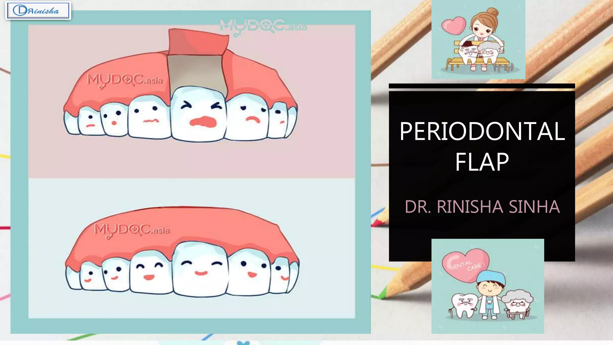
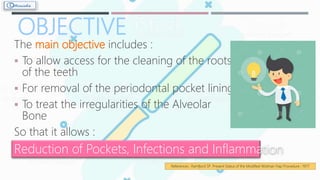
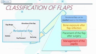
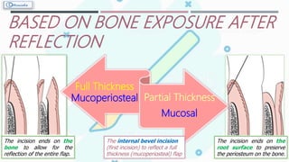
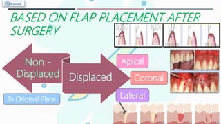
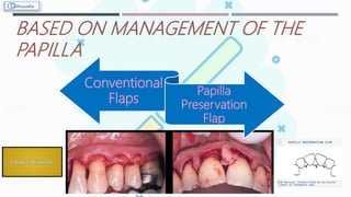
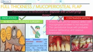
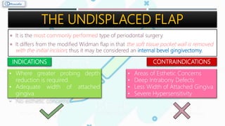
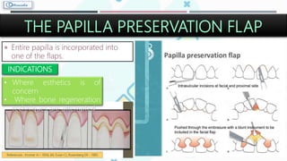
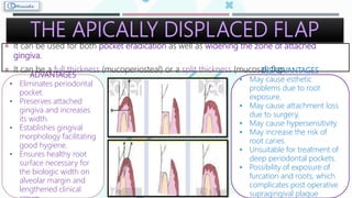
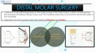
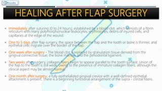
PERIODONTAL
FLAP
DR. RINISHA SINHA
CONTENTS
• Definition
• Objective
• Indications and
Contraindications
• Incisions
• Classification of Flap
 Based on Bone Exposure after
Reflection
 Based on Flap Placement after
Surgery
 Based on Management of the
Papilla
• What after Incision !?
• Mucoperiosteal Flap
• Mucosal Flap
• Different Flap Techniques
 The Conventional Flap
 The Modified Widman Flap
 The Undisplaced Flap
 The Papilla Preservation
Flap
 The Apically Displaced Flap
• Distal Molar Surgery
• Healing After Flap Surgery
• References
DEFINITION
PERIODONTAL FLAP is a section of gingiva and/or
mucosa surgically separated from the underlying
tissues to provide visibility of and access to the
bone and root surface.
 It also allows gingiva to be displaced from one
location to a different location in patients with
any mucogingival involvement.
 According to Webster's - "Flap is a piece of
tissue partly severed from its place of origin for
use in surgical grafting and repair of defects.
References : Newman M, Takei H, Klokkevold P, Carranza F. Carranza’s Clinical Periodontology – 10th edition;; 2006
References : Groove P. B. Webster's Third New International Dictionary ; 1961
OBJECTIVE
The main objective includes :
 To allow access for the cleaning of the roots
of the teeth
 For removal of the periodontal pocket lining
 To treat the irregularities of the Alveolar
Bone
So that it allows :
Reduction of Pockets, Infections and Inflammation
References : Ramfjord SP. Present Status of the Modified Widman Flap Procedure ; 1977
•Intra bony pockets
•Irregular bony contours
•Deep craters
•For regenerative procedures, such as bone
grafting and GTR
•Persistent inflammation and Periodontal
pockets even after phase 1 therapy
•Grade II or III furcation involvement
•Root resection / hemi - section
•Intrabony pockets on distal areas of last molars
-Uncontrolled medical conditions such as:
- Unstable angina
- Uncontrolled diabetes
- Uncontrolled hypertension
-Myocardial infarction
-Poor plaque control
-Gingival overgrowth
-Unrealistic patient expectations or desires
INCISIONS
HORIZONTAL INCISION
VERTICALINCISION
INTERNAL
BEVEL
INCISION
INTERDENTAL
INCISION
CREVICULAR
INCISION
References : Borchard R, Erpenstein H. Incisions and tissue management in periodontal surgery; 2004
CLASSIFICATION OF FLAPS
Periodontal flaps can be
classified based on the following:
Bone exposure after
flap reflection
Placement of the flap
after surgery
Management of the
papilla
BASED ON BONE EXPOSURE AFTER
REFLECTION
Full Thickness
Mucoperiosteal Partial Thickness
Mucosal
The internal bevel incision
(first incision) to reflect a full
thickness (mucoperiosteal) flap
The incision ends on the
root surface to preserve
the periosteum on the bone.
The incision ends on the
bone to allow for the
reflection of the entire flap.
BASED ON FLAP PLACEMENT AFTER
SURGERY
Non -
Displaced Displaced
Apical
Coronal
LateralTo Original Place
BASED ON MANAGEMENT OF THE
PAPILLA
Conventional
Flaps
Papilla
Preservation
Flap
ELEVATION
OF THE
FLAP
FLAP
RETRACTION
BLUNT DISSECTION WITH PERIOSTEAL
ELEVATOR
FOR THE REFLECTION OF FULL
THICKNESS FLAP
SHARP DISSECTION WITH SURGICAL
SCALPEL
FOR THE REFLECTION OF PARTIAL
THICKNESS FLAP
Retraction should be passive
without any tension.
Force should not be necessary to
keep the flap retracted.
The edge of the retractor always
be kept on bone.
FULL THICKNESS / MUCOPERIOSTEAL FLAP
Periosteum is reflected to “expose the underlying
bone”.
INDICATIONS
IN RESECTIVE OSSEOUS SURGERY
CONTRAINDICATIONS
• Where treatment for osseous defect with
mucogingival problem is not required.
• Thin periodontal tissue with probable
osseous dehiscence and osseous
fenestration.
• Where alveolar bone is thin.
Blunt dissection with
periosteal elevator to
separate the
mucoperiosteum from the
bone by moving it
mesially, distally, apically
PARTIAL THICKNESS FLAP / SPLIT THICKNESS FLAP
/ MUCOSAL FLAP
Periosteum covers the
Bone.
INDICATIONS CONTRAINDICATIONS
When the flap has to be positioned
apically
When operator does not
desire to expose the bone
Sharp dissection with
Surgical Scalpel
References : Staffileno H. Palatal flap surgery - 1969
FLAP
TECHNIQUES
THE
MODIFIED
WIDMAN
FLAP
THE
UNDISPLACED
FLAP
THE
APICALLY
DISPLACED
FLAP
DISTAL MOLAR
SURGERY
FLAPS FOR
REGENERATIVE
SURGERY
THE
CONVENTIONAL
FLAP
THE
PALATAL
FLAP
THE PAPILLA
PRESERVATION
FLAP
THE CONVENTIONAL FLAP
 The incisions for the facial and the
lingual or palatal flap reach the tip of
the interdental papilla, thereby splitting
the papilla into a facial half and a lingual
or palatal half.
INDICATIONS
• When the interdental areas
are too narrow to permit
the preservation of flap.
• When there is a need for
displacing flaps.
The Conventional or Traditional flap technique
A. Internal bevel incision, splitting the papilla, and the
vertical incisions are drawn in interrupted lines.
B. The flap has been elevated, and the wedge of tissue
next to tooth is still in place.
C. All marginal tissue has been removed, exposing
underlying bone.
D. Tissue returned to its original position. Proximal
• It includes the Modified Widman
flap, the Undisplaced flap, the
Apically Displaced flap.
THE MODIFIED WIDMAN FLAP
 By Ramfjord and Nissle (1974)
 The main goals of the procedure include optimum mechanical sub gingival
root planing with direct vision.
INDICATIONS
Effective with pocket depths of 5‐7
mm
CONTRAINDICATIONS
• Lack of or very thin and narrow attached gingiva
• Osseous surgical procedureADVANTAGES
• Root cleaning with direct
vision.
• Tissue friendly
• Healing by primary intention
• Minimal crestal bone
resorption
• Lack of post operative
discomfortReferences : Ramfjord SP, Nissle RR. The modified Widman Flap - 1974
INTERNAL BEVEL INCISION = 0.5 – 2 mm
FLAP REFLECTION
CREVICULAR INCISION
HORIZONTAL INCISION
ROOT PLANING
COMPLETE COVERAGE OF INTERDENTAL DEFECTS
THE UNDISPLACED FLAP
 It is the most commonly performed type of periodontal surgery.
 It differs from the modified Widman flap in that the soft tissue pocket wall is removed
with the initial incision; thus it may be considered an internal bevel gingivectomy.
INDICATIONS
• Where greater probing depth
reduction is required.
• Adequate width of attached
gingiva.
• No esthetic concerns.
CONTRAINDICATIONS
• Areas of Esthetic Concerns
• Deep Intrabony Defects
• Less Width of Attached Gingiva
• Severe Hypersensitivity
THE PAPILLA PRESERVATION FLAP
 Entire papilla is incorporated into
one of the flaps.
INDICATIONS
• Where esthetics is of
concern
• Where bone regeneration
techniques are attempted
References : Kromer H – 1956; 66; Evian CI, Rosenberg ER - 1985
THE APICALLY DISPLACED FLAP
 It can be used for both pocket eradication as well as widening the zone of attached
gingiva.
 It can be a full thickness (mucoperiosteal) or a split thickness (mucosal) flap.
ADVANTAGES
• Eliminates periodontal
pocket.
• Preserves attached
gingiva and increases
its width.
• Establishes gingival
morphology facilitating
good hygiene.
• Ensures healthy root
surface necessary for
the biologic width on
alveolar margin and
lengthened clinical
DISADVANTAGES
• May cause esthetic
problems due to root
exposure.
• May cause attachment loss
due to surgery.
• May cause hypersensitivity.
• May increase the risk of
root caries.
• Unsuitable for treatment of
deep periodontal pockets.
• Possibility of exposure of
furcation and roots, which
complicates post operative
supragingival plaque
DISTAL MOLAR SURGERY
 Treatment of periodontal pockets on the distal surface of terminal molars is often complicated by
the presence of bulbous fibrous tissue over the maxillary tuberosity or prominent retromolar pads in
the mandible.
 Operations for this purpose were described by Robinson and Braden.
TRIANGULAR
SQUARE,
PARALLEL or
H - DESIGN
 Immediately after suturing (0 to 24 hours), established by a blood clot, which consists of a fibrin
reticulum with many polymorphonuclear leukocytes, erythrocytes, debris of injured cells, and
capillaries at the edge of the wound.
 One to 3 days after flap surgery, the space between the flap and the tooth or bone is thinner, and
epithelial cells migrate over the border of the flap.
 One week after surgery ‐ The blood clot is replaced by granulation tissue derived from the
gingival connective tissue, the bone marrow, and the periodontal ligament.
 Two weeks after surgery, collagen fibers begin to appear parallel to the tooth surface. Union of
the flap to the tooth is still weak, owing to the presence of immature collagen fibers, although the
clinical aspect may be almost normal.
 One month after surgery, a fully epithelialized gingival crevice with a well‐defined epithelial
attachment is present. There is a beginning functional arrangement of the supra - crestal fibers.
HEALING AFTER FLAP SURGERY
REFERENCES Carranza’s Clinical
Periodontology :
10th Edition
 Periobasics : A
textbook of
Periodontics
and
Implantology
 Journal of
Pharmaceuticals and
Scientific Innovation
 William Becker, urton E.
Becker, Clifford Ochsenbein,
Gloria Kerry, John Prichard :
A Review – 1987
 Kromer H – 1956; 66; Evian
CI, Rosenberg ER – 1985
 Newman M, Takei H,
Klokkevold P – 2006
Periodontal flap

Periodontal flap

  • 1.
  • 2.
    CONTENTS • Definition • Objective •Indications and Contraindications • Incisions • Classification of Flap  Based on Bone Exposure after Reflection  Based on Flap Placement after Surgery  Based on Management of the Papilla • What after Incision !? • Mucoperiosteal Flap • Mucosal Flap • Different Flap Techniques  The Conventional Flap  The Modified Widman Flap  The Undisplaced Flap  The Papilla Preservation Flap  The Apically Displaced Flap • Distal Molar Surgery • Healing After Flap Surgery • References
  • 3.
    DEFINITION PERIODONTAL FLAP isa section of gingiva and/or mucosa surgically separated from the underlying tissues to provide visibility of and access to the bone and root surface.  It also allows gingiva to be displaced from one location to a different location in patients with any mucogingival involvement.  According to Webster's - "Flap is a piece of tissue partly severed from its place of origin for use in surgical grafting and repair of defects. References : Newman M, Takei H, Klokkevold P, Carranza F. Carranza’s Clinical Periodontology – 10th edition;; 2006 References : Groove P. B. Webster's Third New International Dictionary ; 1961
  • 4.
    OBJECTIVE The main objectiveincludes :  To allow access for the cleaning of the roots of the teeth  For removal of the periodontal pocket lining  To treat the irregularities of the Alveolar Bone So that it allows : Reduction of Pockets, Infections and Inflammation References : Ramfjord SP. Present Status of the Modified Widman Flap Procedure ; 1977
  • 5.
    •Intra bony pockets •Irregularbony contours •Deep craters •For regenerative procedures, such as bone grafting and GTR •Persistent inflammation and Periodontal pockets even after phase 1 therapy •Grade II or III furcation involvement •Root resection / hemi - section •Intrabony pockets on distal areas of last molars -Uncontrolled medical conditions such as: - Unstable angina - Uncontrolled diabetes - Uncontrolled hypertension -Myocardial infarction -Poor plaque control -Gingival overgrowth -Unrealistic patient expectations or desires
  • 6.
    INCISIONS HORIZONTAL INCISION VERTICALINCISION INTERNAL BEVEL INCISION INTERDENTAL INCISION CREVICULAR INCISION References :Borchard R, Erpenstein H. Incisions and tissue management in periodontal surgery; 2004
  • 7.
    CLASSIFICATION OF FLAPS Periodontalflaps can be classified based on the following: Bone exposure after flap reflection Placement of the flap after surgery Management of the papilla
  • 8.
    BASED ON BONEEXPOSURE AFTER REFLECTION Full Thickness Mucoperiosteal Partial Thickness Mucosal The internal bevel incision (first incision) to reflect a full thickness (mucoperiosteal) flap The incision ends on the root surface to preserve the periosteum on the bone. The incision ends on the bone to allow for the reflection of the entire flap.
  • 9.
    BASED ON FLAPPLACEMENT AFTER SURGERY Non - Displaced Displaced Apical Coronal LateralTo Original Place
  • 10.
    BASED ON MANAGEMENTOF THE PAPILLA Conventional Flaps Papilla Preservation Flap
  • 11.
    ELEVATION OF THE FLAP FLAP RETRACTION BLUNT DISSECTIONWITH PERIOSTEAL ELEVATOR FOR THE REFLECTION OF FULL THICKNESS FLAP SHARP DISSECTION WITH SURGICAL SCALPEL FOR THE REFLECTION OF PARTIAL THICKNESS FLAP Retraction should be passive without any tension. Force should not be necessary to keep the flap retracted. The edge of the retractor always be kept on bone.
  • 12.
    FULL THICKNESS /MUCOPERIOSTEAL FLAP Periosteum is reflected to “expose the underlying bone”. INDICATIONS IN RESECTIVE OSSEOUS SURGERY CONTRAINDICATIONS • Where treatment for osseous defect with mucogingival problem is not required. • Thin periodontal tissue with probable osseous dehiscence and osseous fenestration. • Where alveolar bone is thin. Blunt dissection with periosteal elevator to separate the mucoperiosteum from the bone by moving it mesially, distally, apically
  • 13.
    PARTIAL THICKNESS FLAP/ SPLIT THICKNESS FLAP / MUCOSAL FLAP Periosteum covers the Bone. INDICATIONS CONTRAINDICATIONS When the flap has to be positioned apically When operator does not desire to expose the bone Sharp dissection with Surgical Scalpel References : Staffileno H. Palatal flap surgery - 1969
  • 14.
  • 15.
    THE CONVENTIONAL FLAP The incisions for the facial and the lingual or palatal flap reach the tip of the interdental papilla, thereby splitting the papilla into a facial half and a lingual or palatal half. INDICATIONS • When the interdental areas are too narrow to permit the preservation of flap. • When there is a need for displacing flaps. The Conventional or Traditional flap technique A. Internal bevel incision, splitting the papilla, and the vertical incisions are drawn in interrupted lines. B. The flap has been elevated, and the wedge of tissue next to tooth is still in place. C. All marginal tissue has been removed, exposing underlying bone. D. Tissue returned to its original position. Proximal • It includes the Modified Widman flap, the Undisplaced flap, the Apically Displaced flap.
  • 16.
    THE MODIFIED WIDMANFLAP  By Ramfjord and Nissle (1974)  The main goals of the procedure include optimum mechanical sub gingival root planing with direct vision. INDICATIONS Effective with pocket depths of 5‐7 mm CONTRAINDICATIONS • Lack of or very thin and narrow attached gingiva • Osseous surgical procedureADVANTAGES • Root cleaning with direct vision. • Tissue friendly • Healing by primary intention • Minimal crestal bone resorption • Lack of post operative discomfortReferences : Ramfjord SP, Nissle RR. The modified Widman Flap - 1974
  • 17.
    INTERNAL BEVEL INCISION= 0.5 – 2 mm FLAP REFLECTION CREVICULAR INCISION HORIZONTAL INCISION ROOT PLANING COMPLETE COVERAGE OF INTERDENTAL DEFECTS
  • 18.
    THE UNDISPLACED FLAP It is the most commonly performed type of periodontal surgery.  It differs from the modified Widman flap in that the soft tissue pocket wall is removed with the initial incision; thus it may be considered an internal bevel gingivectomy. INDICATIONS • Where greater probing depth reduction is required. • Adequate width of attached gingiva. • No esthetic concerns. CONTRAINDICATIONS • Areas of Esthetic Concerns • Deep Intrabony Defects • Less Width of Attached Gingiva • Severe Hypersensitivity
  • 19.
    THE PAPILLA PRESERVATIONFLAP  Entire papilla is incorporated into one of the flaps. INDICATIONS • Where esthetics is of concern • Where bone regeneration techniques are attempted References : Kromer H – 1956; 66; Evian CI, Rosenberg ER - 1985
  • 20.
    THE APICALLY DISPLACEDFLAP  It can be used for both pocket eradication as well as widening the zone of attached gingiva.  It can be a full thickness (mucoperiosteal) or a split thickness (mucosal) flap. ADVANTAGES • Eliminates periodontal pocket. • Preserves attached gingiva and increases its width. • Establishes gingival morphology facilitating good hygiene. • Ensures healthy root surface necessary for the biologic width on alveolar margin and lengthened clinical DISADVANTAGES • May cause esthetic problems due to root exposure. • May cause attachment loss due to surgery. • May cause hypersensitivity. • May increase the risk of root caries. • Unsuitable for treatment of deep periodontal pockets. • Possibility of exposure of furcation and roots, which complicates post operative supragingival plaque
  • 21.
    DISTAL MOLAR SURGERY Treatment of periodontal pockets on the distal surface of terminal molars is often complicated by the presence of bulbous fibrous tissue over the maxillary tuberosity or prominent retromolar pads in the mandible.  Operations for this purpose were described by Robinson and Braden. TRIANGULAR SQUARE, PARALLEL or H - DESIGN
  • 22.
     Immediately aftersuturing (0 to 24 hours), established by a blood clot, which consists of a fibrin reticulum with many polymorphonuclear leukocytes, erythrocytes, debris of injured cells, and capillaries at the edge of the wound.  One to 3 days after flap surgery, the space between the flap and the tooth or bone is thinner, and epithelial cells migrate over the border of the flap.  One week after surgery ‐ The blood clot is replaced by granulation tissue derived from the gingival connective tissue, the bone marrow, and the periodontal ligament.  Two weeks after surgery, collagen fibers begin to appear parallel to the tooth surface. Union of the flap to the tooth is still weak, owing to the presence of immature collagen fibers, although the clinical aspect may be almost normal.  One month after surgery, a fully epithelialized gingival crevice with a well‐defined epithelial attachment is present. There is a beginning functional arrangement of the supra - crestal fibers. HEALING AFTER FLAP SURGERY
  • 23.
    REFERENCES Carranza’s Clinical Periodontology: 10th Edition  Periobasics : A textbook of Periodontics and Implantology  Journal of Pharmaceuticals and Scientific Innovation  William Becker, urton E. Becker, Clifford Ochsenbein, Gloria Kerry, John Prichard : A Review – 1987  Kromer H – 1956; 66; Evian CI, Rosenberg ER – 1985  Newman M, Takei H, Klokkevold P – 2006