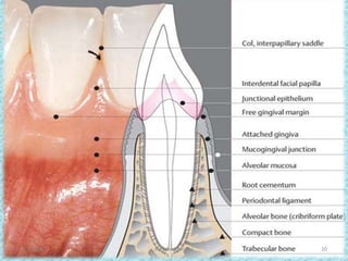
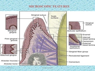
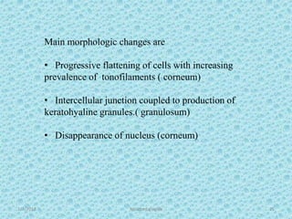
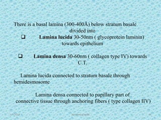
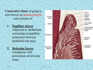
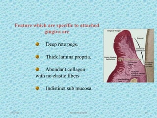
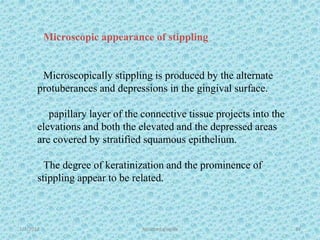
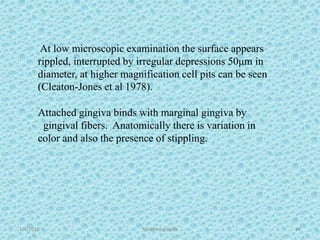
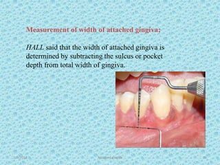
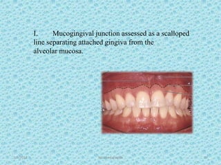
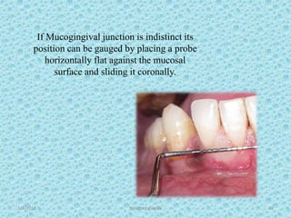
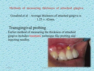
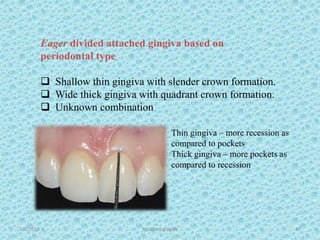
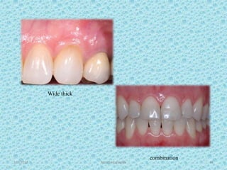
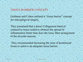
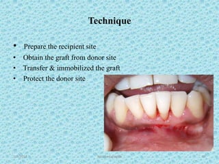
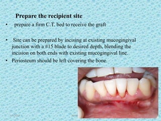
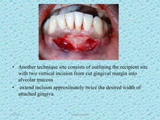
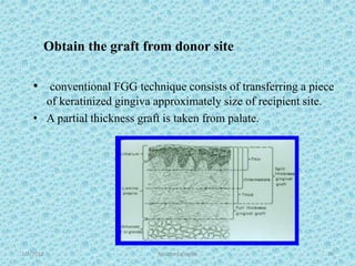
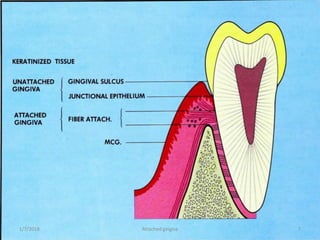
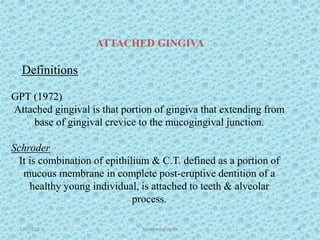
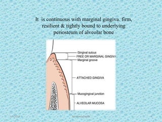
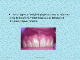
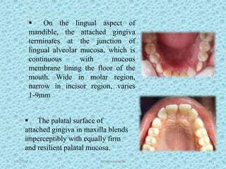
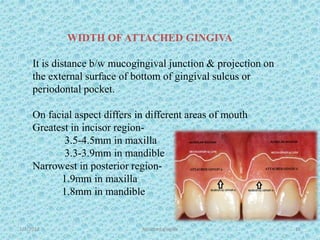
The document discusses attached gingiva, defining it as the portion of gingiva that extends from the base of the gingival crevice to the mucogingival junction. It describes the width and thickness of attached gingiva, noting it varies between 1-9mm wide and has an average thickness of 1.25mm. Microscopically, attached gingiva has a keratinized, cellular epithelium and dense connective tissue. It functions to act as a buffer zone, bear trauma and forces from occlusion, and prevent attachment loss and recession.
INTRODUCTION
DEFINITION
TYPES OF TRAUMA FROM OCCLUSION
GLICKMAN CONCEPT
WAERHAUG CONCEPT
STAGES OF TISSUE RESPONSE TO INJURY
CLINICAL AND RADIOGRAPHIC FEATURES OF TFO
CLINICAL DIAGNOSIS OF TFO
TFO AND IMPLANTS
TREATMENT OF TFO
CONCLUSION
REFRENCES
In periodontology, classifications are widely used to categorize defects due to periodontitis according to their etiology, diagnosis, treatment and prognosis.
Several classifications have been proposed in the literature in order to facilitate the diagnosis of gingival recessions.
Phase I periodontal therapy is the first in the chronologic sequence of procedures that constitute periodontal treatment. It is also referred to as cause related therapy or non-surgical periodontal therapy.
Dentists play an important role in the diagnosis and management of desquamative gingivitis. The importance of being able to recognise and properly diagnose this condition is accentuated by the fact that a serious and life threatening disease may initially manifest as desquamative gingivitis.
Aberrant Frenum !!
No worries... When Frenectomy is here.
Hello Periodontists,
Here's the entire process of Frenectomy in a nutshell and various ways to encounter the same.
Lets Shoot ...
The periodontal dressing is a physical barrier that is placed in the surgical site to protect the healing tissues from the forces produced during mastication, for comfort and close adaptation.
Furcation involvement is a common sequela of severe chronic periodontal disease. Its effective management has a profound influence on the outcome of periodontal therapy.
seminar on gingiva
contents:
Introduction
Definition
Development of gingiva
Macroscopic anatomy
Microscopic anatomy
Blood supply
Lymphatic drainage
Nerve supply
Correlation of clinical and microscopic features
Repair/healing of gingiva
Age changes
Gingival diseases
Clinical considerations
Conclusion
References
mucogingival surgery or plastic surgery of muco-gingival tissue is a surgical procedure targeted to correct and eliminate anatomic, developmental and traumatic alterations of gingiva.
The defense mechanism of gingiva includes GCF, Saliva, epithelial barrier and connective tissue cells. All these protect the periodontium from bacterial invasion.
Indian Dental Academy: will be one of the most relevant and exciting training center with best faculty and flexible training programs for dental professionals who wish to advance in their dental practice,Offers certified courses in Dental implants,Orthodontics,Endodontics,Cosmetic Dentistry, Prosthetic Dentistry, Periodontics and General Dentistry.
macroscopic/ clinical features of gingiva
With video clips
Short video descriptions
Lecture for 3rd BDS students
Periodontology
Periodontics aspect
Clinical features of the gingiva
Indian Dental Academy: will be one of the most relevant and exciting training center with best faculty and flexible training programs for dental professionals who wish to advance in their dental practice,Offers certified courses in Dental implants,Orthodontics,Endodontics,Cosmetic Dentistry, Prosthetic Dentistry, Periodontics and General Dentistry.
• Introduction
• Definitions
• Macroscopic Features
• Microscopic Features
• Blood supply
• Nerve supply
• Lymphatic drainage
• Role of epithelium in defence mechanism
• Oxygen consumption of gingiva
• Correlation of Macroscopic with microscopic features
• Conclusion
Tom Selleck Health: A Comprehensive Look at the Iconic Actor’s Wellness Journeygreendigital
Tom Selleck, an enduring figure in Hollywood. has captivated audiences for decades with his rugged charm, iconic moustache. and memorable roles in television and film. From his breakout role as Thomas Magnum in Magnum P.I. to his current portrayal of Frank Reagan in Blue Bloods. Selleck's career has spanned over 50 years. But beyond his professional achievements. fans have often been curious about Tom Selleck Health. especially as he has aged in the public eye.
Follow us on: Pinterest
Introduction
Many have been interested in Tom Selleck health. not only because of his enduring presence on screen but also because of the challenges. and lifestyle choices he has faced and made over the years. This article delves into the various aspects of Tom Selleck health. exploring his fitness regimen, diet, mental health. and the challenges he has encountered as he ages. We'll look at how he maintains his well-being. the health issues he has faced, and his approach to ageing .
Early Life and Career
Childhood and Athletic Beginnings
Tom Selleck was born on January 29, 1945, in Detroit, Michigan, and grew up in Sherman Oaks, California. From an early age, he was involved in sports, particularly basketball. which played a significant role in his physical development. His athletic pursuits continued into college. where he attended the University of Southern California (USC) on a basketball scholarship. This early involvement in sports laid a strong foundation for his physical health and disciplined lifestyle.
Transition to Acting
Selleck's transition from an athlete to an actor came with its physical demands. His first significant role in "Magnum P.I." required him to perform various stunts and maintain a fit appearance. This role, which he played from 1980 to 1988. necessitated a rigorous fitness routine to meet the show's demands. setting the stage for his long-term commitment to health and wellness.
Fitness Regimen
Workout Routine
Tom Selleck health and fitness regimen has evolved. adapting to his changing roles and age. During his "Magnum, P.I." days. Selleck's workouts were intense and focused on building and maintaining muscle mass. His routine included weightlifting, cardiovascular exercises. and specific training for the stunts he performed on the show.
Selleck adjusted his fitness routine as he aged to suit his body's needs. Today, his workouts focus on maintaining flexibility, strength, and cardiovascular health. He incorporates low-impact exercises such as swimming, walking, and light weightlifting. This balanced approach helps him stay fit without putting undue strain on his joints and muscles.
Importance of Flexibility and Mobility
In recent years, Selleck has emphasized the importance of flexibility and mobility in his fitness regimen. Understanding the natural decline in muscle mass and joint flexibility with age. he includes stretching and yoga in his routine. These practices help prevent injuries, improve posture, and maintain mobilit
- Video recording of this lecture in English language: https://youtu.be/lK81BzxMqdo
- Video recording of this lecture in Arabic language: https://youtu.be/Ve4P0COk9OI
- Link to download the book free: https://nephrotube.blogspot.com/p/nephrotube-nephrology-books.html
- Link to NephroTube website: www.NephroTube.com
- Link to NephroTube social media accounts: https://nephrotube.blogspot.com/p/join-nephrotube-on-social-media.html
Title: Sense of Smell
Presenter: Dr. Faiza, Assistant Professor of Physiology
Qualifications:
MBBS (Best Graduate, AIMC Lahore)
FCPS Physiology
ICMT, CHPE, DHPE (STMU)
MPH (GC University, Faisalabad)
MBA (Virtual University of Pakistan)
Learning Objectives:
Describe the primary categories of smells and the concept of odor blindness.
Explain the structure and location of the olfactory membrane and mucosa, including the types and roles of cells involved in olfaction.
Describe the pathway and mechanisms of olfactory signal transmission from the olfactory receptors to the brain.
Illustrate the biochemical cascade triggered by odorant binding to olfactory receptors, including the role of G-proteins and second messengers in generating an action potential.
Identify different types of olfactory disorders such as anosmia, hyposmia, hyperosmia, and dysosmia, including their potential causes.
Key Topics:
Olfactory Genes:
3% of the human genome accounts for olfactory genes.
400 genes for odorant receptors.
Olfactory Membrane:
Located in the superior part of the nasal cavity.
Medially: Folds downward along the superior septum.
Laterally: Folds over the superior turbinate and upper surface of the middle turbinate.
Total surface area: 5-10 square centimeters.
Olfactory Mucosa:
Olfactory Cells: Bipolar nerve cells derived from the CNS (100 million), with 4-25 olfactory cilia per cell.
Sustentacular Cells: Produce mucus and maintain ionic and molecular environment.
Basal Cells: Replace worn-out olfactory cells with an average lifespan of 1-2 months.
Bowman’s Gland: Secretes mucus.
Stimulation of Olfactory Cells:
Odorant dissolves in mucus and attaches to receptors on olfactory cilia.
Involves a cascade effect through G-proteins and second messengers, leading to depolarization and action potential generation in the olfactory nerve.
Quality of a Good Odorant:
Small (3-20 Carbon atoms), volatile, water-soluble, and lipid-soluble.
Facilitated by odorant-binding proteins in mucus.
Membrane Potential and Action Potential:
Resting membrane potential: -55mV.
Action potential frequency in the olfactory nerve increases with odorant strength.
Adaptation Towards the Sense of Smell:
Rapid adaptation within the first second, with further slow adaptation.
Psychological adaptation greater than receptor adaptation, involving feedback inhibition from the central nervous system.
Primary Sensations of Smell:
Camphoraceous, Musky, Floral, Pepperminty, Ethereal, Pungent, Putrid.
Odor Detection Threshold:
Examples: Hydrogen sulfide (0.0005 ppm), Methyl-mercaptan (0.002 ppm).
Some toxic substances are odorless at lethal concentrations.
Characteristics of Smell:
Odor blindness for single substances due to lack of appropriate receptor protein.
Behavioral and emotional influences of smell.
Transmission of Olfactory Signals:
From olfactory cells to glomeruli in the olfactory bulb, involving lateral inhibition.
Primitive, less old, and new olfactory systems with different path
Flu Vaccine Alert in Bangalore Karnatakaaddon Scans
As flu season approaches, health officials in Bangalore, Karnataka, are urging residents to get their flu vaccinations. The seasonal flu, while common, can lead to severe health complications, particularly for vulnerable populations such as young children, the elderly, and those with underlying health conditions.
Dr. Vidisha Kumari, a leading epidemiologist in Bangalore, emphasizes the importance of getting vaccinated. "The flu vaccine is our best defense against the influenza virus. It not only protects individuals but also helps prevent the spread of the virus in our communities," he says.
This year, the flu season is expected to coincide with a potential increase in other respiratory illnesses. The Karnataka Health Department has launched an awareness campaign highlighting the significance of flu vaccinations. They have set up multiple vaccination centers across Bangalore, making it convenient for residents to receive their shots.
To encourage widespread vaccination, the government is also collaborating with local schools, workplaces, and community centers to facilitate vaccination drives. Special attention is being given to ensuring that the vaccine is accessible to all, including marginalized communities who may have limited access to healthcare.
Residents are reminded that the flu vaccine is safe and effective. Common side effects are mild and may include soreness at the injection site, mild fever, or muscle aches. These side effects are generally short-lived and far less severe than the flu itself.
Healthcare providers are also stressing the importance of continuing COVID-19 precautions. Wearing masks, practicing good hand hygiene, and maintaining social distancing are still crucial, especially in crowded places.
Protect yourself and your loved ones by getting vaccinated. Together, we can help keep Bangalore healthy and safe this flu season. For more information on vaccination centers and schedules, residents can visit the Karnataka Health Department’s official website or follow their social media pages.
Stay informed, stay safe, and get your flu shot today!
The prostate is an exocrine gland of the male mammalian reproductive system
It is a walnut-sized gland that forms part of the male reproductive system and is located in front of the rectum and just below the urinary bladder
Function is to store and secrete a clear, slightly alkaline fluid that constitutes 10-30% of the volume of the seminal fluid that along with the spermatozoa, constitutes semen
A healthy human prostate measures (4cm-vertical, by 3cm-horizontal, 2cm ant-post ).
It surrounds the urethra just below the urinary bladder. It has anterior, median, posterior and two lateral lobes
It’s work is regulated by androgens which are responsible for male sex characteristics
Generalised disease of the prostate due to hormonal derangement which leads to non malignant enlargement of the gland (increase in the number of epithelial cells and stromal tissue)to cause compression of the urethra leading to symptoms (LUTS
Lung Cancer: Artificial Intelligence, Synergetics, Complex System Analysis, S...Oleg Kshivets
RESULTS: Overall life span (LS) was 2252.1±1742.5 days and cumulative 5-year survival (5YS) reached 73.2%, 10 years – 64.8%, 20 years – 42.5%. 513 LCP lived more than 5 years (LS=3124.6±1525.6 days), 148 LCP – more than 10 years (LS=5054.4±1504.1 days).199 LCP died because of LC (LS=562.7±374.5 days). 5YS of LCP after bi/lobectomies was significantly superior in comparison with LCP after pneumonectomies (78.1% vs.63.7%, P=0.00001 by log-rank test). AT significantly improved 5YS (66.3% vs. 34.8%) (P=0.00000 by log-rank test) only for LCP with N1-2. Cox modeling displayed that 5YS of LCP significantly depended on: phase transition (PT) early-invasive LC in terms of synergetics, PT N0—N12, cell ratio factors (ratio between cancer cells- CC and blood cells subpopulations), G1-3, histology, glucose, AT, blood cell circuit, prothrombin index, heparin tolerance, recalcification time (P=0.000-0.038). Neural networks, genetic algorithm selection and bootstrap simulation revealed relationships between 5YS and PT early-invasive LC (rank=1), PT N0—N12 (rank=2), thrombocytes/CC (3), erythrocytes/CC (4), eosinophils/CC (5), healthy cells/CC (6), lymphocytes/CC (7), segmented neutrophils/CC (8), stick neutrophils/CC (9), monocytes/CC (10); leucocytes/CC (11). Correct prediction of 5YS was 100% by neural networks computing (area under ROC curve=1.0; error=0.0).
CONCLUSIONS: 5YS of LCP after radical procedures significantly depended on: 1) PT early-invasive cancer; 2) PT N0--N12; 3) cell ratio factors; 4) blood cell circuit; 5) biochemical factors; 6) hemostasis system; 7) AT; 8) LC characteristics; 9) LC cell dynamics; 10) surgery type: lobectomy/pneumonectomy; 11) anthropometric data. Optimal diagnosis and treatment strategies for LC are: 1) screening and early detection of LC; 2) availability of experienced thoracic surgeons because of complexity of radical procedures; 3) aggressive en block surgery and adequate lymph node dissection for completeness; 4) precise prediction; 5) adjuvant chemoimmunoradiotherapy for LCP with unfavorable prognosis.
Report Back from SGO 2024: What’s the Latest in Cervical Cancer?bkling
Are you curious about what’s new in cervical cancer research or unsure what the findings mean? Join Dr. Emily Ko, a gynecologic oncologist at Penn Medicine, to learn about the latest updates from the Society of Gynecologic Oncology (SGO) 2024 Annual Meeting on Women’s Cancer. Dr. Ko will discuss what the research presented at the conference means for you and answer your questions about the new developments.
Couples presenting to the infertility clinic- Do they really have infertility...Sujoy Dasgupta
Dr Sujoy Dasgupta presented the study on "Couples presenting to the infertility clinic- Do they really have infertility? – The unexplored stories of non-consummation" in the 13th Congress of the Asia Pacific Initiative on Reproduction (ASPIRE 2024) at Manila on 24 May, 2024.
Prix Galien International 2024 Forum ProgramLevi Shapiro
June 20, 2024, Prix Galien International and Jerusalem Ethics Forum in ROME. Detailed agenda including panels:
- ADVANCES IN CARDIOLOGY: A NEW PARADIGM IS COMING
- WOMEN’S HEALTH: FERTILITY PRESERVATION
- WHAT’S NEW IN THE TREATMENT OF INFECTIOUS,
ONCOLOGICAL AND INFLAMMATORY SKIN DISEASES?
- ARTIFICIAL INTELLIGENCE AND ETHICS
- GENE THERAPY
- BEYOND BORDERS: GLOBAL INITIATIVES FOR DEMOCRATIZING LIFE SCIENCE TECHNOLOGIES AND PROMOTING ACCESS TO HEALTHCARE
- ETHICAL CHALLENGES IN LIFE SCIENCES
- Prix Galien International Awards Ceremony
Ethanol (CH3CH2OH), or beverage alcohol, is a two-carbon alcohol
that is rapidly distributed in the body and brain. Ethanol alters many
neurochemical systems and has rewarding and addictive properties. It
is the oldest recreational drug and likely contributes to more morbidity,
mortality, and public health costs than all illicit drugs combined. The
5th edition of the Diagnostic and Statistical Manual of Mental Disorders
(DSM-5) integrates alcohol abuse and alcohol dependence into a single
disorder called alcohol use disorder (AUD), with mild, moderate,
and severe subclassifications (American Psychiatric Association, 2013).
In the DSM-5, all types of substance abuse and dependence have been
combined into a single substance use disorder (SUD) on a continuum
from mild to severe. A diagnosis of AUD requires that at least two of
the 11 DSM-5 behaviors be present within a 12-month period (mild
AUD: 2–3 criteria; moderate AUD: 4–5 criteria; severe AUD: 6–11 criteria).
The four main behavioral effects of AUD are impaired control over
drinking, negative social consequences, risky use, and altered physiological
effects (tolerance, withdrawal). This chapter presents an overview
of the prevalence and harmful consequences of AUD in the U.S.,
the systemic nature of the disease, neurocircuitry and stages of AUD,
comorbidities, fetal alcohol spectrum disorders, genetic risk factors, and
pharmacotherapies for AUD.
micro teaching on communication m.sc nursing.pdfAnurag Sharma
Microteaching is a unique model of practice teaching. It is a viable instrument for the. desired change in the teaching behavior or the behavior potential which, in specified types of real. classroom situations, tends to facilitate the achievement of specified types of objectives.
Explore natural remedies for syphilis treatment in Singapore. Discover alternative therapies, herbal remedies, and lifestyle changes that may complement conventional treatments. Learn about holistic approaches to managing syphilis symptoms and supporting overall health.
Are There Any Natural Remedies To Treat Syphilis.pdf
ATTACHED GINGIVA
1. 1/7/2018 Attached gingiva 1
Dr Mushahida Anjum JR-1
Deptt. Of Periodontology
Dental College Azamgarh
ATTACHED GINGIVA
2. Introduction
Definition
Width of attached gingiva
Thickness of attached gingiva
Microscopic features
Stippling
Measurement of width
Measurement of thickness
Function
Methods of increasing attached gingiva
CONTENTS
1/7/2018 Attached gingiva 2
3. INTRODUCTION
Historically a certain amount of attached gingiva has
been considered important to maintain gingival health
and prevent gingival recession.
Mucogingival surgery or periodontal plastic surgery
still remains a valid alternative for increasing the
amount of attached gingiva.
1/7/2018 Attached gingiva 3
4. 1/7/2018 Attached gingiva 4
Orban and sicher - oral cavity is lined by
three different kind of mucosa.
Masticatory mucosa - hard palate and
gingiva of alveolar process
Lining mucosa - lips, cheeks and
vestibular fornix
Specialized mucosa covering the
dorsum of tongue.
Masticatory mucosa-
Gingiva & covering of
hard palate
Specialized mucosa-
Dorsum of tongue
Oral mucous membrane-
Lining of oral cavity
Oral
mucosa
5. 1/7/2018 Attached gingiva 5
Gingiva is the part of the oral mucosa that covers the
alveolar processes of the jaws and surrounds the
necks of the teeth - Carranza 10th ed
The fibrous investing tissue, covered by keratinized
epithelium, which immediately surrounds a tooth
and is contiguous with its periodontal ligament and
with the mucosal tissues of the mouth. - AAP 1992
GINGIVA
6. 1/7/2018 Attached gingiva 6
Anatomically gingiva is divided
into:
Free
Attached and
Interdental gingiva.
8. ATTACHED GINGIVA
Definitions
GPT (1972)
Attached gingival is that portion of gingiva that extending from
base of gingival crevice to the mucogingival junction.
Schroder
It is combination of epithilium & C.T. defined as a portion of
mucous membrane in complete post-eruptive dentition of a
healthy young individual, is attached to teeth & alveolar
process.
1/7/2018 Attached gingiva 8
9. 1/7/2018 Attached gingiva 9
It is continuous with marginal gingiva. firm,
resilient & tightly bound to underlying
periosteum of alveolar bone
10. Facial aspect of attached gingiva extends to relatively
loose & movable alveolar mucosa & is demarcated
by mucogingival junction
1/7/2018 Attached gingiva 10
11. On the lingual aspect of
mandible, the attached gingiva
terminates at the junction of
lingual alveolar mucosa, which is
continuous with mucous
membrane lining the floor of the
mouth. Wide in molar region,
narrow in incisor region, varies
1-9mm
The palatal surface of
attached gingiva in maxilla blends
imperceptibly with equally firm
and resilient palatal mucosa.
1/7/2018 Attached gingiva 11
12. Classification
There are 3 types of attached gingiva
Type 1: there is a minimum of 5mm of attached gingiva
covering edentulous ridge from lingual & buccal tangent to
buccal side of proposed implant site.
(flap can be apically positioned to increase zone of keratinized tissue on facial aspect)
Type 2: there is keratinized tissue on top of ridge & at lingual /
palatal tangent to proposed implant site.
1/7/2018 Attached gingiva 12
13. 1/7/2018 Attached gingiva 13
• Divided into two classes
class 1; Enough lingual keratinized gingiva at proposed
implant site
(a free gingival graft is recommended on facial aspect to increase zone of
keratinized tissue)
class 2; most keratinized tissue will be eliminated on lingual
side if gingiva is festooned around implant
(A gingival graft is performed on buccal side & apically positioned flap on
lingual side to increase zone of keratinized gingiva)
14. Type 3: keratinized tissue of alveolar ridge is present only on
lingual /palatal side of propsed implant side
(A free gingival graft is used to increase zone of keratinized tissue)
1/7/2018 Attached gingiva 14
15. WIDTH OF ATTACHED GINGIVA
It is distance b/w mucogingival junction & projection on
the external surface of bottom of gingival sulcus or
periodontal pocket.
On facial aspect differs in different areas of mouth
Greatest in incisor region-
3.5-4.5mm in maxilla
3.3-3.9mm in mandible
Narrowest in posterior region-
1.9mm in maxilla
1.8mm in mandible
1/7/2018 Attached gingiva 15
16. 1/7/2018 Attached gingiva 16
presence of an ‘adequate’ zone of gingiva was considered
critical for maintenance of marginal tissue health & for
prevention of continuous loss of connective tissue
attachment.
Inadequate zone of gingiva
1) facilitate subgingival plaque formation
because of improper pocket closure resulting from
mobility of marginal tissue. [Friedman (1962)]
2) favour attachment loss & soft tissue recession
b/c of less tissue resistance to apical spread of
plaque-associated gingival lesion. [stern et al(1976)]
17. 1/7/2018 Attached gingiva 17
Adequate or sufficient dimension
dimension of gingiva varied
Bowers et al 1963 suggested <1mm may be
sufficient
Corn et al 1962 suggested apico-coronal height of
keratinized tissue ought to exceed 3 mm
De trey and Bernimoulin 1980,Friedman 1962
suggested
Any dimension of gingiva which is compatible with
gingival health, prevents retraction of gingival margin
during movement of mucosa.
18. 1/7/2018 Attached gingiva 18
Lang & loe 1972
Based on observation 2mm keratinized gingiva is
adequate of which 1mm must be attached.
On basis of observation it seems justified to assume
that the anatomical width of the attached gingiva is in
direct proportion to the amount of past tooth eruption.
19. 1/7/2018 Attached gingiva 19
THICKNESS OF THE GINGIVA
In 1977 Goslind et al, reported that gingival
thickness varied considerably among subjects,
within individual thickness varied among areas.
The average thickness of the attached gingiva was
1.25mm +/-0.42mm.
The thickness increased in the mandible from anterior
to posterior and was inversely proportional to the
width.
The mean thickness was 1.41mm
22. 1/7/2018 Attached gingiva 22
Histologically, the attached gingiva is better suited than
non-keratinized mucosa to withstand mechanical irritations.
The epithelium of attached gingiva is keratinized,
predominantly cellular in nature, & contains prominent
epithelial ridges.
The connective tissue is less cellular, composed primarily
of collagen fibers & ground substance.
23. 1/7/2018 Attached gingiva 23
Epithelium of attached gingiva
is lined by four layers.
Stratum Basale.
Stratum spinosum
Stratum granulosm
Stratum corneum.
24. 1/7/2018 Attached gingiva 24
Proliferation of keratinocytes takes place by mitosis in
basal layer & less frequently in supra basal layers.
Differentiation involves process of keratinazation,
which consists of progression of biochemical &
morphologic events
25. 1/7/2018 Attached gingiva 25
Main morphologic changes are
• Progressive flattening of cells with increasing
prevalence of tonofilaments ( corneum)
• Intercellular junction coupled to production of
keratohyaline granules.( granulosum)
• Disappearance of nucleus (corneum)
26. 1/7/2018 Attached gingiva 26
There is a basal lamina (300-400Å) below stratum basale
divided into
Lamina lucida 30-50nm ( glycoprotein laminin)
towards epithelium
Lamina densa 30-60nm ( collagen type IV) towards
C.T.
Lamina lucida connected to stratum basale through
hemidesmosome
Lamina densa connected to papillary part of
connective tissue through anchoring fibers ( type collagen IIV)
27. 1/7/2018 Attached gingiva 27
Connective tissue of gingiva,
also known as lamina propria
and consists of:
1. Papillary layers
subjacent to epithelium
consisting of papillary
projection between
epithelial rete pegs.
2. Reticular layers
contiguous with
periosteum of alveolar
bone.
28. 1/7/2018 Attached gingiva 28
CELLS
Principal cell type of gingival epithelium is
keratinocytes.
Other cells found in epithelium are clear cells or
non keratinocytes. include langerhans cells,
Merkel cells & melanocytes
29. 1/7/2018 Attached gingiva 29
Feature which are specific to attached
gingiva are
Deep rete pegs.
Thick lamina propria.
Abundant collagen
with no elastic fibers
Indistinct sub mucosa.
30. 1/7/2018 Attached gingiva 30
Attached gingiva is tough, inflexible and resistant to
abrasion.
Collagenous nature of connective tissue and its
adherence to underlying muco-periosteum determine
the firmness of attached gingiva.
Elongated papilla provides good mechanical attachment
and prevents epithelium being striped under shear forces.
Thick network of closely packed collagen fibers resist
the loading. Thus attached gingiva can bear the
compressive and shear forces.
31. 1/7/2018 Attached gingiva 31
SURFACE TEXTURE OF ATTACHED GINGIVA
stippling
Stippling is a form of adaptive specialization or
reinforcement for function.
The attached gingiva presents a textured surface like that
of an orange peel and is referred to as being stippled.
Stippling is best viewed by drying the gingiva in natural
light.
32. Surface texture is also related to the presence & degree
of epithelial keratinization.
Keratinization is considered a protective adaptatation
for function.
It increase when gingiva is stimulated by tooth
brushing
1/7/2018 Attached gingiva 32
33. Microscopic appearance of stippling
Microscopically stippling is produced by the alternate
protuberances and depressions in the gingival surface.
papillary layer of the connective tissue projects into the
elevations and both the elevated and the depressed areas
are covered by stratified squamous epithelium.
The degree of keratinization and the prominence of
stippling appear to be related.
1/7/2018 Attached gingiva 33
34. At low microscopic examination the surface appears
rippled, interrupted by irregular depressions 50μm in
diameter, at higher magnification cell pits can be seen
(Cleaton-Jones et al 1978).
1/7/2018 Attached gingiva 34
Attached gingiva binds with marginal gingiva by
gingival fibers. Anatomically there is variation in
color and also the presence of stippling.
35. 1/7/2018 Attached gingiva 35
Measurement of width of attached gingiva;
HALL said that the width of attached gingiva is
determined by subtracting the sulcus or pocket
depth from total width of gingiva.
37. 1/7/2018 Attached gingiva 37
I. Mucogingival junction assessed as a scalloped
line separating attached gingiva from the
alveolar mucosa.
38. 1/7/2018 Attached gingiva 38
Visual method
AG = total width of gingiva ̶ pocket depth
This is done by stretching the lip or cheek to
demarcate the mucogingival line while pocket is
being probed.
39. 1/7/2018 Attached gingiva 39
Roll test / functional method
II. Assessed as a borderline between movable and
immovable tissue.
Tissue mobility waas assessed by
running a horizontally positioned
probe from the vestibule toward
the gingival margin using light force.
40. 1/7/2018 Attached gingiva 40
III. Assessed visually after staining the mucogingival
Junction with potassium iodide solution. Attached
gingiva is iodine-negative.
41. 1/7/2018 Attached gingiva 41
If mucogingival junction is distinct
this is done by stretching the lip or
cheek to demarcate Mucogingival
junction while pocket is being
probed.
42. 1/7/2018 Attached gingiva 42
If Mucogingival junction is indistinct its
position can be gauged by placing a probe
horizontally flat against the mucosal
surface and sliding it coronally.
43. 1/7/2018 Attached gingiva 43
Methods of measuring thickness of attached gingiva
Gosalind et al – Average thickness of attached gingiva is
1.25 ± .42mm.
Transgingival probing
Earlier method of measuring the thickness of attached
gingiva includes traumatic technique like probing and
injecting needles.
44. 1/7/2018 Attached gingiva 44
Now a day’s new methods include
measuring atraumatically with the
help of newer device called
“KRUPP SDM”
45. 1/7/2018 Attached gingiva 45
This device uses pulse echo principle with aids of pulse
generator and measurement frequency of 5MHz, a
piezoelectric crystal is allowed to oscillate.
Ultrasonic pulses are transmitted through the sound
permeable gingiva. On reaching bone or teeth surface, it is
reflected.
46. 1/7/2018 Attached gingiva 46
A transducer probe of 4mm diameter moistened with
saliva is applied to measure site with slight pressure to
produce acoustic coupling.
By timing received echo with respect to transmission of
pulse, thickness is digitally displayed.
47. 1/7/2018 Attached gingiva 47
Eager divided attached gingiva based on
periodontal type
Shallow thin gingiva with slender crown formation.
Wide thick gingiva with quadrant crown formation.
Unknown combination
Thin gingiva – more recession as
compared to pockets
Thick gingiva – more pockets as
compared to recession
49. 1/7/2018 Attached gingiva 49
Clinical implication of thickness of attached gingiva
Gingiva thickness is genetically determined and associated
with tooth form.
Therefore surrounding soft tissue should carefully be
considered when tooth form or size has to be altered.
Successful clinical outcome of both regenerative and
periodontal surgical procedures, highly rely on the
thickness of attached gingiva covering it.
Claffey et al said that in case of thin gingiva, there is
increased amount of recession following non-surgical
periodontal treatment.
51. Functions
Acts as buffer zone/ neutral zone
b/w movable & immovable zone
Esthetic
Bears trauma from occlusion
Reduce plaque b/c of proper margin closure
Reduce inflammation around tooth
Bears masticatory forces
Braces gingiva firmly against tooth
Maintain oral hygiene by removing food away
from gingival margin
Prevent transmission of frenal pull
1/7/2018 Attached gingiva 51
52. 1/7/2018 Attached gingiva 52
Prevent attachment loss & soft tissue recession b/c
of more tissue resistance
Maintain vestibular depth
Prevent root caries, chemical erosion
Hygiene aids are more comfortable to use within the
keratinized tissue as it’s more resistant to abrasion.
Provides tight collar around implants.
53. 1/7/2018 Attached gingiva 53
Dissipating physiological forces exerted by the
muscular fibers of the alveolar mucosa on the gingival
tissue
Increase resistance to
external injury and
contribute in stabilization of
gingival margin.
Against frictional forces.
54. 1/7/2018 Attached gingiva 541/7/2018 Attached gingiva 54
keratinized attached gingiva around implants:
The needs for keratinized gingiva around dental
implant are more controversial, the color,
contour and texture of soft tissue drape should be
similar around implants.
Absence of keratinized mucosa increases the
suseptibility of peri-implant lesions and plaque
induced destruction.
55. 1/7/2018 Attached gingiva 55
Amount of keratinized gingiva did not affect the
success rate of implants.
However, implants with less than 2mm of keratinized
gingiva exhibited increased bleeding upon probing,
redness, and inflammation, which may contribute to
later failure.
56. 1/7/2018 Attached gingiva 56
Clinical significance of attached gingiva
The structure of the gingival tissues is the basis for the healthy
gingival function.
The presence of thick keratinized gingival covering serves as
an effective barrier that resists damage from the physical forces
of mastication and thermal and chemical stimuli.
The integrity of the gingival connective tissue also seems to
counteract the forces applied to the gingiva by the muscles of
mastication and facial expression and also frenal pull.
57. 1/7/2018 Attached gingiva 57
Patient experiencing
discomfort during tooth brushing
and chewing – Deep periodontal
Pockets.
In cases where orthodontic
treatment planned and final
position is expected to result in
recession.
Indication to increase width of attached gingiva
58. 1/7/2018 Attached gingiva 58
To improve aesthetic – The
coverage of denuded root
surface for aesthetic which
increase the attached gingiva.
For teeth that serve as an
abutment for fixed or removable
partial denture, as well area in
relation to denture.
59. 1/7/2018 Attached gingiva 59
TISSUE BARRIER CONCEPT
Goldman and Cohen outlined a “tissue barrier” concept
for mucogingival surgery.
They postulated that a dense Collagenous band of
connective tissue retard or obstruct the spread of
inflammation better than does the loose fiber arrangement
of the alveolar mucosa.
They recommended increasing the zone of keratinized
tissue to achieve an adequate tissue barrier
60. 1/7/2018 Attached gingiva 60
GENERAL PRINCIPLES FOR MUCOGINGIVAL
SURGERY
1. Existing keratinized gingiva should always be maintained.
2. Exposing bone to increasing the zone of keratinized gingiva is
contraindicated (wilderman1964).
3. When an adequate zone of keratinized gingiva exists,
vestibular depth is not a factor.
62. METHOD OF INCREASING THE WIDTH OF
ATTACHED GINGIVA
• The earliest of these technique
are the vestibular extension operations
– 1. Denudation techniques. (Ochsenbein 1960, Corn 1962,
Wilderman 1964) - Removal of all soft tissue within an
area extending from below the the gingival margin to a
3mm apical to the mucogingival junction leaving the
alveolar bone completely exposed.
1/7/2018 Attached gingiva 62
63. 1/7/2018 Attached gingiva 63
Stripping of buccal half of the mucoperiosteum by
sharp dissection ½ to 1 cm apical to muco-buccal fold
bone is covered with periosteum.
Insertion of surgical cement.
2. Periosteal retention
procedure or Split flap
procedure (Staffileno et al.
1962, 1966, Wilderman 1963,
Pfeifer 1965)
64. 1/7/2018 Attached gingiva 64
A mucogingival flap consists of epithelium & a layer of
C.T. was reflected & then excised.
Gingival fibers attached to tooth were also removed.
No exposure of bone & wound was covered by a layer
of C.T. of varied thickness.
Results showed an average increase in attached
gingiva tissue of 2.1mm. MGJ moved apically the same
distance
65. 1/7/2018 Attached gingiva 65
3. Free grafts have been used
for gingival augmentation
(Nabers 1966, Sullivan &
Atkins 1968, Hawley
&Staffileno 1970, Edel 1974).
If alveolar mucosa separated from periosteum & then
stripping the periosteum from bone, called double flap
procedure.
This same periosteum would be sutured over the bone to
act as biological pack
66. 1/7/2018 Attached gingiva 66
The FGGs had been utilized to increase amounts of
keratinized tissue and obtain root coverage.
This extremely predictable procedure has been used to
treat various types of mucogingival defects, Including
recession, the aberrant frenum, and the shallow vestibule.
However, probably the procedure’s most common use is to
increase the amount of keratinized tissue.
FREE GINGIVAL GRAFT
67. Technique
• Prepare the recipient site
• Obtain the graft from donor site
• Transfer & immobilized the graft
• Protect the donor site
1/7/2018 Attached gingiva 67
68. 1/7/2018 Attached gingiva 68
Prepare the recipient site
• prepare a firm C.T. bed to receive the graft
• Site can be prepared by incising at existing mucogingival
junction with a #15 blade to desired depth, blending the
incision on both ends with existing mucogingival line.
• Periosteum should be left covering the bone.
69. 1/7/2018 Attached gingiva 69
• Another technique site consists of outlining the recipient site
with two vertical incision from cut gingival margin into
alveolar mucosa
• extend incision approximately twice the desired width of
attached gingiva.
70. 1/7/2018 Attached gingiva 70
Obtain the graft from donor site
• conventional FGG technique consists of transferring a piece
of keratinized gingiva approximately size of recipient site.
• A partial thickness graft is taken from palate.
71. 1/7/2018 Attached gingiva 71
separate graft with blade
place the template over the donor site, & make a shallow
incision around it with a #15 blade.
insert the blade to desired thickness at one edge of the graft
72. 1/7/2018 Attached gingiva 72
Transfer & immobilize the graft
• Remove the sponge from recipient site
• Position the graft & adapt it firmly to recipient site b/c dead
space impairs vascularization & jeopardize the graft.
• suturing take place
73. 1/7/2018 Attached gingiva 73
Protect the donor site
• Cover the donor site with a periodontal pack
• Sutures may be used in order to stabilize the blood clot during
early phases of healing.
• Fabrication of an acrylic or
plastic stent before surgical
procedure is most effective.
74. Apically displaced flap
• An internal bevel incision is made
• It should be no more than about 1mm from crest of gingiva &
directed to crest of the bone.
• Crevicular incision are made followed by initial elevation of
flap, then interdental incision is performed, & wedges of tissue
that contains pocket wall is removed.
• Vertical incisions are made extending beyond mucogingival
junction.
• Full thickness flap is elevated by blunt dissection with
periosteal elevator.
• Split thickness elevated using sharp dissection with BP blade.
1/7/2018 Attached gingiva 74
75. 1/7/2018 Attached gingiva 75
• After removal of granulation tissue, scaling & root
planing, & osseous surgery if required, flap is displaced
apically.
• Sling suture in full thickness, direct loop in split thickness
flap.
• A dry foil is placed over the flap before covering it with
pack.
76. Modified apically repositioned flap (MARF)
1/7/2018 Attached gingiva 76
It is indicated to be used should
present with minimal or no
inflammation, physiologic
sulcus depth, & at least .5 mm of
attached gingiva.
A horizontal beveled incision is
made with a # 15 blade
approximately .5mm coronal to
MGJ into attached gingiva.
77. 1/7/2018 Attached gingiva 77
• Blade makes a contact with
periosteum at a point slightly apical
to alveolar crest.
• Incision should be parallel to MGJ
so that approx .5mm of attached
gingiva remains along the coronal
portion.
• A split thickness flap is elevated,
dissection is extended in apical
direction as far as deemed necessary.
( 5-6mm usually sufficient )
78. 1/7/2018 Attached gingiva 78
• Flap is moved apically & secured to periosteum with
inturrepted sutures using 6-0 gut.
• With a moist gauge, gentle digital pressure is applied for 3-5
minutes to maintain close contact with underlying
periosteum.
79. 1/7/2018 Attached gingiva 79
CONCLUSION
The adequate width attached gingiva cover the
essential component for Maintaining Healthy
Periodontium .
Adequate keratinized gingiva provides a firm and
stable Base for maintaining good oral hygiene,
restorative and esthetic procedure.
Restoring dentist should be aware of the biology of
keratinized Gingiva and methods for increasing the
attached gingiva for a successful treatment Outcome.