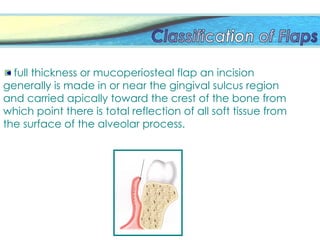
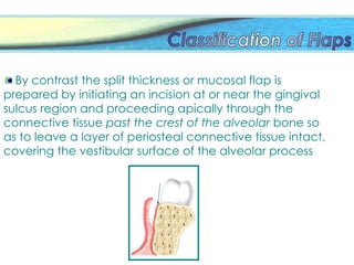
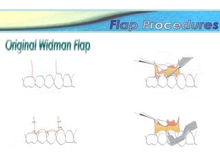
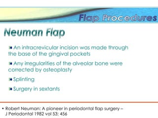
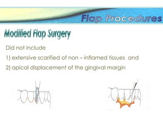
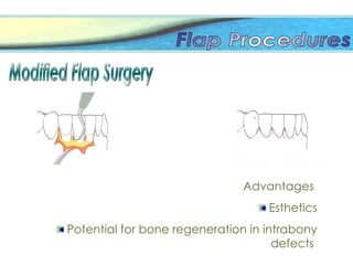
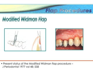
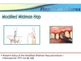
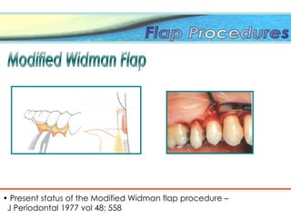
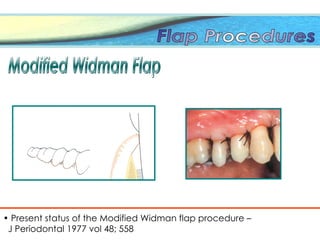
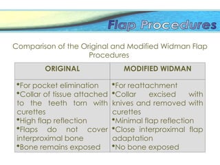
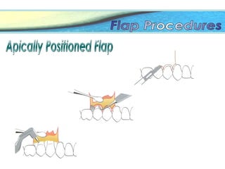
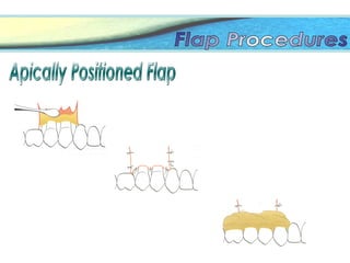
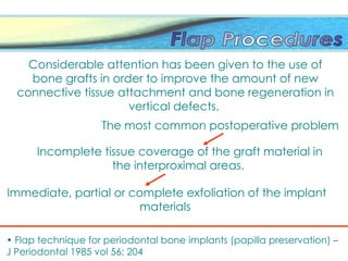
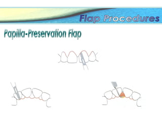
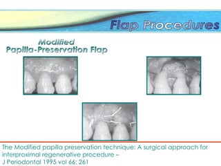
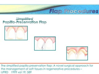
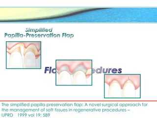
It is one of the most ubiquitous diseases
known to man
No race is immune; no region is free from the
its widespread incidence
Essential pathogenesis of this disease is not
well understood and has not revealed to
permit changes in the therapeutic
approaches.
Surgery has been defined as the act and art of
treating diseases or injuries by manual operation
ROBICSEK
(1884)
YOUNGER
(1902)
KIRKLAND
(1931)
NORBERG
(1926)
LEONARD
WIDMAN
(1916)
KRONFELD
(1935)
SCHLUGER
(1949)
NYMAN et al
(1982)
GOLDMAN
(1950)
RAMFJORD;
NISSLE
(1974)
HIRSCHFELD
(1952)
TAKEI et al
(1985)
FRIEDMAN
(1955;1962)
• “Flap is a piece of tissue parity severed from its place
of origin for use in surgical grafting and repair of body
defects”
 “Flat portion of tissue, either skin, mucosa or
mucoperiosteum which is partially severed from its
deeper surroundings.”
• Webster’s Dictionary
 F. J Harty Rogston Concise Illustrated Dental Dictionary
 A Periodontal flap is defined as, “a section of
the gingiva and or oral mucosa, surgically
elevated from the underlying tissues to provide
visibility of the bone and root surface.”
 Carranza’s Clinical periodontology ninth edition
Widman, Cieszynski, and
Neuman “pyorrhea Alveolar and its
Treatment Published in
Berlin in 1912”
Widman in 1916 flap surgery for pocket
elimination
The English edition of widman’s
article in 1918 gives a detailed
description of a mucoperiosteal
flap design
Zentler in 1918 use of a crevicular
mucoperiosteal flap in a
manner similar to what
Neuman
Kirkland in 1931 flap procedure for the purpose
of reattachment
Periodontal flaps can be classified based on the
following:
Bone exposure after flap reflection.
Placement of the flap after surgery
Management of the papilla
Presence / absence of releasing incisions
Full thickness
(mucoperiosteal)
all the soft tissue,
including the periosteum,
is reflected to expose the
underlying bone
Partial thickness (mucosal)
flaps /split thickness flap
includes only the epithelium
and a layer of the underlying
connective tissue. The bone
remains covered by a layer of
connective tissue, including the
periosteum
Bone exposure after flap reflection.
full thickness or mucoperiosteal flap an incision
generally is made in or near the gingival sulcus region
and carried apically toward the crest of the bone from
which point there is total reflection of all soft tissue from
the surface of the alveolar process.
By contrast the split thickness or mucosal flap is
prepared by initiating an incision at or near the gingival
sulcus region and proceeding apically through the
connective tissue past the crest of the alveolar bone so
as to leave a layer of periosteal connective tissue intact,
covering the vestibular surface of the alveolar process
Full thickness
(mucoperiosteal)
Partial thickness (mucosal)
flaps /split thickness flap
Significant difference and Advantages between full
thickness and partial thickness:
The epithelial tissue repairs itself similarly in both procedures.
In both flap procedures there is no significant
variation as to the repair of the connective tissue
The difference in repair between the two flaps
becomes manifest in the region of the alveolar process
In the full thickness flap-the resorbtive
activity at the six to eight-day period affects the entire
layers of circumferential lamellae and a portion of the
Haversian systems that are immediately subjacent to
those lamellae, so it is a distinct quantitative difference
as to the amount of bone that is resorbed.
J Periodontal 1972; 141
Twenty-one day period, where now definite
osteogenesis is characteristic of the alveolar process
associated with the split thickness flap, it is at this time
that one can observe that very little change that took
place by resorption at the crest of the process and only
some on the vestibular surface
Histologically The full thickness flap is a few days behind
the split thickness flap in the repair rate
There are many more osteoclasts and Osteoblasts in
action during their respective times of activity with the
full thickness flap. This again is related to the degree of
damage or trauma by surgery.
Reflecting a split thickness flap achieves thinness with
body and permits its reapposition at the gingival margin
region with it being better contoured and much more
adaptable than the heavy-bodied full thickness
mucoperiosteal flap
Alveolar crest reduction following full and partial
thickness flap:
Kohler and Ramfjord, after full thickness flaps, found
only slight crestal bone loss
Donnenfeld, Marks, and Glickman reported a loss of
crestal alveolar bone after full thickness flaps (1.05 to 1.2
mm)
Pfeifer, in a 21-day study on four patients, reported very
little osteoclastic activity in response to a partial thickness
flap
J Periodontal 1972; 141
Based on flap placement after surgery
undisplaced flaps
Displaced flaps
Displaced flaps have the important
advantage of preserving the outer portion
of the pocket wall and transforming it into
attached gingiva
Based on management of the papilla
conventional
papilla preservation flaps.
conventional flap is used when,
the interdental spaces are too narrow, thereby
precluding the papilla, and
When the flap is to be displaced.
Presence / absence of releasing incisions
Flap with releasing incisions
Envelope flap
Depending on direction of transfer and geometry
Mode of transfer
Rotational: All rotational flaps share the
common characteristic of movement around a
pivot point. The radius of the arc of rotation is
the line of greatest tension. The greater the
rotation, the greater the actual shortening of the
flap.
Mode of transfer
Advancement: Advanced flaps reach their
final site without rotation or any lateral
movement. They can consist of one or more
pedicles. Consists of two straight-line, vertical
incisions with or without 100 to 110 degree back
cuts
Both the advanced flap and the rotational flap can be
further classified according to the geometry of the
flap.
Geometry:
Transposition, A rectangular segment of gingiva and
mucosa is used.
Rotational, A semicircular segment of gingiva or
mucosa is used.
Prevention of flap
necrosis
Prevention of flap
tearing
According to Laskin(1980)
Incision should start adjacent to the operative
area
Incision should avoid transection of major nerves
and vessels
An adequate blood supply
Avoid incisions in an area of thinned mucosa
Releasing incisions if access is inadequate
Laskin, D.M. Oral and Maxillofacial surgery. Vol 1
Three important objectives:
• it removes the pocket lining:
• it conceives the relatively uninvolved positioned
becomes attached gingival. Which, if apical
positioned, becomes attached gingiva and
• it produces a sharp thin flap margin for
adaptation to the bone- tooth junction
Horizontal Incisions
1. The internal bevel incision
2. The cervical incision
3. Interdental incisions
The internal bevel incision
Observations by Cattermole and
Wade (1978),
• Initial greater inflammation in the
linear incision segments than
scalloped incision segments.
• Overall no significant difference
Cervical incision
Interdental incisions
Cut back incision
These three incisions allow the removal of the
gingival around the tooth (i.e. the pocket
epithelium and the adjacent granulomatous
tissue) A curette or a large scalar (U 15/30) can
be used for this purpose.
If no vertical incisions are made the
flap is called an envelope flap.
 Carranza’s Clinical periodontology ninth edition
Vertical incisions must extend beyond the
mucogingival line reaching the alveolar
mucosa to allow for the release of the flap
to be displaced
Vertical Incisions
Vertical or oblique releasing incision can be used on
one or both ends of the horizontal incision depending
on the design and purpose of the flap
vertical incisions in the lingual and
palatal areas are avoided
Vertical Incisions
Incorrect
correct
Flap reflection
Indications
Increasing depth of the periodontal pockets
Increasing width of the tooth surfaces
The presence of root fissures, root concavities,
furcations, and defective margins of dental
restorations in the subgingival area
Reduced accessibility and the presence of one or
several of the above mentioned conditions may
prevent proper debridement of shallow pockets
• Badersten et al. 1981, Lindhe et al., 1982
At the completion of treatment,
No sub or supragingival dental deposits.
No pathologic pockets (no bleeding on probing to
the bottom of the pockets)
No plaque retaining aberrations of gingival
morphology.
No plaque-retaining parts of restorations in relation
to the gingival margin.
Contraindications
•Uncooperative Patient
• Cardiovascular disease
(Fay & O’Neil 1984).
• Blood disorders
• Hormonal disorders
• Smoking
(Siana et al., 1989),
The main advantages of the “original Widman flap”
Healing with primary intention and
That it was possible to reestablish a proper contour of
the alveolar bone in sites with angular bony defects.
• Clinical periodontology and Implantology - Jan Lindhe fourth edition
• Robert Neuman: A pioneer in periodontal flap surgery –
J Periodontal 1982 vol 53; 456
An intracrevicular incision was made through
the base of the gingival pockets
Any irregularities of the alveolar bone were
corrected by osteoplasty
Splinting
Surgery in sextants
Did not include
1) extensive scarified of non – inflamed tissues and
2) apical displacement of the gingival margin
Advantages
Esthetics
Potential for bone regeneration in intrabony
defects
• Present status of the Modified Widman flap procedure –
J Periodontal 1977 vol 48; 558
• Present status of the Modified Widman flap procedure –
J Periodontal 1977 vol 48; 558
• Present status of the Modified Widman flap procedure –
J Periodontal 1977 vol 48; 558
• Present status of the Modified Widman flap procedure –
J Periodontal 1977 vol 48; 558
The ultrastucture of the reformed epithelium with
hemidesmosomes, basement lamina and several layers
of elongated epithelial cells parallel to the tooth
surface.
Listgarten; Frank et al
• Present status of the Modified Widman flap procedure –
J Periodontal 1977 vol 48; 558
Advantages and disadvantages of the modified widman
flap
Establishing an intimate post operative adaptation of
healthy connective tissue and normal epithelium to
contacting tooth surface
Access for proper instrumentation
Advantages and disadvantages of the modified widman
flap
One apparent disadvantage of the modified
Widman flap surgery is the flat or concave interproximal
architecture
 If meticulous oral hygiene is maintained, the
interproximal tissues will regenerate over a few months
with gain rather than loss of attachment
The Michigan Study
In 1961 the first report from the first longitudinal study
Scaling and root planning, oral hygiene
instruction, and occlusal adjustment.
Subgingival
curettage
Pocket elimination
surgery
The greatest gain was obtained with the modified
Widman flap, followed by subgingival curettage, and
then pockets elimination surgery. When severe pockets
were treated, the modified Widman flap produced a
gain that was significantly better than that obtained
with the other two techniques.
Subgingival curettage, Modified Widman flap, or
Pocket elimination surgery
when gain in attachment was considered, there were no
differences among techniques.
Comparison of the Original and Modified Widman Flap
Procedures
ORIGINAL MODIFIED WIDMAN
For pocket elimination
Collar of tissue attached
to the teeth torn with
curettes
High flap reflection
Flaps do not cover
interproximal bone
Bone remains exposed
For reattachment
Collar excised with
knives and removed with
curettes
Minimal flap reflection
Close interproximal flap
adaptation
No bone exposed
Minimum pocket depth postoperatively.
If optimal soft tissue coverage of the alveolar bone is
obtained the post sugical bone loss is minimal.
The postoperative position of the gingiva margin may
be controlled and the entire mucogingival complex may
be maintained.
Advantages
Treatment of periodontal pockets on the distal surface
of distal molars is complicated by a presence of
bulbous tissues over the tuberosity or by a prominent
retromolar pad.
Gingivectomy
procedure
Distal wedge procedure
(Robinson 1966)
Incomplete tissue coverage of the graft material in
the interproximal areas.
Considerable attention has been given to the use of
bone grafts in order to improve the amount of new
connective tissue attachment and bone regeneration in
vertical defects.
The most common postoperative problem
Immediate, partial or complete exfoliation of the implant
materials
• Flap technique for periodontal bone implants (papilla preservation) –
J Periodontal 1985 vol 56; 204
The Modified papilla preservation technique: A surgical approach for
interproximal regenerative procedure –
J Periodontal 1995 vol 66; 261
The Modified papilla preservation technique: A surgical approach for
interproximal regenerative procedure –
J Periodontal 1995 vol 66; 261
Horizontal and vertical mattress suturing is done
The simplified papilla preservation flap: A novel surgical approach for
the management of soft tissues in regenerative procedures –
IJPRD 1999 vol 19; 589
The simplified papilla preservation flap: A novel surgical approach for
the management of soft tissues in regenerative procedures –
IJPRD 1999 vol 19; 589
The simplified papilla preservation flap: A novel surgical approach for
the management of soft tissues in regenerative procedures –
IJPRD 1999 vol 19; 589
The simplified papilla preservation flap: A novel surgical approach for
the management of soft tissues in regenerative procedures –
IJPRD 1999 vol 19; 589
Grupe and Warren
(1956)
Norberg (1926);
Bernimoulin et al (1975)
Tarnow (1986)
This design minimizes the amount of vascular
embarrassment and sloughing of the coronal edge of
the flap because the base of the flap is wider
preserving an adequate blood supply.
A surgical Modification for Implant Fixture
Installation
Vertical incisions can be placed to ease the outer buccal
flap elevation with out any significant compromise in blood
supply.
• The overlapped flap: A surgical modification for implant fixture
installation –
IJPRD 1990 vol 10; 209
Langer and Langer
(1990)
• The overlapped flap: A surgical modification for implant fixture
installation –
IJPRD 1990 vol 10; 209
• The overlapped flap: A surgical modification for implant fixture
installation –
IJPRD 1990 vol 10; 209
• The overlapped flap: A surgical modification for implant fixture
installation –
IJPRD 1990 vol 10; 209
The eversed crestal flap: A surgical modification in endosseous implant
procedures –
Qunt int 1994 vol 25; 229
Landsberg (1994)
Sulcular flaps remain the most frequently used in endodontic
surgery
(Beer et al 2000)
The main disadvantage of these are recession and
especially, unpredictable shrinkage of the papilla during
healing
(zimmermann et al 2001)
• Papilla base incision: a new approach to recession free healing of the
interdental papilla after endodontic surgery –
Int Endo Jol 2003 vol 35; 453
A new approach to recession – free healing of the
interdental papilla after endodontic surgery
Velvart. P
Hemorrhage associated with surgery is a common
problem which requires proper management.
Definitive data regarding surgical blood loss was
unavailable until 1924 first studied operative
hemorrhage during general surgery.
Gatch and Little in 1924 first studied operative
hemorrhage during general surgery
•Baab, D. A., Ammons, W. F., Selipsky, H. Blood loss during periodontal
surgery. J Periodontol 1977; 48: 693
Mclvor and Wengraf studied blood loss
calorimetrically during gingivectomies and / or
isolated periodontal flap procedures on 14 patients.
(12 – 62 ml)
Berdon 12 published the first report on hemorrhage
during periodontal surgery. Using a
cyanmethemoglobin comparison technique. He
established that approximately 5 ml to 149 ml of blood
was lost.
Ariaudo (1970) estimated that full mouth periodontal
flap procedures resulted in 350ml blood loss.
•Baab, D. A., Ammons, W. F., Selipsky, H. Blood loss during periodontal
surgery. J Periodontol 1977; 48: 693
Factors affecting blood loss
• Systemic factors
 Age
 Blood pressure
 Bleeding time
Duration of Surgery
extent of surgical field
degree of inflammation
no of teeth involved
anesthesia
length of incision
• Local factors
• Surgical technique
Flap survival
• Periodontal reconstructive flaps classification and surgical
considerations – IJPRD 1991 vol 11; 481
The final pattern should be larger than the area to be
reconstructed.
Flap survival
The surgeon should consider all possible designs and
factors that may increase flap survival.
All phases of flap transformation should be considered
including possible shortening of the flap and the desired
angles and vectors of movement
Specific attention should be given to the length of the
pattern to avoid tension or sinking of the flap.
• Periodontal reconstructive flaps classification and surgical
considerations – IJPRD 1991 vol 11; 481
Flap survival
• Periodontal reconstructive flaps classification and surgical
considerations – IJPRD 1991 vol 11; 481
Flap necrosis
Atraumatic technique
Atraumatic and gentle surgical techniques should be
practiced throughout the surgical procedure
Hot sponges
(66 degrees)
Promote
coagulation
Increase capillary
bleeding
Increase tissue
damage
Incidence of
wound infection
Tissue necrosis
When the outline crosses two dissimilar surfaces, for
example,
Gingiva and mucosa, the surgeon should place the
mucosa under tension and commence incisions from the
less firm surface, from mucosa to gingiva.
The initial phase of Atraumatic surgery consists of an
outline of the recipient and donor sites as well as the
transfer phases
The optimal time for suture removal is when the
tensile strength of the healing wound exceeds the
strength of the suture and is sufficient to maintain
the approximation without assistance.
Wound closure
The relationship between the sutures and wound edges is
important
Tension on the
sutures
Postoperative
swelling
Reduce
circulation
Further edge
separation
Surgical injury creates the environment and stimulus
for cellular differentiation
Primary wound
healing
Secondary wound
healing
Tertiary wound
healing
The migration takes place at a rate of 0.5 mm per
day
Epithelium
Epithelial cells begin to proliferate at wound margins
at 1-2 days.
A replaced flap may be sealed to the tooth in 2-4
days.
Epithelialization and the formation of a junctional
epithelium are complete by the end of the
second week
Connective tissue
Fibroblasts begin to proliferate after day 2 with evidence of
collagen synthesis in the wound by day 4.
In the most uncomplicated periodontal surgery, restoration
of gingival connective tissue will be complete in 4-6 weeks
Alveolar bone
Reactive bone resorption Osteoclasts appear in the
wound at about day 4 and display peak osteoclastic
activity at day 10
Osteoblasts begin to appear and proliferate in the
wound during the second week, and they display
peak osteoblastic activity by the end of the third
week.
Bone remodeling and maturation is a feature of the third
postoper­
ative month.
Proliferating cementoblasts appear adjacent to root
surfaces at the end of month one and proceed with
cementogenesis during the months two and three.
Cementum
Cementogenesis delayed in onset; it is also slow to
exert its effect on the overall outcome of
periodontal healing
These events are critical to the formation of a new
attachment apparatus and some forms of new
attachment
New techniques in periodontal surgery are being developed
which aim to conserve rather than discard periodontal tissue.
Surgical procedures should be designed with these facts in
mind:
• Use of full thickness and partial thickness
• Reflecting flap as much gingiva as possible should be retained
• Flap should be reflected in relaxed manner
• Vertical releasing incisions should be given where they are
necessary
• Prevent perforation of the flap
• Attention should be paid during suturing of the flaps
PERIODONTAL FLAP SURGERY  study perio(2).ppt

PERIODONTAL FLAP SURGERY study perio(2).ppt

  • 2.
    It is oneof the most ubiquitous diseases known to man No race is immune; no region is free from the its widespread incidence Essential pathogenesis of this disease is not well understood and has not revealed to permit changes in the therapeutic approaches.
  • 4.
    Surgery has beendefined as the act and art of treating diseases or injuries by manual operation
  • 6.
  • 7.
  • 9.
    • “Flap isa piece of tissue parity severed from its place of origin for use in surgical grafting and repair of body defects”  “Flat portion of tissue, either skin, mucosa or mucoperiosteum which is partially severed from its deeper surroundings.” • Webster’s Dictionary  F. J Harty Rogston Concise Illustrated Dental Dictionary
  • 10.
     A Periodontalflap is defined as, “a section of the gingiva and or oral mucosa, surgically elevated from the underlying tissues to provide visibility of the bone and root surface.”  Carranza’s Clinical periodontology ninth edition
  • 11.
    Widman, Cieszynski, and Neuman“pyorrhea Alveolar and its Treatment Published in Berlin in 1912” Widman in 1916 flap surgery for pocket elimination The English edition of widman’s article in 1918 gives a detailed description of a mucoperiosteal flap design
  • 12.
    Zentler in 1918use of a crevicular mucoperiosteal flap in a manner similar to what Neuman Kirkland in 1931 flap procedure for the purpose of reattachment
  • 13.
    Periodontal flaps canbe classified based on the following: Bone exposure after flap reflection. Placement of the flap after surgery Management of the papilla Presence / absence of releasing incisions
  • 14.
    Full thickness (mucoperiosteal) all thesoft tissue, including the periosteum, is reflected to expose the underlying bone Partial thickness (mucosal) flaps /split thickness flap includes only the epithelium and a layer of the underlying connective tissue. The bone remains covered by a layer of connective tissue, including the periosteum Bone exposure after flap reflection.
  • 15.
    full thickness ormucoperiosteal flap an incision generally is made in or near the gingival sulcus region and carried apically toward the crest of the bone from which point there is total reflection of all soft tissue from the surface of the alveolar process.
  • 16.
    By contrast thesplit thickness or mucosal flap is prepared by initiating an incision at or near the gingival sulcus region and proceeding apically through the connective tissue past the crest of the alveolar bone so as to leave a layer of periosteal connective tissue intact, covering the vestibular surface of the alveolar process
  • 17.
    Full thickness (mucoperiosteal) Partial thickness(mucosal) flaps /split thickness flap
  • 18.
    Significant difference andAdvantages between full thickness and partial thickness: The epithelial tissue repairs itself similarly in both procedures. In both flap procedures there is no significant variation as to the repair of the connective tissue The difference in repair between the two flaps becomes manifest in the region of the alveolar process
  • 19.
    In the fullthickness flap-the resorbtive activity at the six to eight-day period affects the entire layers of circumferential lamellae and a portion of the Haversian systems that are immediately subjacent to those lamellae, so it is a distinct quantitative difference as to the amount of bone that is resorbed. J Periodontal 1972; 141
  • 20.
    Twenty-one day period,where now definite osteogenesis is characteristic of the alveolar process associated with the split thickness flap, it is at this time that one can observe that very little change that took place by resorption at the crest of the process and only some on the vestibular surface
  • 21.
    Histologically The fullthickness flap is a few days behind the split thickness flap in the repair rate There are many more osteoclasts and Osteoblasts in action during their respective times of activity with the full thickness flap. This again is related to the degree of damage or trauma by surgery. Reflecting a split thickness flap achieves thinness with body and permits its reapposition at the gingival margin region with it being better contoured and much more adaptable than the heavy-bodied full thickness mucoperiosteal flap
  • 22.
    Alveolar crest reductionfollowing full and partial thickness flap: Kohler and Ramfjord, after full thickness flaps, found only slight crestal bone loss Donnenfeld, Marks, and Glickman reported a loss of crestal alveolar bone after full thickness flaps (1.05 to 1.2 mm) Pfeifer, in a 21-day study on four patients, reported very little osteoclastic activity in response to a partial thickness flap J Periodontal 1972; 141
  • 23.
    Based on flapplacement after surgery undisplaced flaps Displaced flaps Displaced flaps have the important advantage of preserving the outer portion of the pocket wall and transforming it into attached gingiva
  • 24.
    Based on managementof the papilla conventional papilla preservation flaps. conventional flap is used when, the interdental spaces are too narrow, thereby precluding the papilla, and When the flap is to be displaced.
  • 25.
    Presence / absenceof releasing incisions Flap with releasing incisions Envelope flap
  • 26.
    Depending on directionof transfer and geometry Mode of transfer Rotational: All rotational flaps share the common characteristic of movement around a pivot point. The radius of the arc of rotation is the line of greatest tension. The greater the rotation, the greater the actual shortening of the flap.
  • 27.
    Mode of transfer Advancement:Advanced flaps reach their final site without rotation or any lateral movement. They can consist of one or more pedicles. Consists of two straight-line, vertical incisions with or without 100 to 110 degree back cuts
  • 28.
    Both the advancedflap and the rotational flap can be further classified according to the geometry of the flap. Geometry: Transposition, A rectangular segment of gingiva and mucosa is used. Rotational, A semicircular segment of gingiva or mucosa is used.
  • 29.
  • 30.
    According to Laskin(1980) Incisionshould start adjacent to the operative area Incision should avoid transection of major nerves and vessels An adequate blood supply Avoid incisions in an area of thinned mucosa Releasing incisions if access is inadequate Laskin, D.M. Oral and Maxillofacial surgery. Vol 1
  • 31.
    Three important objectives: •it removes the pocket lining: • it conceives the relatively uninvolved positioned becomes attached gingival. Which, if apical positioned, becomes attached gingiva and • it produces a sharp thin flap margin for adaptation to the bone- tooth junction Horizontal Incisions 1. The internal bevel incision 2. The cervical incision 3. Interdental incisions
  • 32.
    The internal bevelincision Observations by Cattermole and Wade (1978), • Initial greater inflammation in the linear incision segments than scalloped incision segments. • Overall no significant difference
  • 33.
  • 34.
    These three incisionsallow the removal of the gingival around the tooth (i.e. the pocket epithelium and the adjacent granulomatous tissue) A curette or a large scalar (U 15/30) can be used for this purpose. If no vertical incisions are made the flap is called an envelope flap.  Carranza’s Clinical periodontology ninth edition
  • 35.
    Vertical incisions mustextend beyond the mucogingival line reaching the alveolar mucosa to allow for the release of the flap to be displaced Vertical Incisions Vertical or oblique releasing incision can be used on one or both ends of the horizontal incision depending on the design and purpose of the flap vertical incisions in the lingual and palatal areas are avoided
  • 36.
  • 37.
  • 38.
    Indications Increasing depth ofthe periodontal pockets Increasing width of the tooth surfaces The presence of root fissures, root concavities, furcations, and defective margins of dental restorations in the subgingival area Reduced accessibility and the presence of one or several of the above mentioned conditions may prevent proper debridement of shallow pockets • Badersten et al. 1981, Lindhe et al., 1982
  • 39.
    At the completionof treatment, No sub or supragingival dental deposits. No pathologic pockets (no bleeding on probing to the bottom of the pockets) No plaque retaining aberrations of gingival morphology. No plaque-retaining parts of restorations in relation to the gingival margin.
  • 41.
    Contraindications •Uncooperative Patient • Cardiovasculardisease (Fay & O’Neil 1984). • Blood disorders • Hormonal disorders • Smoking (Siana et al., 1989),
  • 44.
    The main advantagesof the “original Widman flap” Healing with primary intention and That it was possible to reestablish a proper contour of the alveolar bone in sites with angular bony defects. • Clinical periodontology and Implantology - Jan Lindhe fourth edition
  • 46.
    • Robert Neuman:A pioneer in periodontal flap surgery – J Periodontal 1982 vol 53; 456 An intracrevicular incision was made through the base of the gingival pockets Any irregularities of the alveolar bone were corrected by osteoplasty Splinting Surgery in sextants
  • 47.
    Did not include 1)extensive scarified of non – inflamed tissues and 2) apical displacement of the gingival margin
  • 48.
    Advantages Esthetics Potential for boneregeneration in intrabony defects
  • 49.
    • Present statusof the Modified Widman flap procedure – J Periodontal 1977 vol 48; 558
  • 50.
    • Present statusof the Modified Widman flap procedure – J Periodontal 1977 vol 48; 558
  • 51.
    • Present statusof the Modified Widman flap procedure – J Periodontal 1977 vol 48; 558
  • 52.
    • Present statusof the Modified Widman flap procedure – J Periodontal 1977 vol 48; 558
  • 53.
    The ultrastucture ofthe reformed epithelium with hemidesmosomes, basement lamina and several layers of elongated epithelial cells parallel to the tooth surface. Listgarten; Frank et al • Present status of the Modified Widman flap procedure – J Periodontal 1977 vol 48; 558 Advantages and disadvantages of the modified widman flap Establishing an intimate post operative adaptation of healthy connective tissue and normal epithelium to contacting tooth surface Access for proper instrumentation
  • 54.
    Advantages and disadvantagesof the modified widman flap One apparent disadvantage of the modified Widman flap surgery is the flat or concave interproximal architecture  If meticulous oral hygiene is maintained, the interproximal tissues will regenerate over a few months with gain rather than loss of attachment
  • 55.
    The Michigan Study In1961 the first report from the first longitudinal study Scaling and root planning, oral hygiene instruction, and occlusal adjustment. Subgingival curettage Pocket elimination surgery
  • 56.
    The greatest gainwas obtained with the modified Widman flap, followed by subgingival curettage, and then pockets elimination surgery. When severe pockets were treated, the modified Widman flap produced a gain that was significantly better than that obtained with the other two techniques. Subgingival curettage, Modified Widman flap, or Pocket elimination surgery when gain in attachment was considered, there were no differences among techniques.
  • 57.
    Comparison of theOriginal and Modified Widman Flap Procedures ORIGINAL MODIFIED WIDMAN For pocket elimination Collar of tissue attached to the teeth torn with curettes High flap reflection Flaps do not cover interproximal bone Bone remains exposed For reattachment Collar excised with knives and removed with curettes Minimal flap reflection Close interproximal flap adaptation No bone exposed
  • 58.
    Minimum pocket depthpostoperatively. If optimal soft tissue coverage of the alveolar bone is obtained the post sugical bone loss is minimal. The postoperative position of the gingiva margin may be controlled and the entire mucogingival complex may be maintained. Advantages
  • 61.
    Treatment of periodontalpockets on the distal surface of distal molars is complicated by a presence of bulbous tissues over the tuberosity or by a prominent retromolar pad. Gingivectomy procedure Distal wedge procedure (Robinson 1966)
  • 63.
    Incomplete tissue coverageof the graft material in the interproximal areas. Considerable attention has been given to the use of bone grafts in order to improve the amount of new connective tissue attachment and bone regeneration in vertical defects. The most common postoperative problem Immediate, partial or complete exfoliation of the implant materials • Flap technique for periodontal bone implants (papilla preservation) – J Periodontal 1985 vol 56; 204
  • 66.
    The Modified papillapreservation technique: A surgical approach for interproximal regenerative procedure – J Periodontal 1995 vol 66; 261
  • 67.
    The Modified papillapreservation technique: A surgical approach for interproximal regenerative procedure – J Periodontal 1995 vol 66; 261 Horizontal and vertical mattress suturing is done
  • 68.
    The simplified papillapreservation flap: A novel surgical approach for the management of soft tissues in regenerative procedures – IJPRD 1999 vol 19; 589
  • 69.
    The simplified papillapreservation flap: A novel surgical approach for the management of soft tissues in regenerative procedures – IJPRD 1999 vol 19; 589
  • 70.
    The simplified papillapreservation flap: A novel surgical approach for the management of soft tissues in regenerative procedures – IJPRD 1999 vol 19; 589
  • 71.
    The simplified papillapreservation flap: A novel surgical approach for the management of soft tissues in regenerative procedures – IJPRD 1999 vol 19; 589
  • 72.
  • 73.
  • 75.
  • 76.
    This design minimizesthe amount of vascular embarrassment and sloughing of the coronal edge of the flap because the base of the flap is wider preserving an adequate blood supply. A surgical Modification for Implant Fixture Installation Vertical incisions can be placed to ease the outer buccal flap elevation with out any significant compromise in blood supply. • The overlapped flap: A surgical modification for implant fixture installation – IJPRD 1990 vol 10; 209 Langer and Langer (1990)
  • 77.
    • The overlappedflap: A surgical modification for implant fixture installation – IJPRD 1990 vol 10; 209
  • 78.
    • The overlappedflap: A surgical modification for implant fixture installation – IJPRD 1990 vol 10; 209
  • 79.
    • The overlappedflap: A surgical modification for implant fixture installation – IJPRD 1990 vol 10; 209
  • 81.
    The eversed crestalflap: A surgical modification in endosseous implant procedures – Qunt int 1994 vol 25; 229 Landsberg (1994)
  • 83.
    Sulcular flaps remainthe most frequently used in endodontic surgery (Beer et al 2000) The main disadvantage of these are recession and especially, unpredictable shrinkage of the papilla during healing (zimmermann et al 2001) • Papilla base incision: a new approach to recession free healing of the interdental papilla after endodontic surgery – Int Endo Jol 2003 vol 35; 453 A new approach to recession – free healing of the interdental papilla after endodontic surgery
  • 84.
  • 85.
    Hemorrhage associated withsurgery is a common problem which requires proper management. Definitive data regarding surgical blood loss was unavailable until 1924 first studied operative hemorrhage during general surgery. Gatch and Little in 1924 first studied operative hemorrhage during general surgery •Baab, D. A., Ammons, W. F., Selipsky, H. Blood loss during periodontal surgery. J Periodontol 1977; 48: 693
  • 86.
    Mclvor and Wengrafstudied blood loss calorimetrically during gingivectomies and / or isolated periodontal flap procedures on 14 patients. (12 – 62 ml) Berdon 12 published the first report on hemorrhage during periodontal surgery. Using a cyanmethemoglobin comparison technique. He established that approximately 5 ml to 149 ml of blood was lost. Ariaudo (1970) estimated that full mouth periodontal flap procedures resulted in 350ml blood loss. •Baab, D. A., Ammons, W. F., Selipsky, H. Blood loss during periodontal surgery. J Periodontol 1977; 48: 693
  • 87.
    Factors affecting bloodloss • Systemic factors  Age  Blood pressure  Bleeding time Duration of Surgery extent of surgical field degree of inflammation no of teeth involved anesthesia length of incision • Local factors • Surgical technique
  • 88.
    Flap survival • Periodontalreconstructive flaps classification and surgical considerations – IJPRD 1991 vol 11; 481
  • 89.
    The final patternshould be larger than the area to be reconstructed. Flap survival The surgeon should consider all possible designs and factors that may increase flap survival. All phases of flap transformation should be considered including possible shortening of the flap and the desired angles and vectors of movement Specific attention should be given to the length of the pattern to avoid tension or sinking of the flap. • Periodontal reconstructive flaps classification and surgical considerations – IJPRD 1991 vol 11; 481
  • 90.
    Flap survival • Periodontalreconstructive flaps classification and surgical considerations – IJPRD 1991 vol 11; 481 Flap necrosis
  • 91.
    Atraumatic technique Atraumatic andgentle surgical techniques should be practiced throughout the surgical procedure Hot sponges (66 degrees) Promote coagulation Increase capillary bleeding Increase tissue damage Incidence of wound infection Tissue necrosis
  • 92.
    When the outlinecrosses two dissimilar surfaces, for example, Gingiva and mucosa, the surgeon should place the mucosa under tension and commence incisions from the less firm surface, from mucosa to gingiva. The initial phase of Atraumatic surgery consists of an outline of the recipient and donor sites as well as the transfer phases
  • 93.
    The optimal timefor suture removal is when the tensile strength of the healing wound exceeds the strength of the suture and is sufficient to maintain the approximation without assistance. Wound closure The relationship between the sutures and wound edges is important Tension on the sutures Postoperative swelling Reduce circulation Further edge separation
  • 94.
    Surgical injury createsthe environment and stimulus for cellular differentiation Primary wound healing Secondary wound healing Tertiary wound healing
  • 95.
    The migration takesplace at a rate of 0.5 mm per day Epithelium Epithelial cells begin to proliferate at wound margins at 1-2 days. A replaced flap may be sealed to the tooth in 2-4 days. Epithelialization and the formation of a junctional epithelium are complete by the end of the second week
  • 96.
    Connective tissue Fibroblasts beginto proliferate after day 2 with evidence of collagen synthesis in the wound by day 4. In the most uncomplicated periodontal surgery, restoration of gingival connective tissue will be complete in 4-6 weeks
  • 97.
    Alveolar bone Reactive boneresorption Osteoclasts appear in the wound at about day 4 and display peak osteoclastic activity at day 10 Osteoblasts begin to appear and proliferate in the wound during the second week, and they display peak osteoblastic activity by the end of the third week. Bone remodeling and maturation is a feature of the third postoper­ ative month.
  • 98.
    Proliferating cementoblasts appearadjacent to root surfaces at the end of month one and proceed with cementogenesis during the months two and three. Cementum Cementogenesis delayed in onset; it is also slow to exert its effect on the overall outcome of periodontal healing These events are critical to the formation of a new attachment apparatus and some forms of new attachment
  • 99.
    New techniques inperiodontal surgery are being developed which aim to conserve rather than discard periodontal tissue. Surgical procedures should be designed with these facts in mind: • Use of full thickness and partial thickness • Reflecting flap as much gingiva as possible should be retained • Flap should be reflected in relaxed manner • Vertical releasing incisions should be given where they are necessary • Prevent perforation of the flap • Attention should be paid during suturing of the flaps