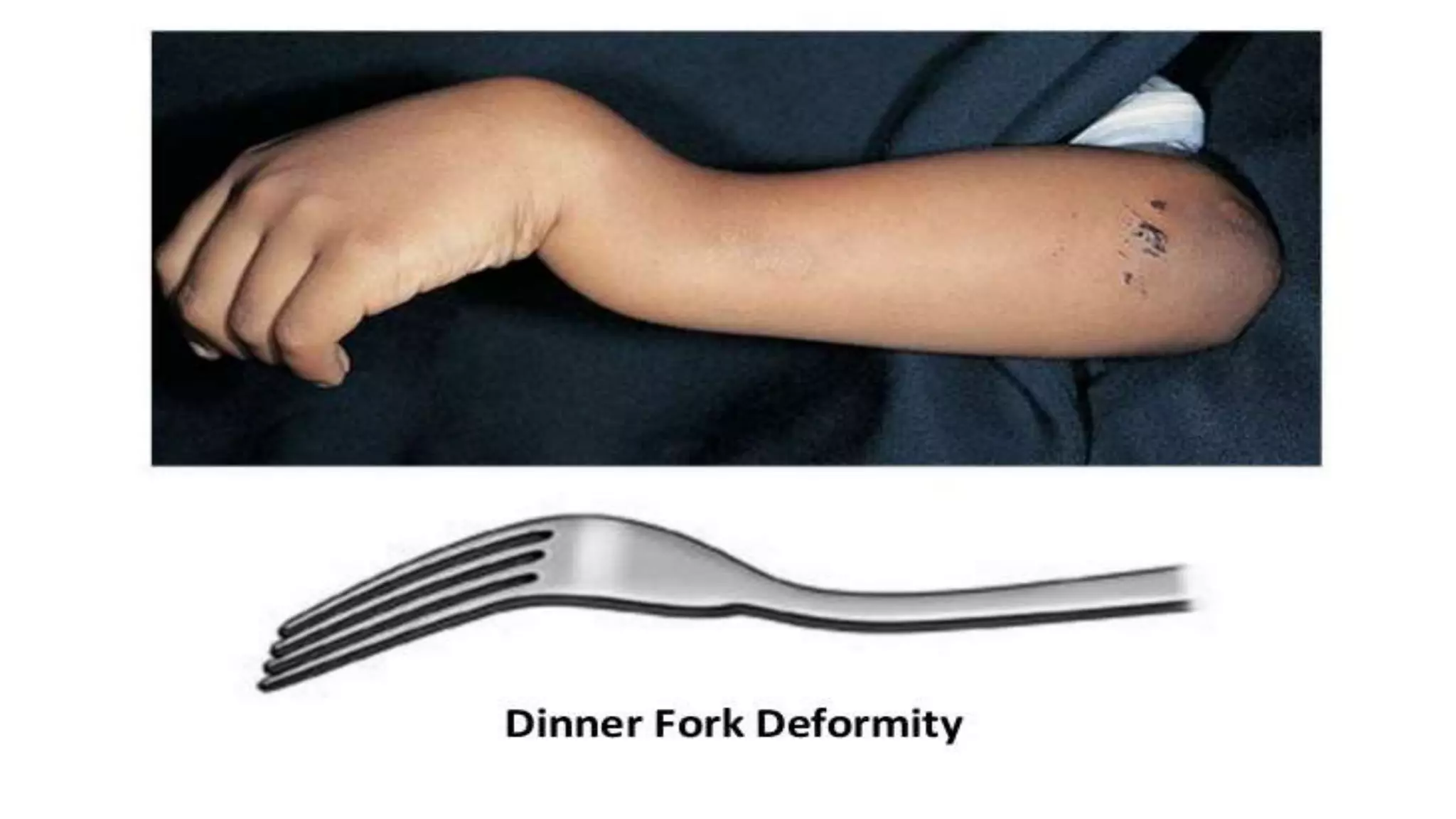
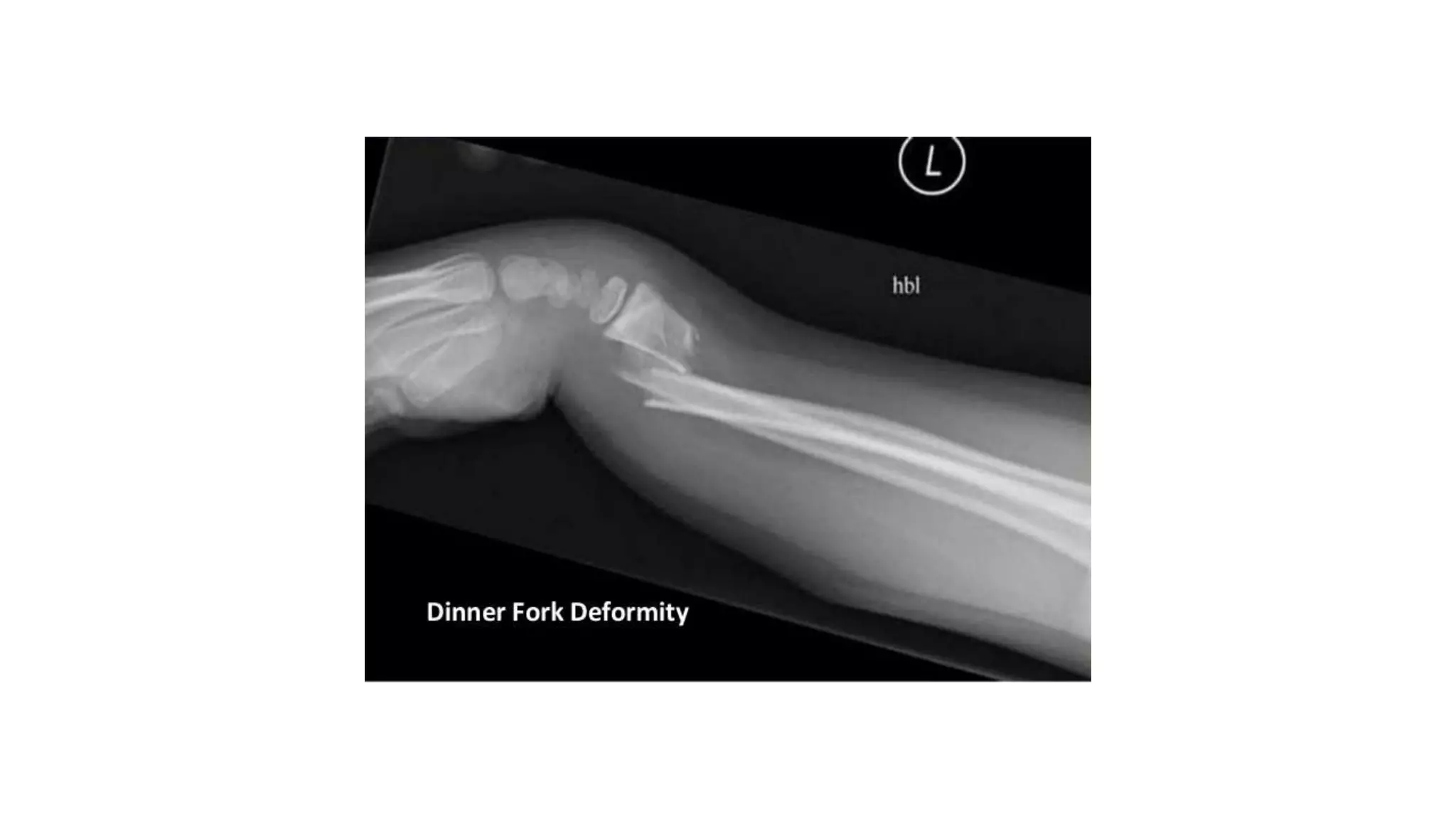
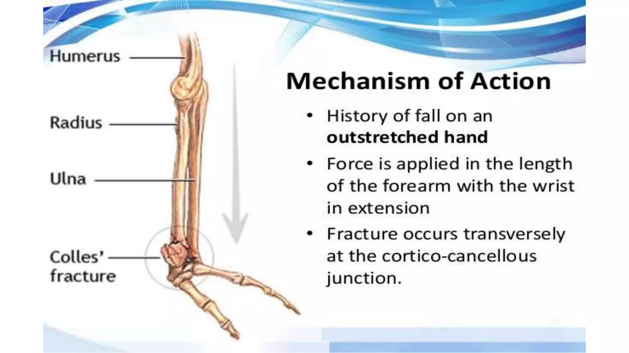
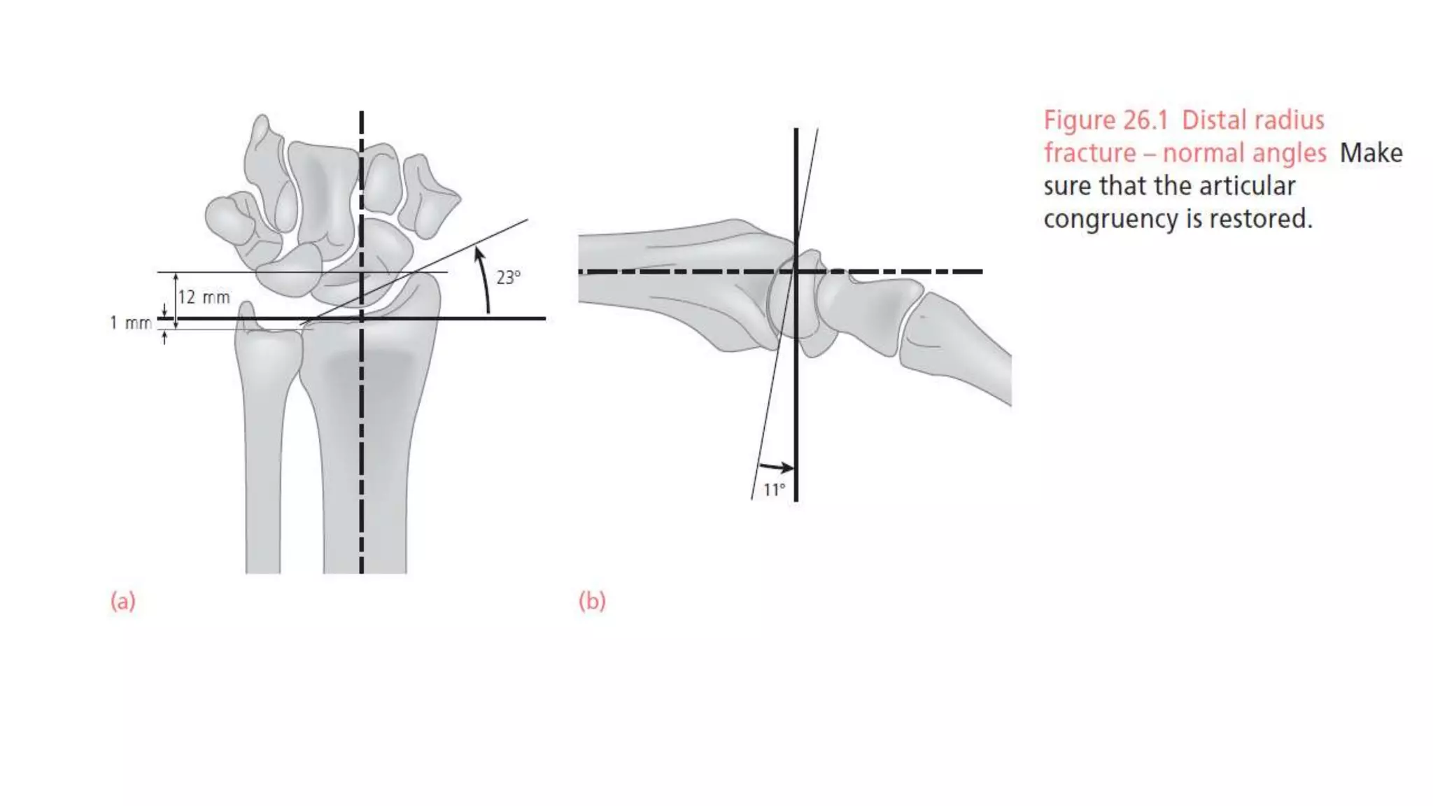
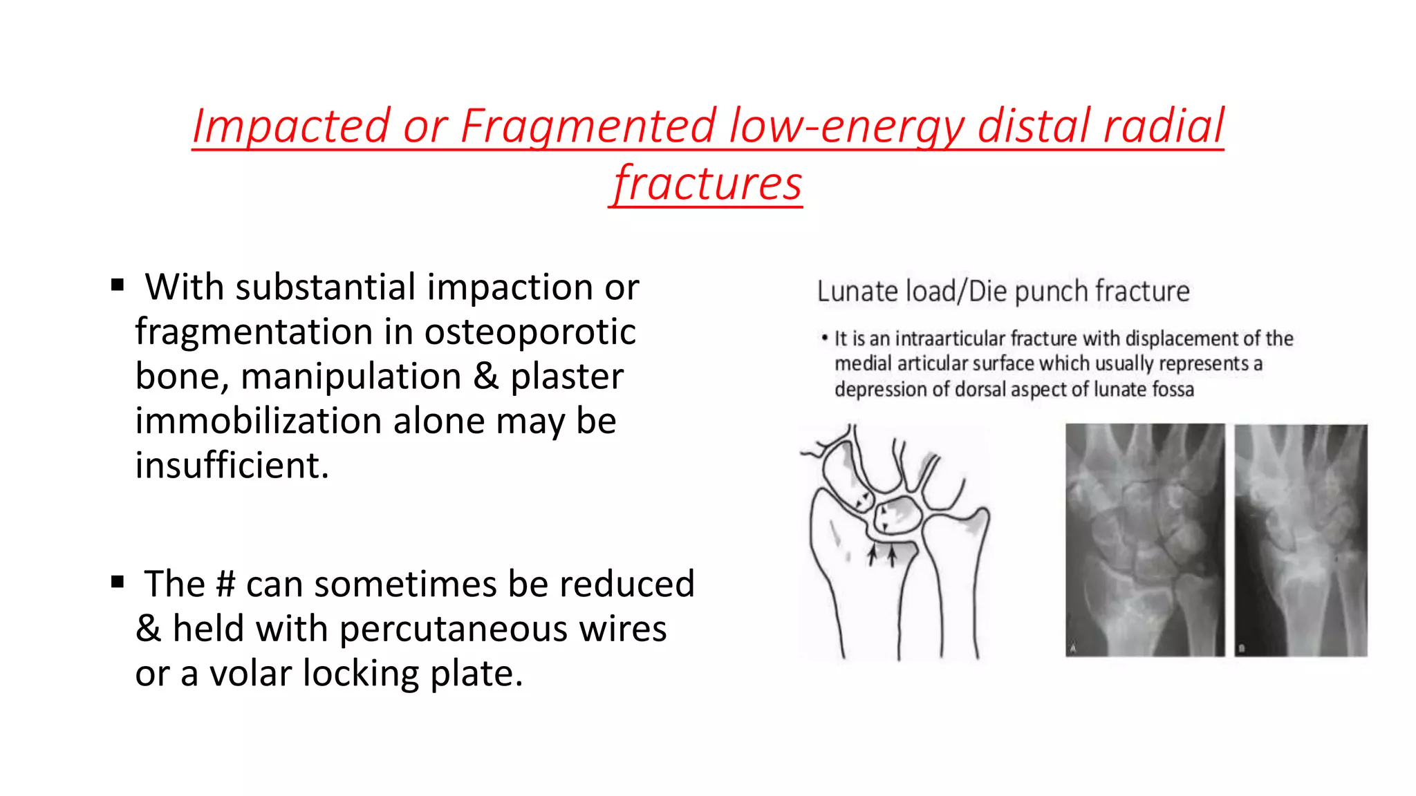
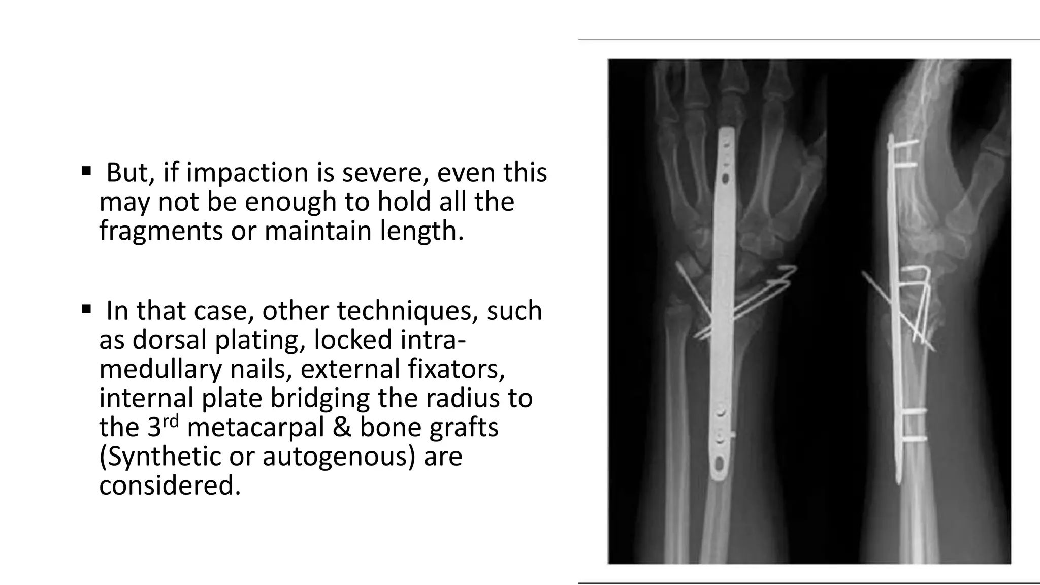
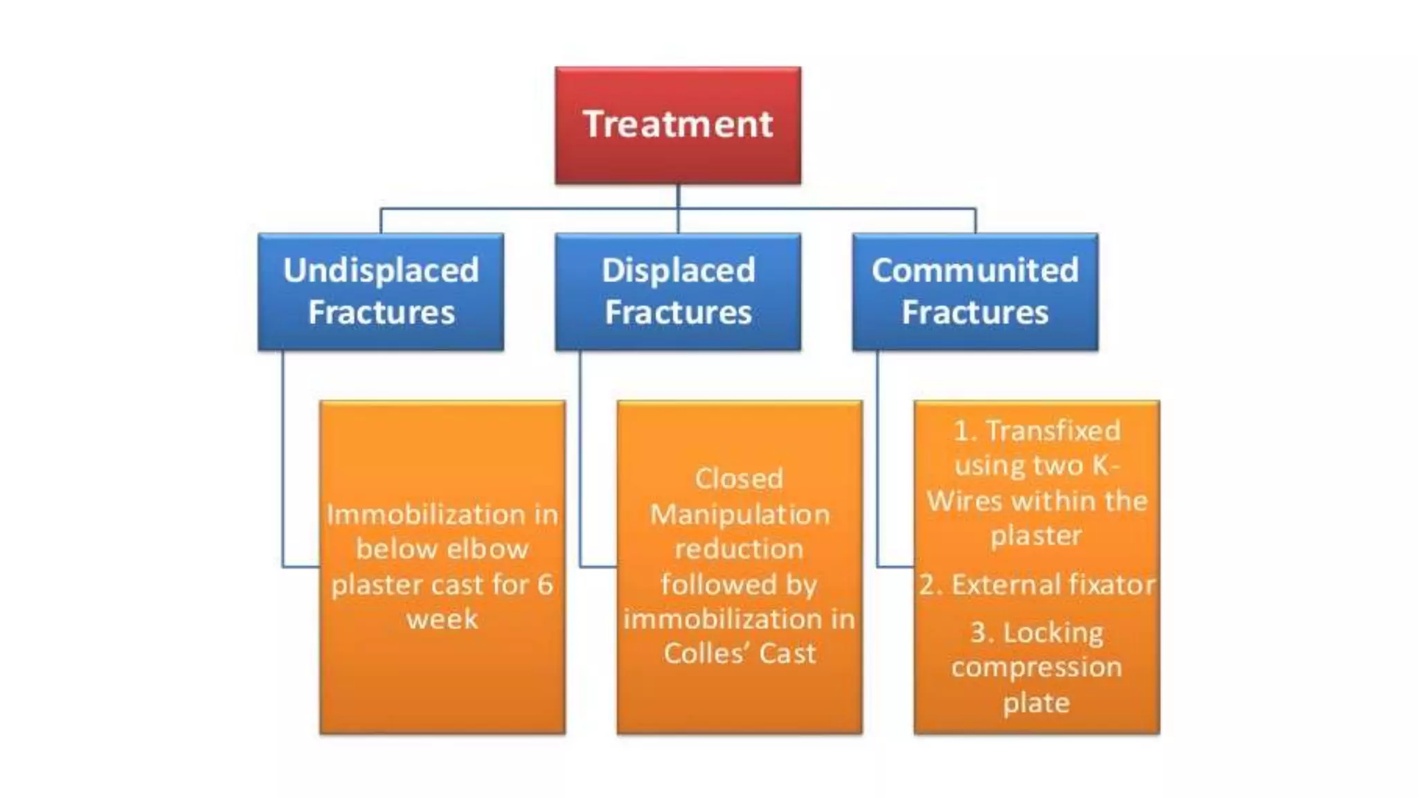
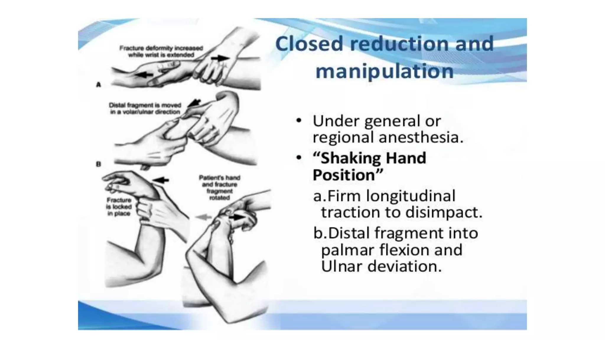
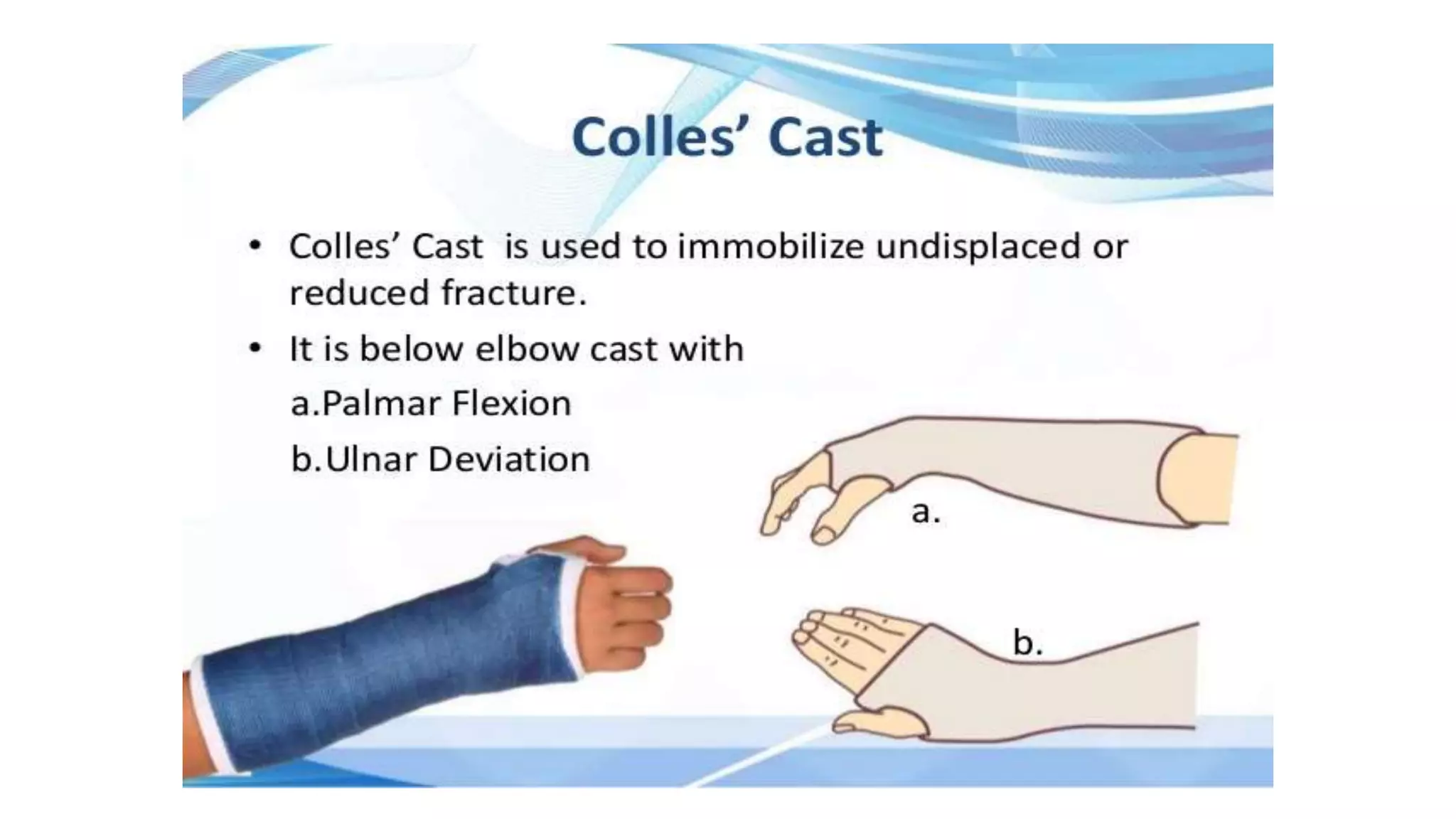
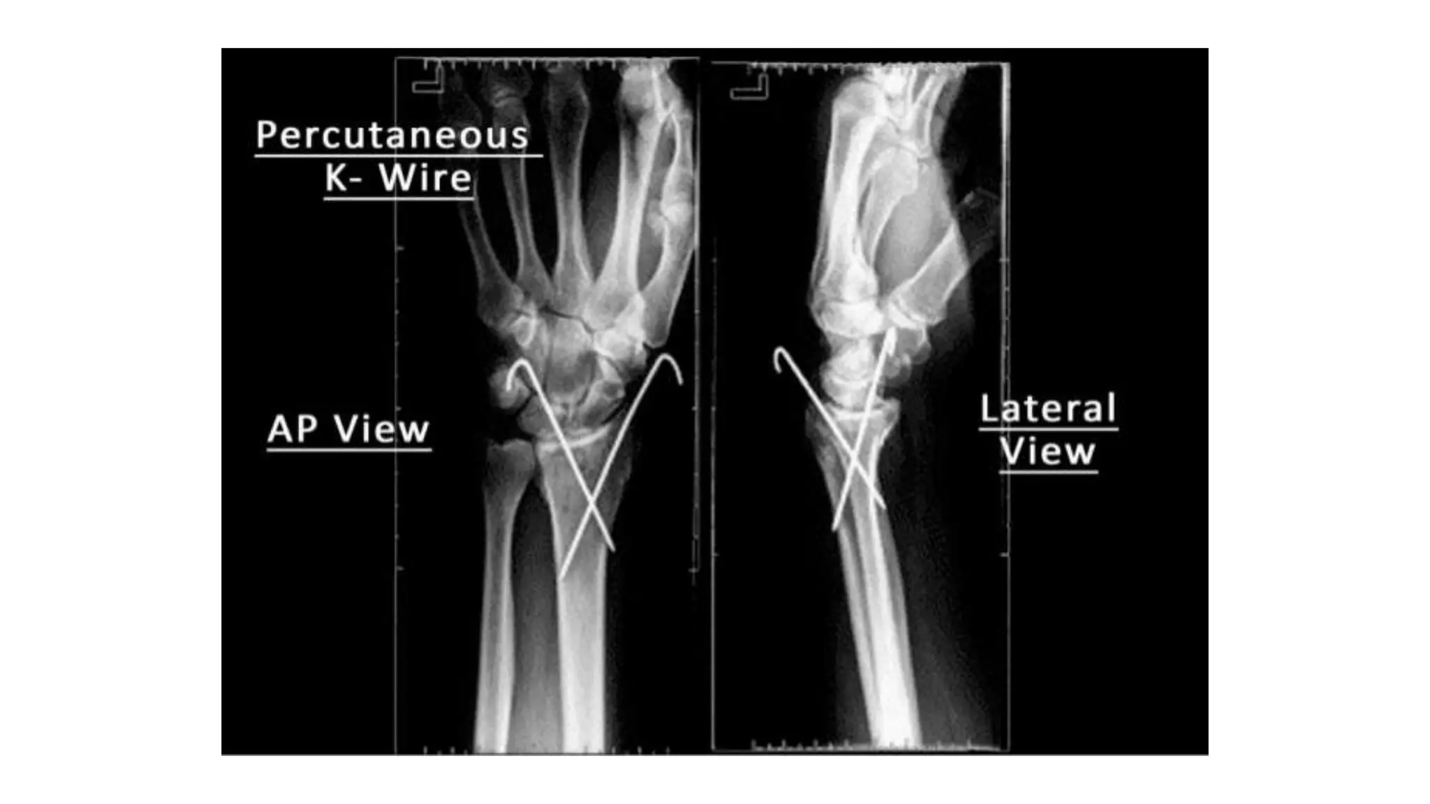
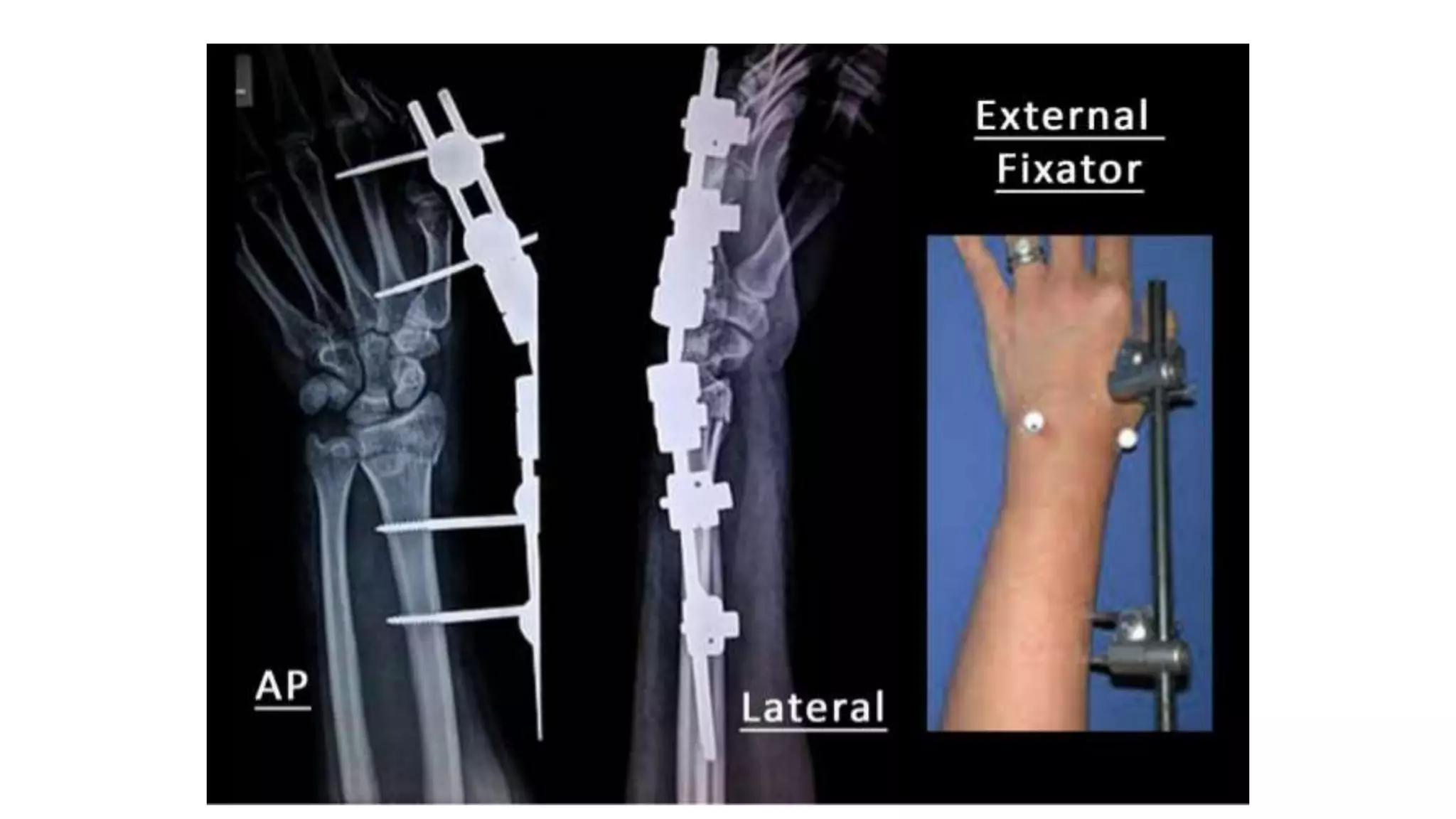
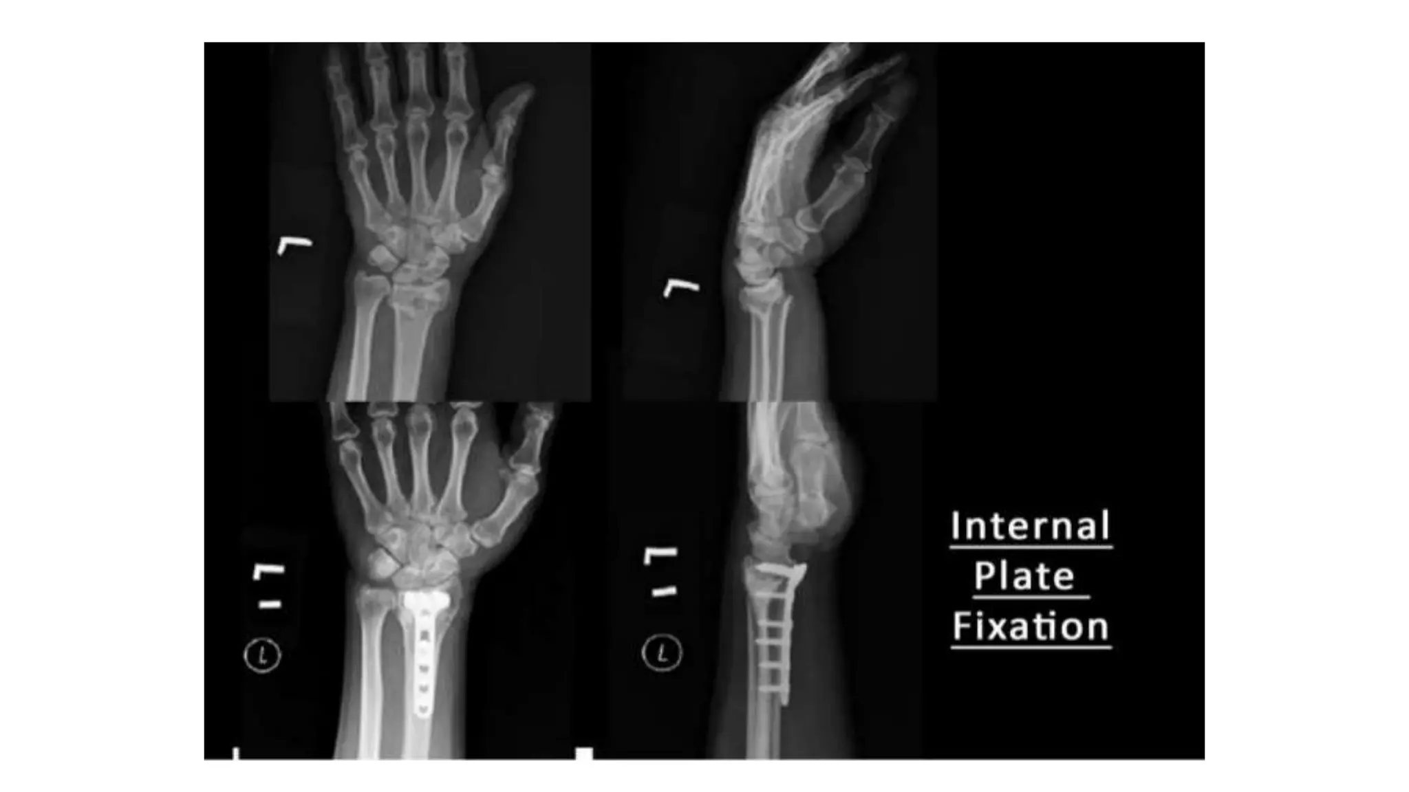
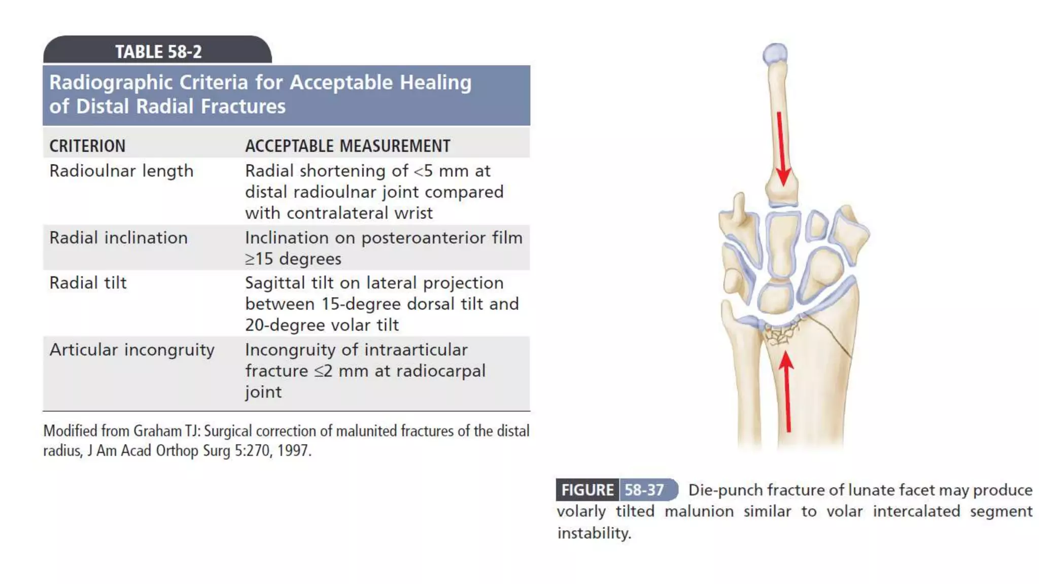
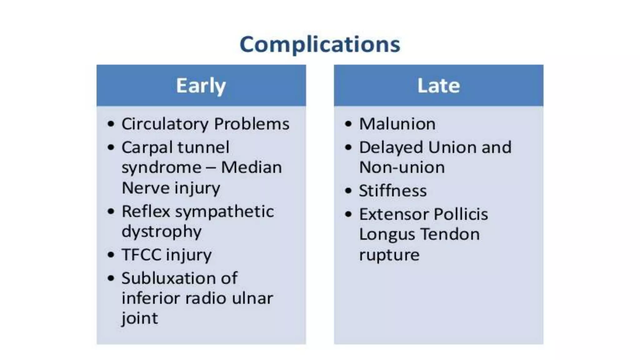
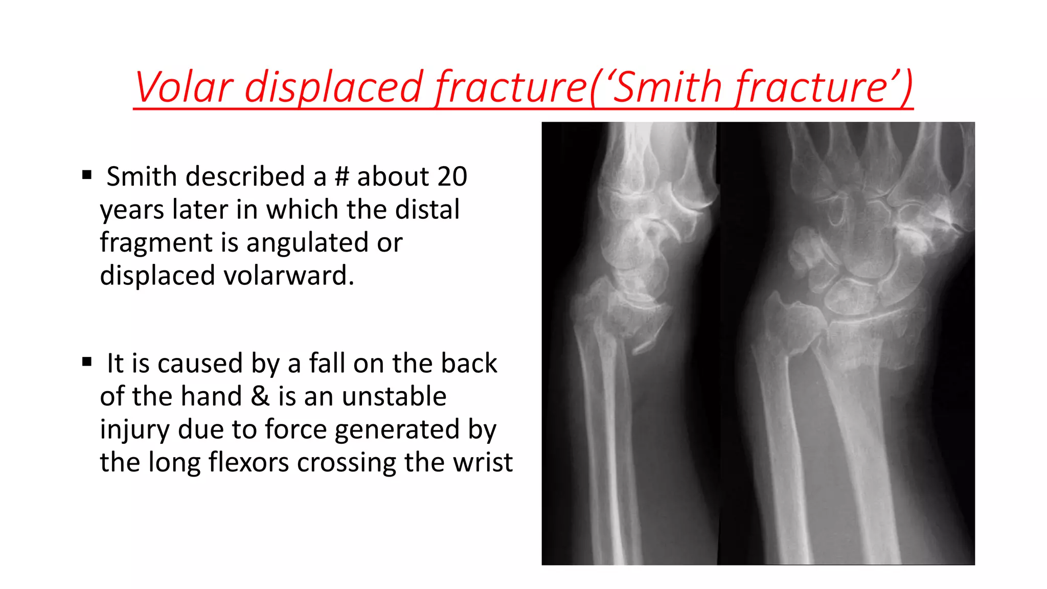
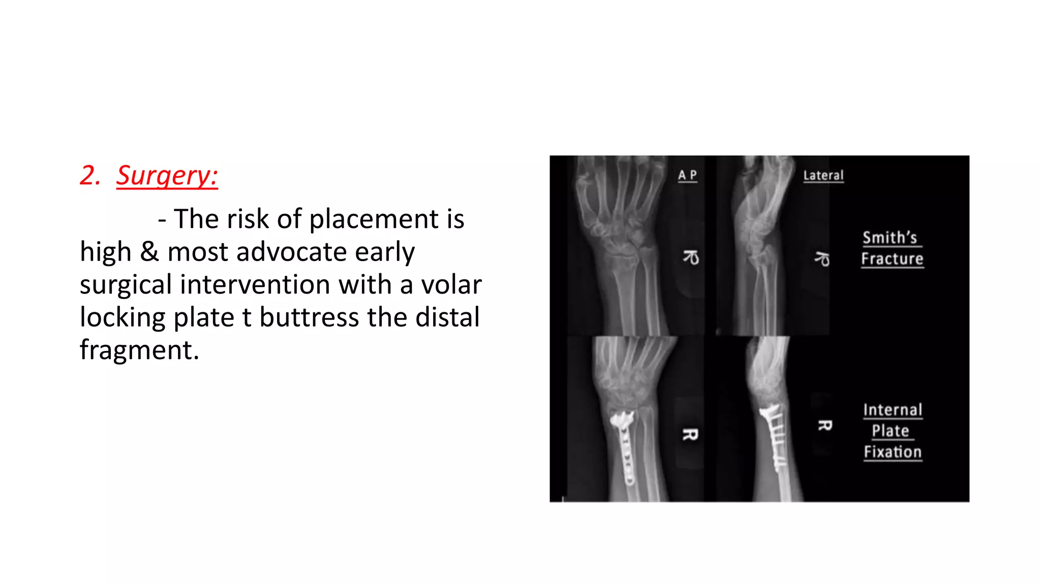
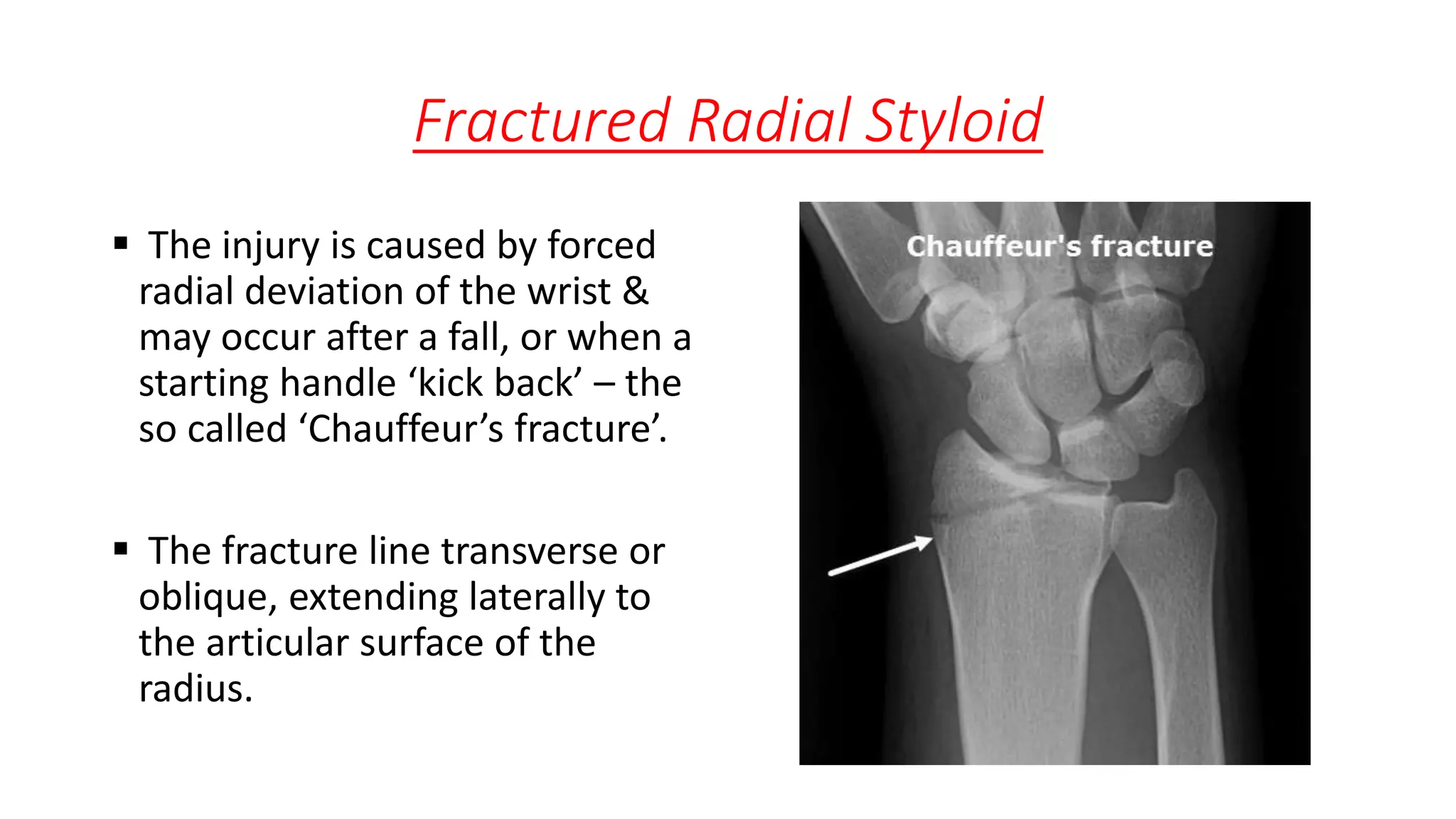
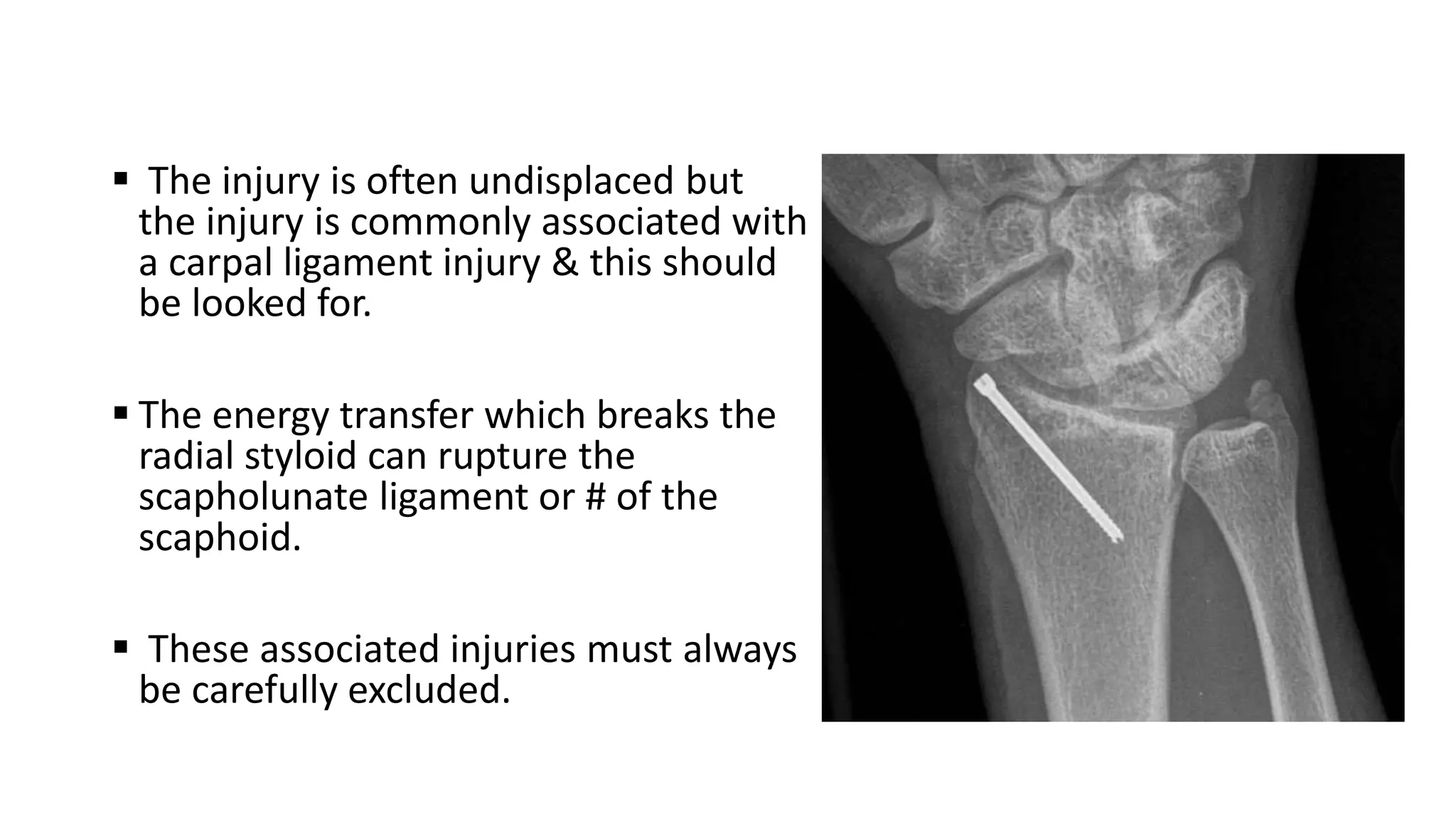
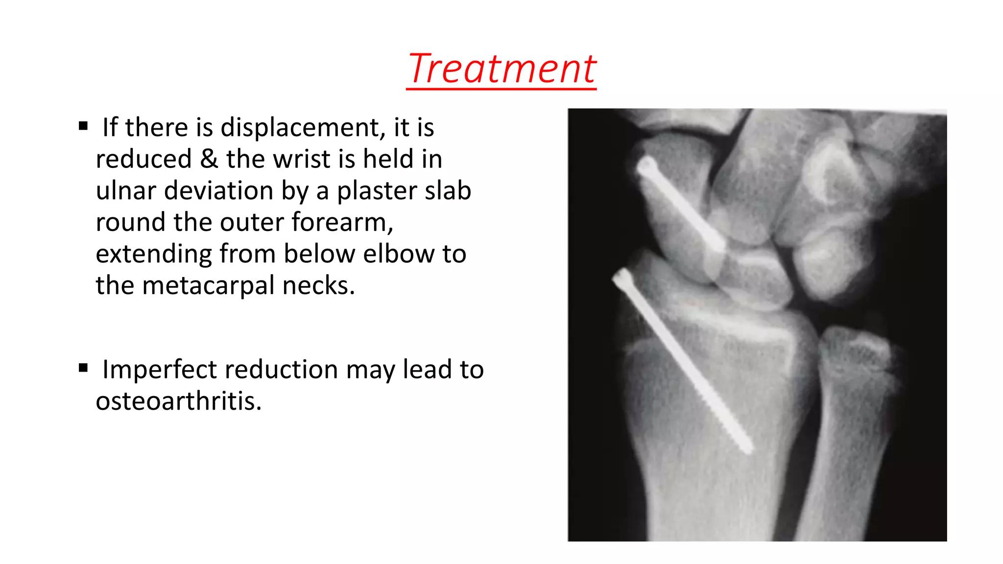
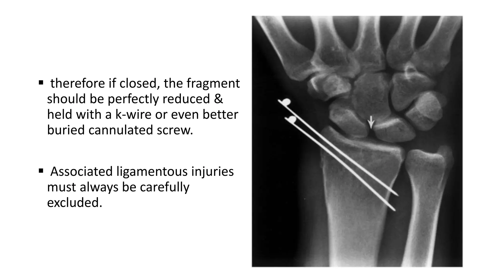
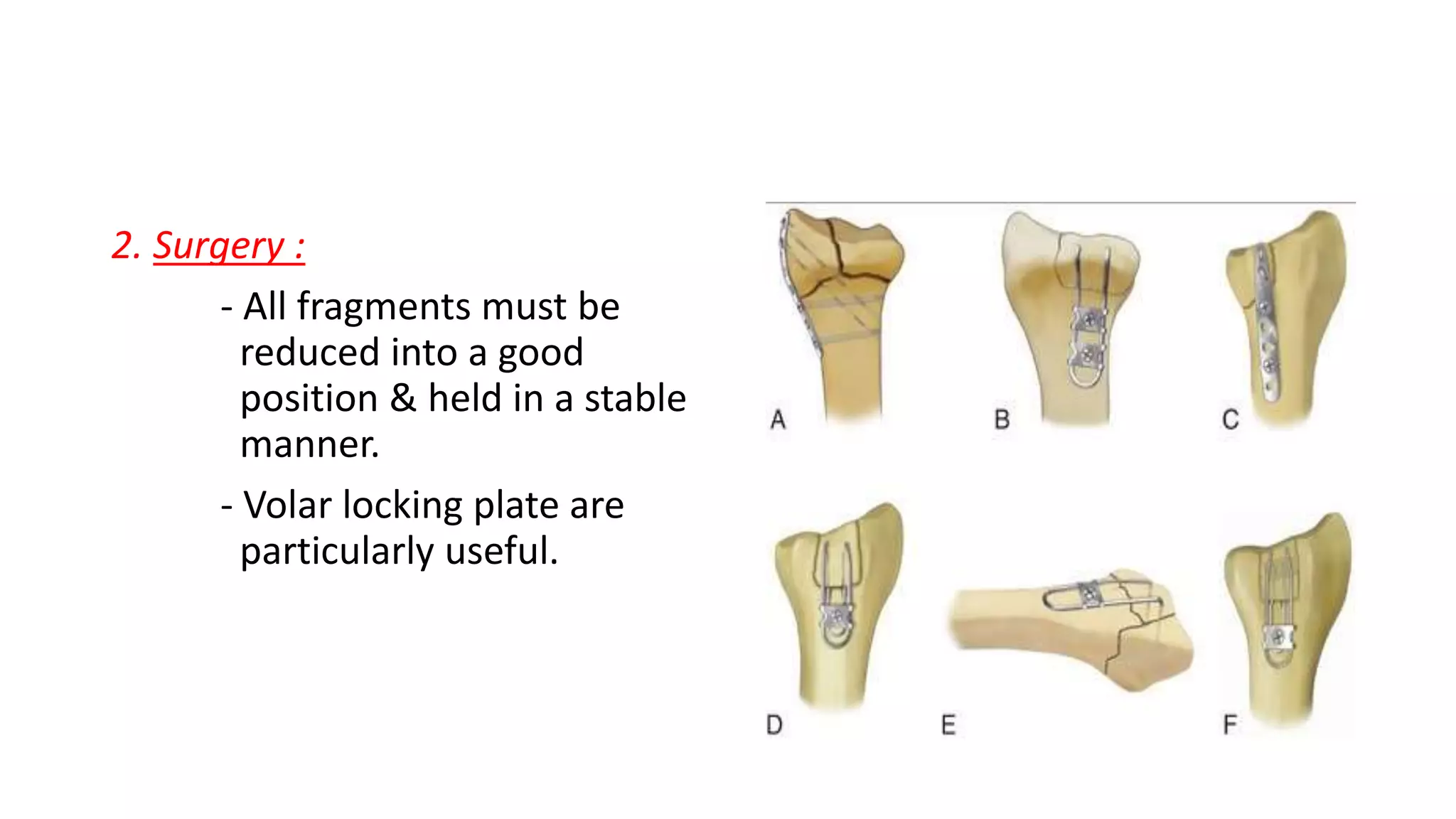
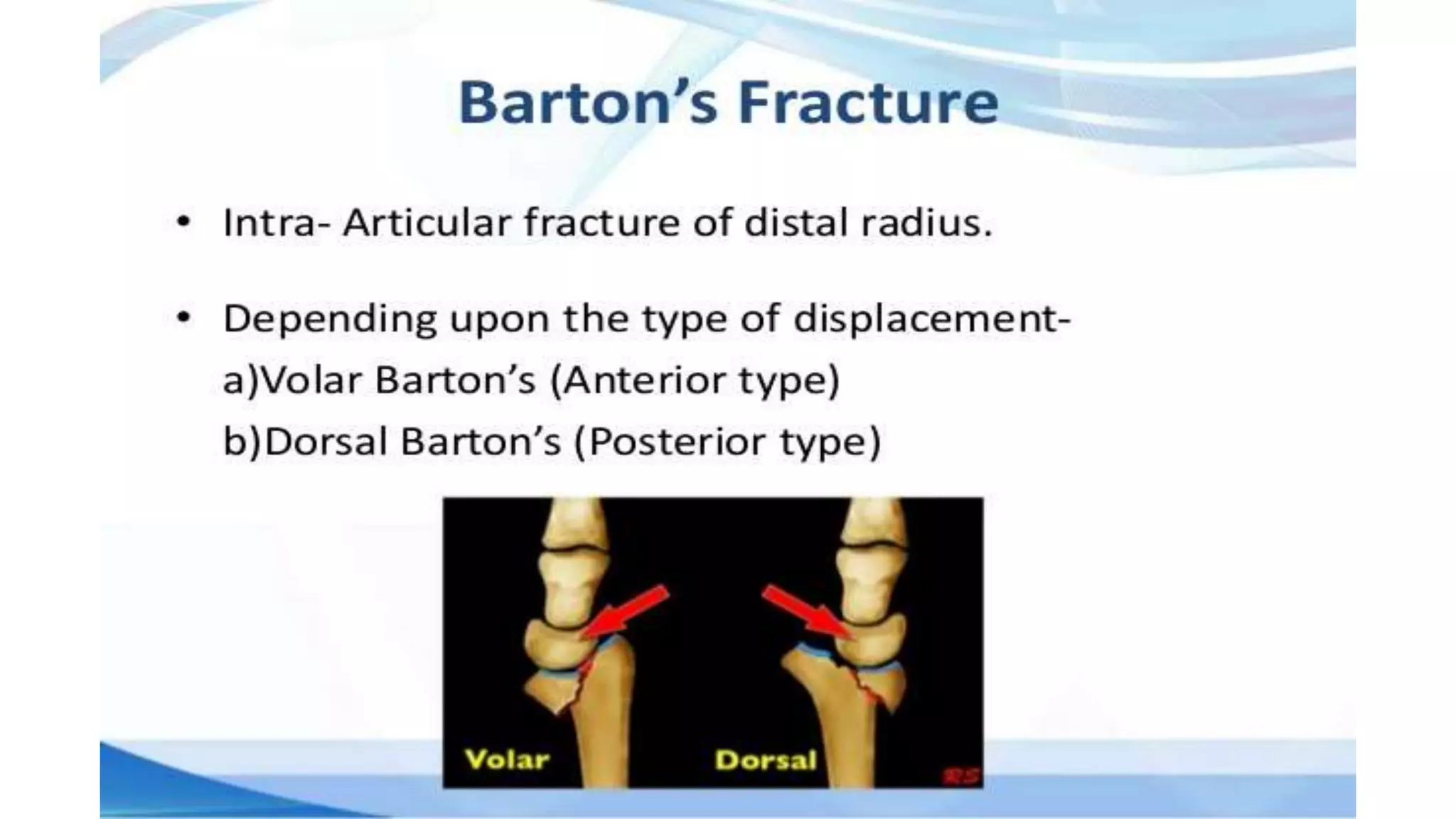
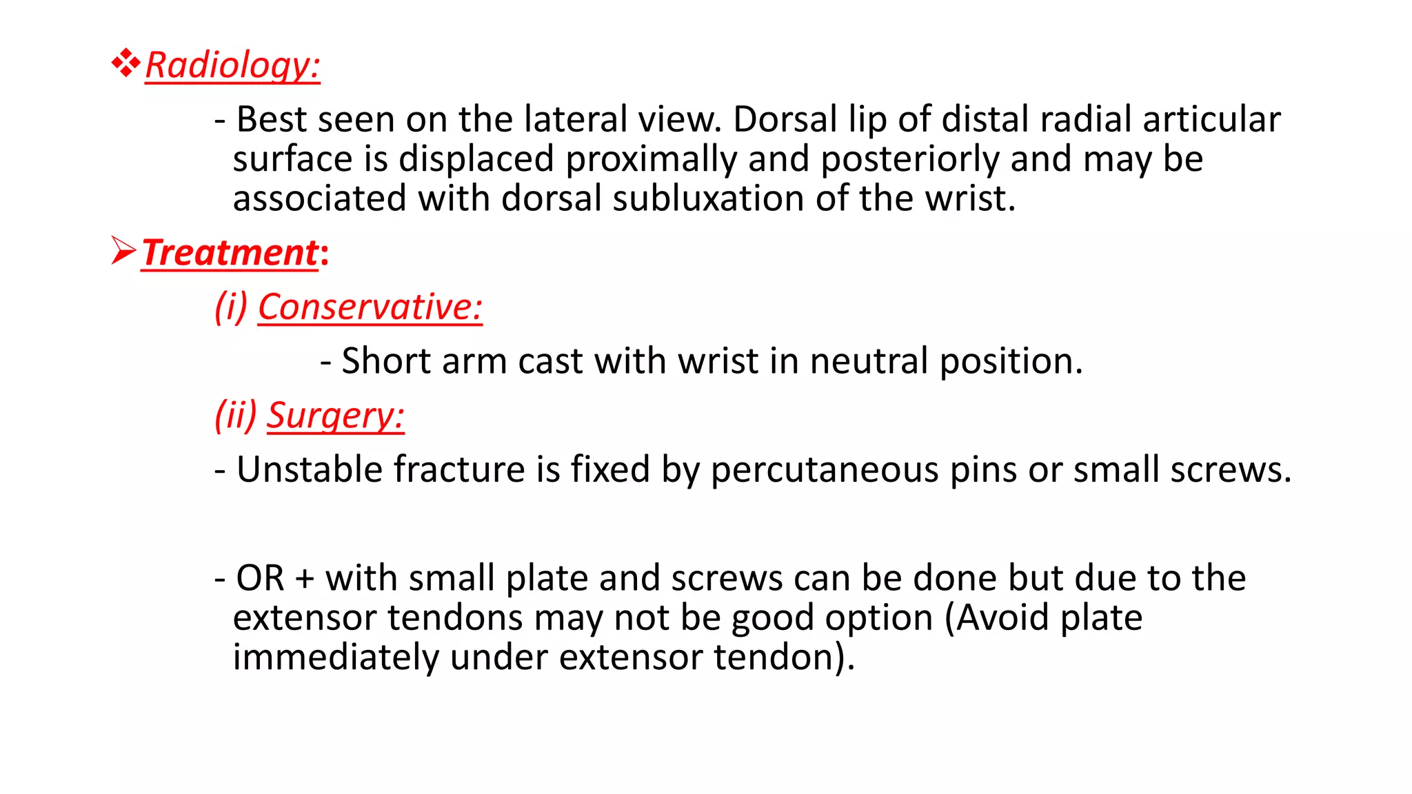
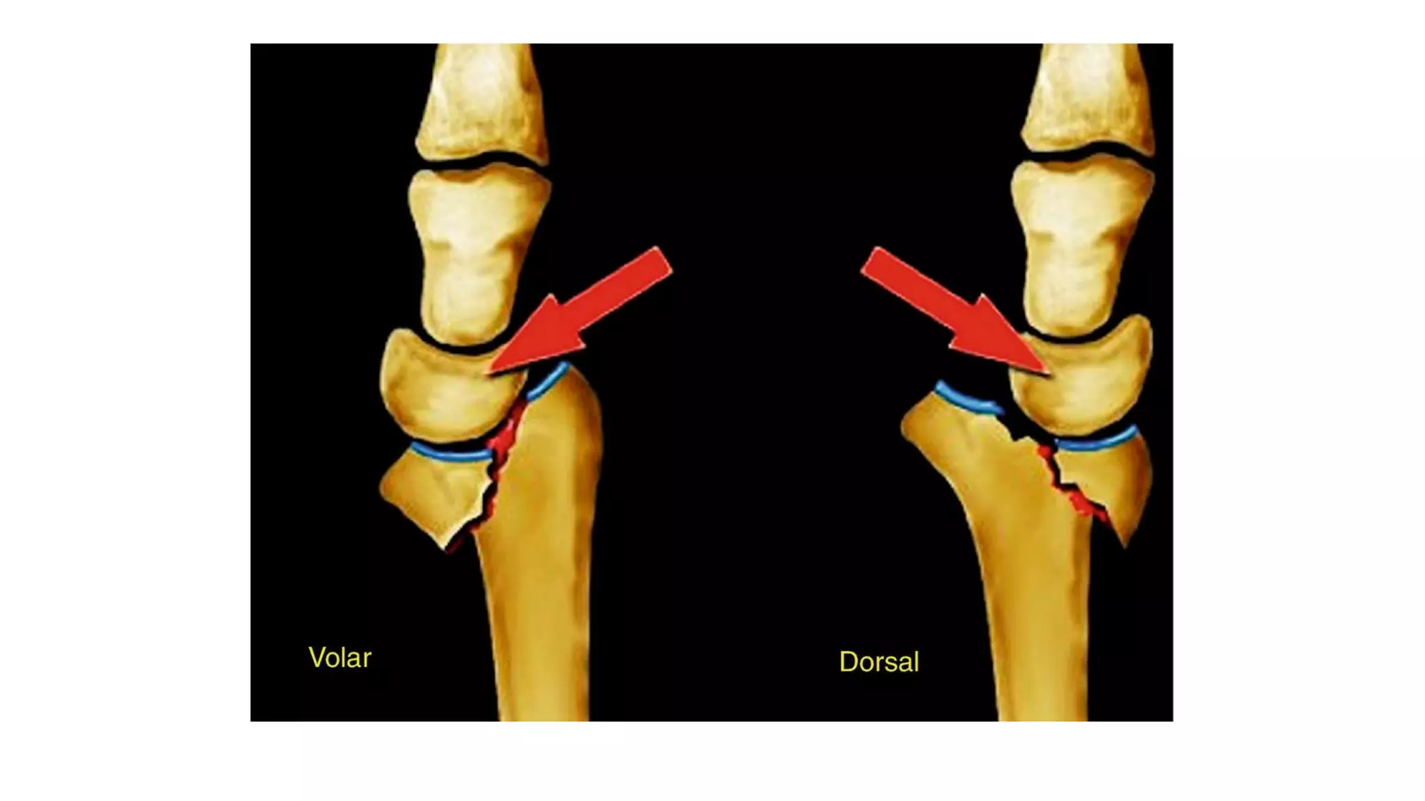
Fracture of the distal Radius
Dr. Ashiqur Rahman
Resident Orthopedics
Dhaka Medical college Hospital
Introduction
 Historically, distal radial # have been classified using eponymous
terms such as Colle’s, Smith’s, or Barton’s fracture.
 But these names can lead to confusion & misunderstanding.
 Treatment options depend on whether the # is intra-articular or
extra-articular & degree of fragmentation of the joint surface & the
metaphysis.
 The wrist can suffer substantial ligamentous injury causing instability
to the carpus or distal radio-ulnar joint(DRUJ).
Low energy dorsally displaced # (‘Colles’ fracture)
 The injury that Abraham Colles described in 1814 (‘Colles fracture’) is
a transverse fracture of the radius just above the wrist, with dorsal
displacement of the distal fragment.
 It is the most common of all fractures in older people, the high
incidence being related to the onset of postmenopausal osteoporosis.
 Thus the patient is usually an older woman who gives a history of
falling on her outstretched hand.
Mechanism of injury & pathological antomy
 Force is applied in the length of the forearm with the wrist in
extension.
 The bone fractures at the cortico-cancellous junction & the distal
fragment collapses into extension, dorsal displacement, radial tilt and
shortening.
Clinical features
- Pain,
- swelling,
- tenderness &
- irregularity of the lower end of the radius.
- ‘Dinner fork’ deformity:
with prominence on the back of the wrist &
a depression in front.
Imaging
- X-ray shows there is a transverse # of the radius @
the cortico-cancellous junction.
- & often the ulnar styloid process is broken off.
- The radial fragment is impacted into radius & backward tilt.
- Sometimes, there is an intra-articular #;
sometimes it is severely fragmented.
- A CT scan in very helpful in planning Rx.
Treatment
 Un-displaced fracture:
- If the # is un-displaced or only very slightly displaced, a dorsal
splint is applied for a day or two until the swelling has resolved,
then the cast is completed.
- An X-ray is taken @ 10 – 14 days to ensure that the fracture has
not slipped; if it has, surgery may be required.
- If not, the cast usually be removed after 05 weeks to allow
mobilization.
Displaced fracture
 Displaced # must be reduced under GA (Hematoma block, Bier’s
block, axillary block or general anesthesia).
 Procedure:
- The hand is grasped & longitudinal traction is applied.
- Sometimes, with extension of the wrist to disimpact the
fragments.
- The distal fragment is then pushed into place by pressing on the
dorsum while manipulating the wrist into flexion, ulnar deviation
& pronation.
 If it is satisfactory, a dorsal plaster slab is applied, extending from just
below the elbow to the metacarpal necks & two-thirds of the way
round the circumference of the wrist.
 The position is then checked by X-ray.
 Extreme positions of flexion & ulnar deviation must be avoided; 20°
in each direction is adequate.
 The arm is kept elevated for the next day or two; shoulder, elbow &
finger exercises are started as soon as possible.
 If the fingers become swollen, cyanosed or painful, there should be
no hesitation in splitting the bandage.
 At approximately 07 days, and if satisfactory then again at about 14
days, fresh X-ray is taken, this is because re-displacement is not
uncommon.
 Another X-ray at 18 – 20 days, just prior to the fracture being too
‘Sticky’ to manipulate again.
 If the position is probably not compatible with a good outcome & if
the risks inherent in surgery are understood, manipulation & fixation
with either percutaneous wires or volar locking plate are undertaken.
 However, in older patient with low functional demands, modest
degrees of displacement should be accepted because:
- Outcome in this pt. is not so dependent on anatomical
perfection.
- Fixation of the fragile bone can be very difficult & is not
without complications.
Impacted or Fragmented low-energy distal radial
fractures
 With substantial impaction or
fragmentation in osteoporotic
bone, manipulation & plaster
immobilization alone may be
insufficient.
 The # can sometimes be reduced
& held with percutaneous wires
or a volar locking plate.
 But, if impaction is severe, even this
may not be enough to hold all the
fragments or maintain length.
 In that case, other techniques, such
as dorsal plating, locked intra-
medullary nails, external fixators,
internal plate bridging the radius to
the 3rd metacarpal & bone grafts
(Synthetic or autogenous) are
considered.
Poor outcome is associated with:
(i) Loss of radial length >3mm.
(ii) Dorsal tilt >15° from neutral depending on age & function.
(iii) Palmar tilt >20° from neutral.
Volar displaced fracture(‘Smith fracture’)
 Smith described a # about 20
years later in which the distal
fragment is angulated or
displaced volarward.
 It is caused by a fall on the back
of the hand & is an unstable
injury due to force generated by
the long flexors crossing the wrist
Clinical features
 Wrist pain
 Garden - spade deformity
Imaging
- There is # through the distal radial metaphysis.
- A lateral view shows that the distal fragment is
displaced or tilted anteriorly.
Treatment
1. Conservative:
- These fractures can be reduced by traction,
supination & extension of the wrist.
- Forearm is immobilized in a cast for 06
weeks.
- X-rays should be taken @ 7 – 10 days to
ensure that the # has not slipped, when a plate
is used.
2. Surgery:
- The risk of placement is
high & most advocate early
surgical intervention with a volar
locking plate t buttress the distal
fragment.
Fractured Radial Styloid
 The injury is caused by forced
radial deviation of the wrist &
may occur after a fall, or when a
starting handle ‘kick back’ – the
so called ‘Chauffeur’s fracture’.
 The fracture line transverse or
oblique, extending laterally to
the articular surface of the
radius.
 The injury is often undisplaced but
the injury is commonly associated with
a carpal ligament injury & this should
be looked for.
 The energy transfer which breaks the
radial styloid can rupture the
scapholunate ligament or # of the
scaphoid.
 These associated injuries must always
be carefully excluded.
Treatment
 If there is displacement, it is
reduced & the wrist is held in
ulnar deviation by a plaster slab
round the outer forearm,
extending from below elbow to
the metacarpal necks.
 Imperfect reduction may lead to
osteoarthritis.
 therefore if closed, the fragment
should be perfectly reduced &
held with a k-wire or even better
buried cannulated screw.
 Associated ligamentous injuries
must always be carefully
excluded.
Fragmented intra-articular fractures
 In the young adult, a fragmented intra-articular fracture is a high-
energy injury.
 A poor outcome will probably result unless intra-articular congruity,
fracture alignment & length are restored & movements started as
soon as possible.
Imaging
(i) X-rays : AP & Lateral
(ii) CT scan
 Are useful to show the fragment alignment.
Treatment
1. Conservative :
- Simple manipulation under anesthesia & cast may be
helpful.
- X-rays are needed at about 07 days.
- If the anatomy is not restored, then either closed
reduction with percutaneous wires & an open
reduction may well be necessary.
2. Surgery :
- All fragments must be
reduced into a good
position & held in a stable
manner.
- Volar locking plate are
particularly useful.
Complications
A. Early :
(i) Circulatory problem:
- Early finger movements & avoiding undue flexion of the
elbow are important to encourage venous drainage.
(ii) Nerve injury:
- Compression of the median nerve in the carpal tunnel is
fairly common.
(iii) Complex regional pain syndrome(CRPS):
- Previously known as Reflex sympathetic dystrophy or
sudeck’s atrophy.
(iii) Ulnar corner pain & instability:
- Due avulsion of ulnar styloid.
(iv) Associated injury to the carpus.
(v) Re-displacement
B. Late:
(i) Malunion
(ii) Delayed union & non-union
(iii) Tendon rupture :
- Rupture of the EPL occurs a few weeks after an
apparently trivial undisplaced # of the lower radius.
- The FPL is vulnerable with a misplaced volar plate.
(iv) Carpal instability
(v) Secondary osteoarthritis
Dorsal Barton’s Fracture
Rim avulsion fractures of the distal radius in which dorsal half of the
distal radius sheaf off with intact volar half & the fragment moves
dorsally carrying the carpus with it.
Mechanism:
- Fall with dorsiflexion and pronation of the distal forearm on a
flexed wrist.
Clinical Features:
- Pain swelling
- tenderness over the wrist
- restricted wrist movements with painful dorsiflexion
Radiology:
- Best seen on the lateral view. Dorsal lip of distal radial articular
surface is displaced proximally and posteriorly and may be
associated with dorsal subluxation of the wrist.
Treatment:
(i) Conservative:
- Short arm cast with wrist in neutral position.
(ii) Surgery:
- Unstable fracture is fixed by percutaneous pins or small screws.
- OR + with small plate and screws can be done but due to the
extensor tendons may not be good option (Avoid plate
immediately under extensor tendon).
Volar Barton fracture
Rim avulsion fracture of the distal radius in which volar half of the
distal radius shear off with intact dorsal half & the fragment moves
forwards carrying the carpus with it.
Mechanism:
• It is due to palmar tensile stress and dorsal shear stress and is usually
combined with Radial styloid fracture.
Clinical Features
• It consists of pain, swelling, tenderness and loss of wrist movements.
Palmar flexion is grossly restricted and painful.
Radiograph:
- Volar rim of distal radial articular surface is displaced forwards.
- May be associated with volar subluxation of the wrist.
Treatment:
(i) Conservative:
- Reduction is simple, but retention is difficult.
- Long arm cast is used.
Surgery:
- If reduction does not remain satisfactorily then fixation
with K wire.
- External fixators and buttress plate, etc. may be done.
- Ellis T-shaped buttress plate fixation is the preferred
method of treatment.
Fracture of the distal radius

Fracture of the distal radius

  • 1.
    Fracture of thedistal Radius Dr. Ashiqur Rahman Resident Orthopedics Dhaka Medical college Hospital
  • 2.
    Introduction  Historically, distalradial # have been classified using eponymous terms such as Colle’s, Smith’s, or Barton’s fracture.  But these names can lead to confusion & misunderstanding.  Treatment options depend on whether the # is intra-articular or extra-articular & degree of fragmentation of the joint surface & the metaphysis.  The wrist can suffer substantial ligamentous injury causing instability to the carpus or distal radio-ulnar joint(DRUJ).
  • 4.
    Low energy dorsallydisplaced # (‘Colles’ fracture)  The injury that Abraham Colles described in 1814 (‘Colles fracture’) is a transverse fracture of the radius just above the wrist, with dorsal displacement of the distal fragment.  It is the most common of all fractures in older people, the high incidence being related to the onset of postmenopausal osteoporosis.  Thus the patient is usually an older woman who gives a history of falling on her outstretched hand.
  • 11.
    Mechanism of injury& pathological antomy  Force is applied in the length of the forearm with the wrist in extension.  The bone fractures at the cortico-cancellous junction & the distal fragment collapses into extension, dorsal displacement, radial tilt and shortening.
  • 14.
    Clinical features - Pain, -swelling, - tenderness & - irregularity of the lower end of the radius. - ‘Dinner fork’ deformity: with prominence on the back of the wrist & a depression in front.
  • 15.
    Imaging - X-ray showsthere is a transverse # of the radius @ the cortico-cancellous junction. - & often the ulnar styloid process is broken off. - The radial fragment is impacted into radius & backward tilt. - Sometimes, there is an intra-articular #; sometimes it is severely fragmented. - A CT scan in very helpful in planning Rx.
  • 17.
    Treatment  Un-displaced fracture: -If the # is un-displaced or only very slightly displaced, a dorsal splint is applied for a day or two until the swelling has resolved, then the cast is completed. - An X-ray is taken @ 10 – 14 days to ensure that the fracture has not slipped; if it has, surgery may be required. - If not, the cast usually be removed after 05 weeks to allow mobilization.
  • 19.
    Displaced fracture  Displaced# must be reduced under GA (Hematoma block, Bier’s block, axillary block or general anesthesia).  Procedure: - The hand is grasped & longitudinal traction is applied. - Sometimes, with extension of the wrist to disimpact the fragments. - The distal fragment is then pushed into place by pressing on the dorsum while manipulating the wrist into flexion, ulnar deviation & pronation.
  • 21.
     If itis satisfactory, a dorsal plaster slab is applied, extending from just below the elbow to the metacarpal necks & two-thirds of the way round the circumference of the wrist.  The position is then checked by X-ray.  Extreme positions of flexion & ulnar deviation must be avoided; 20° in each direction is adequate.
  • 22.
     The armis kept elevated for the next day or two; shoulder, elbow & finger exercises are started as soon as possible.  If the fingers become swollen, cyanosed or painful, there should be no hesitation in splitting the bandage.  At approximately 07 days, and if satisfactory then again at about 14 days, fresh X-ray is taken, this is because re-displacement is not uncommon.  Another X-ray at 18 – 20 days, just prior to the fracture being too ‘Sticky’ to manipulate again.
  • 23.
     If theposition is probably not compatible with a good outcome & if the risks inherent in surgery are understood, manipulation & fixation with either percutaneous wires or volar locking plate are undertaken.  However, in older patient with low functional demands, modest degrees of displacement should be accepted because: - Outcome in this pt. is not so dependent on anatomical perfection. - Fixation of the fragile bone can be very difficult & is not without complications.
  • 24.
    Impacted or Fragmentedlow-energy distal radial fractures  With substantial impaction or fragmentation in osteoporotic bone, manipulation & plaster immobilization alone may be insufficient.  The # can sometimes be reduced & held with percutaneous wires or a volar locking plate.
  • 25.
     But, ifimpaction is severe, even this may not be enough to hold all the fragments or maintain length.  In that case, other techniques, such as dorsal plating, locked intra- medullary nails, external fixators, internal plate bridging the radius to the 3rd metacarpal & bone grafts (Synthetic or autogenous) are considered.
  • 34.
    Poor outcome isassociated with: (i) Loss of radial length >3mm. (ii) Dorsal tilt >15° from neutral depending on age & function. (iii) Palmar tilt >20° from neutral.
  • 37.
    Volar displaced fracture(‘Smithfracture’)  Smith described a # about 20 years later in which the distal fragment is angulated or displaced volarward.  It is caused by a fall on the back of the hand & is an unstable injury due to force generated by the long flexors crossing the wrist
  • 38.
    Clinical features  Wristpain  Garden - spade deformity
  • 39.
    Imaging - There is# through the distal radial metaphysis. - A lateral view shows that the distal fragment is displaced or tilted anteriorly.
  • 40.
    Treatment 1. Conservative: - Thesefractures can be reduced by traction, supination & extension of the wrist. - Forearm is immobilized in a cast for 06 weeks. - X-rays should be taken @ 7 – 10 days to ensure that the # has not slipped, when a plate is used.
  • 41.
    2. Surgery: - Therisk of placement is high & most advocate early surgical intervention with a volar locking plate t buttress the distal fragment.
  • 42.
    Fractured Radial Styloid The injury is caused by forced radial deviation of the wrist & may occur after a fall, or when a starting handle ‘kick back’ – the so called ‘Chauffeur’s fracture’.  The fracture line transverse or oblique, extending laterally to the articular surface of the radius.
  • 43.
     The injuryis often undisplaced but the injury is commonly associated with a carpal ligament injury & this should be looked for.  The energy transfer which breaks the radial styloid can rupture the scapholunate ligament or # of the scaphoid.  These associated injuries must always be carefully excluded.
  • 44.
    Treatment  If thereis displacement, it is reduced & the wrist is held in ulnar deviation by a plaster slab round the outer forearm, extending from below elbow to the metacarpal necks.  Imperfect reduction may lead to osteoarthritis.
  • 45.
     therefore ifclosed, the fragment should be perfectly reduced & held with a k-wire or even better buried cannulated screw.  Associated ligamentous injuries must always be carefully excluded.
  • 46.
    Fragmented intra-articular fractures In the young adult, a fragmented intra-articular fracture is a high- energy injury.  A poor outcome will probably result unless intra-articular congruity, fracture alignment & length are restored & movements started as soon as possible.
  • 47.
    Imaging (i) X-rays :AP & Lateral (ii) CT scan  Are useful to show the fragment alignment.
  • 48.
    Treatment 1. Conservative : -Simple manipulation under anesthesia & cast may be helpful. - X-rays are needed at about 07 days. - If the anatomy is not restored, then either closed reduction with percutaneous wires & an open reduction may well be necessary.
  • 49.
    2. Surgery : -All fragments must be reduced into a good position & held in a stable manner. - Volar locking plate are particularly useful.
  • 50.
    Complications A. Early : (i)Circulatory problem: - Early finger movements & avoiding undue flexion of the elbow are important to encourage venous drainage. (ii) Nerve injury: - Compression of the median nerve in the carpal tunnel is fairly common. (iii) Complex regional pain syndrome(CRPS): - Previously known as Reflex sympathetic dystrophy or sudeck’s atrophy.
  • 51.
    (iii) Ulnar cornerpain & instability: - Due avulsion of ulnar styloid. (iv) Associated injury to the carpus. (v) Re-displacement
  • 52.
    B. Late: (i) Malunion (ii)Delayed union & non-union (iii) Tendon rupture : - Rupture of the EPL occurs a few weeks after an apparently trivial undisplaced # of the lower radius. - The FPL is vulnerable with a misplaced volar plate. (iv) Carpal instability (v) Secondary osteoarthritis
  • 55.
    Dorsal Barton’s Fracture Rimavulsion fractures of the distal radius in which dorsal half of the distal radius sheaf off with intact volar half & the fragment moves dorsally carrying the carpus with it. Mechanism: - Fall with dorsiflexion and pronation of the distal forearm on a flexed wrist. Clinical Features: - Pain swelling - tenderness over the wrist - restricted wrist movements with painful dorsiflexion
  • 56.
    Radiology: - Best seenon the lateral view. Dorsal lip of distal radial articular surface is displaced proximally and posteriorly and may be associated with dorsal subluxation of the wrist. Treatment: (i) Conservative: - Short arm cast with wrist in neutral position. (ii) Surgery: - Unstable fracture is fixed by percutaneous pins or small screws. - OR + with small plate and screws can be done but due to the extensor tendons may not be good option (Avoid plate immediately under extensor tendon).
  • 58.
    Volar Barton fracture Rimavulsion fracture of the distal radius in which volar half of the distal radius shear off with intact dorsal half & the fragment moves forwards carrying the carpus with it. Mechanism: • It is due to palmar tensile stress and dorsal shear stress and is usually combined with Radial styloid fracture. Clinical Features • It consists of pain, swelling, tenderness and loss of wrist movements. Palmar flexion is grossly restricted and painful.
  • 59.
    Radiograph: - Volar rimof distal radial articular surface is displaced forwards. - May be associated with volar subluxation of the wrist. Treatment: (i) Conservative: - Reduction is simple, but retention is difficult. - Long arm cast is used.
  • 60.
    Surgery: - If reductiondoes not remain satisfactorily then fixation with K wire. - External fixators and buttress plate, etc. may be done. - Ellis T-shaped buttress plate fixation is the preferred method of treatment.