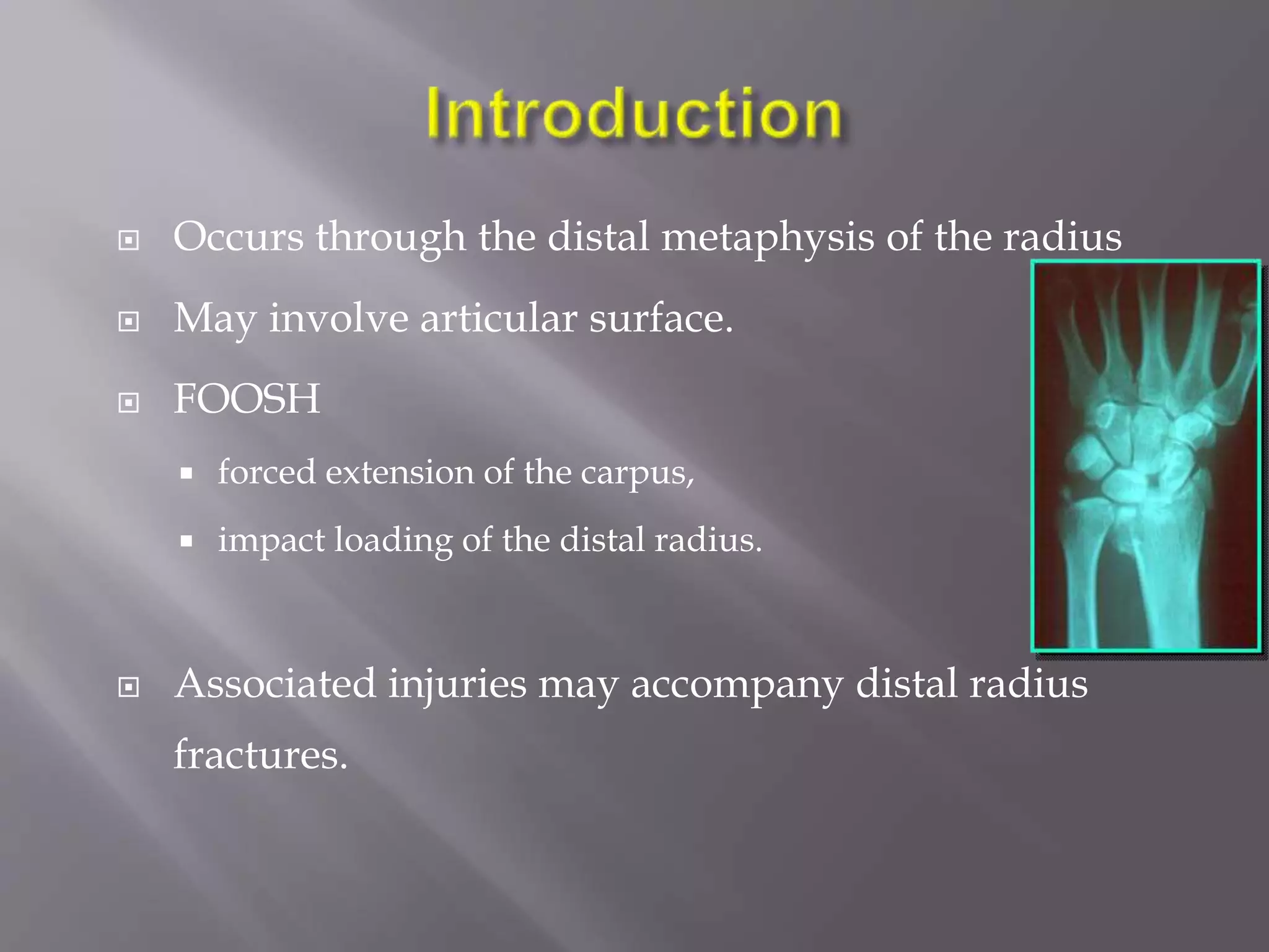
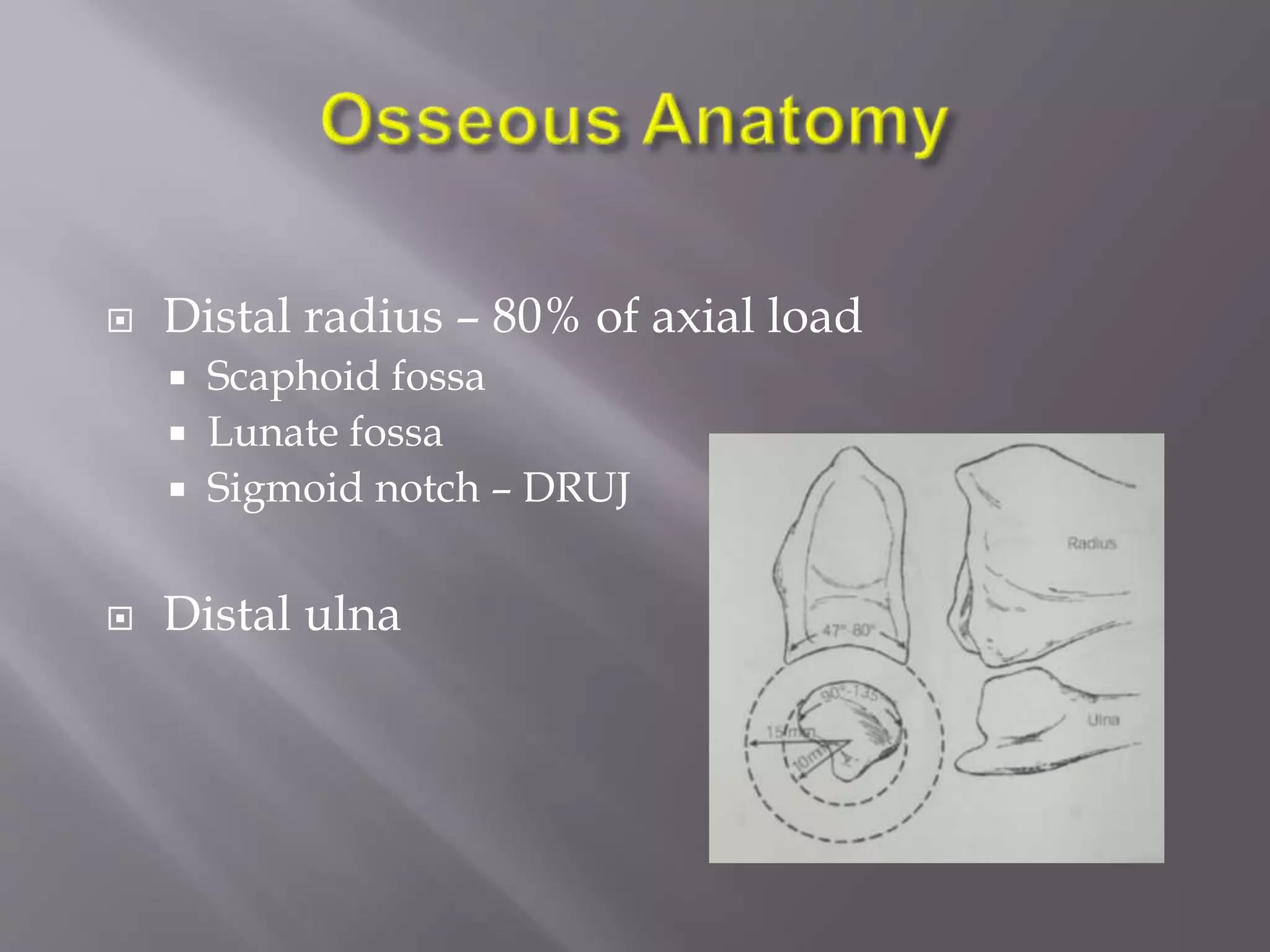
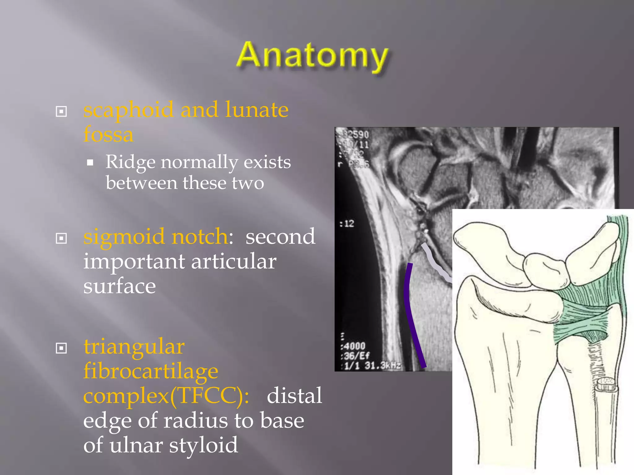
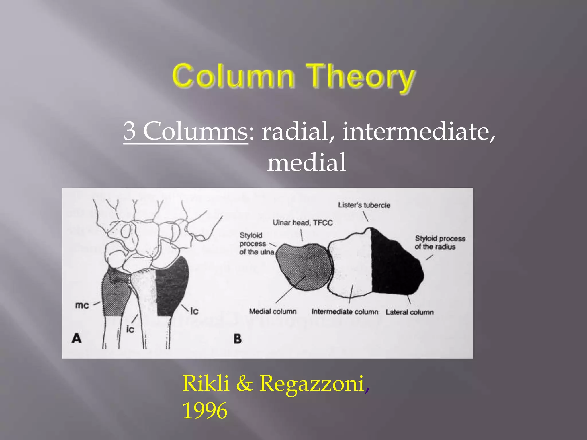
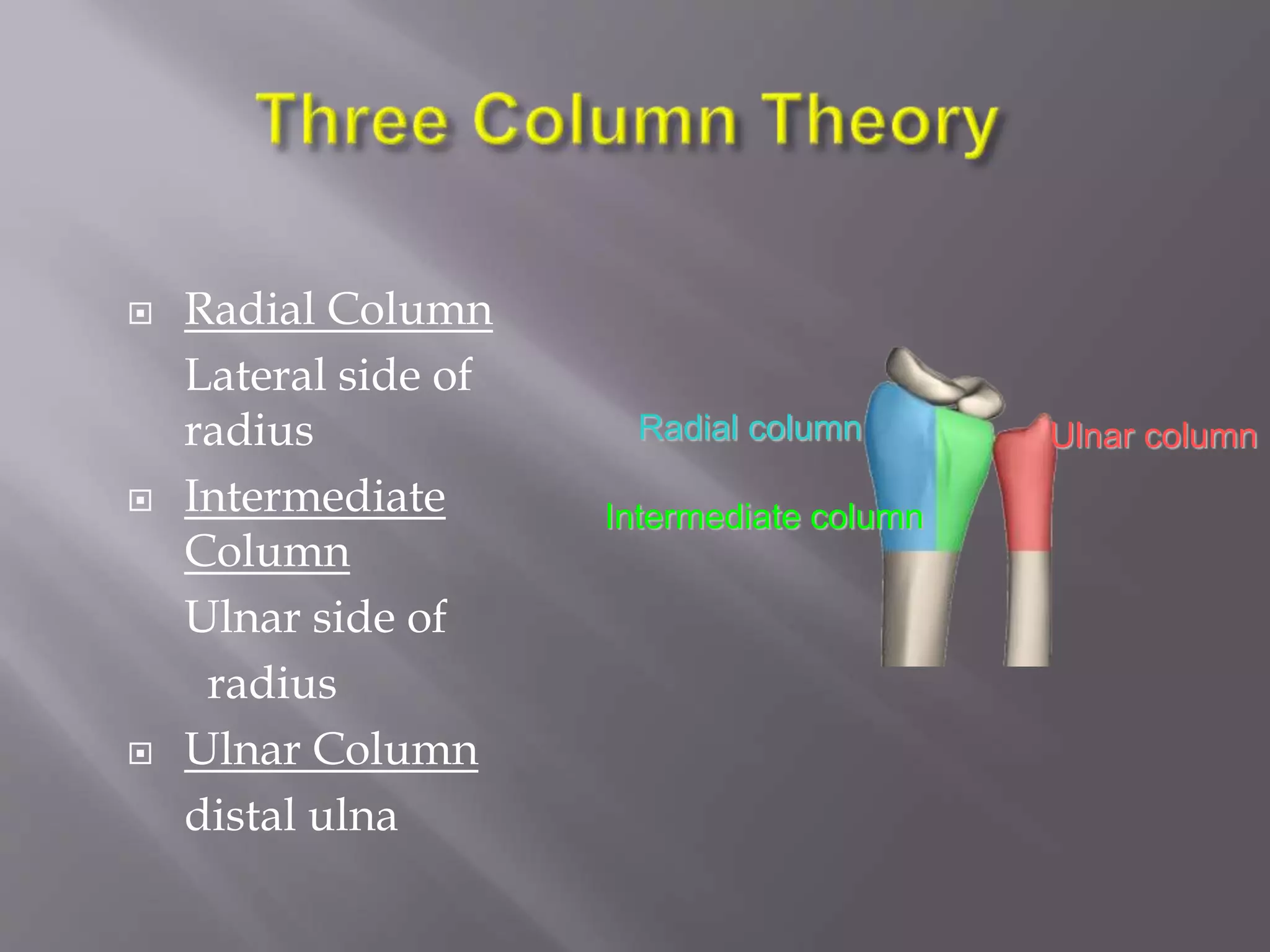
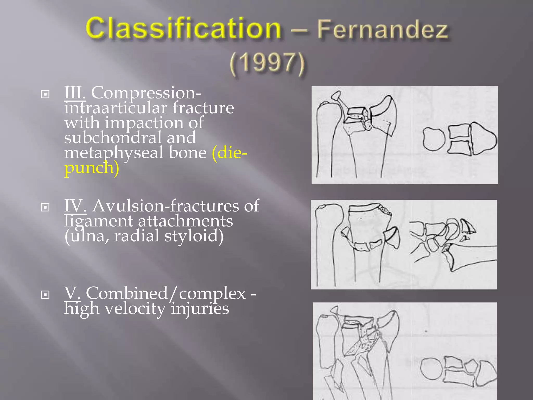
This document discusses the history and treatment of distal radius fractures. Some key points:
- Distal radius fractures are common injuries that were first recognized in the late 18th century, with descriptions of injury patterns evolving over the 19th century.
- Treatment has progressed from casting to external fixation to various internal fixation methods like dorsal, volar, and combined plating approaches.
- Factors like fracture pattern, displacement, comminution, and articular involvement help determine appropriate treatment, whether closed reduction or open reduction with internal fixation.
- The goal of treatment is to restore normal anatomy, allow early motion, and avoid complications like malunion.
Assessent and radiology of distal end radius fractureSusanta85
distal end radius is a common fracture in elderly groups and also in young by high velocity trauma its assessment and radiology should know for its management
paediatric injuries around the elbow
supracondylar elbow injuries
pulled elbow in paediatric age r
radiological signs around elbow in supracondylar fracture humerus
Palestine last event orientationfvgnh .pptxRaedMohamed3
An EFL lesson about the current events in Palestine. It is intended to be for intermediate students who wish to increase their listening skills through a short lesson in power point.
Unit 8 - Information and Communication Technology (Paper I).pdfThiyagu K
This slides describes the basic concepts of ICT, basics of Email, Emerging Technology and Digital Initiatives in Education. This presentations aligns with the UGC Paper I syllabus.
Synthetic Fiber Construction in lab .pptxPavel ( NSTU)
Synthetic fiber production is a fascinating and complex field that blends chemistry, engineering, and environmental science. By understanding these aspects, students can gain a comprehensive view of synthetic fiber production, its impact on society and the environment, and the potential for future innovations. Synthetic fibers play a crucial role in modern society, impacting various aspects of daily life, industry, and the environment. ynthetic fibers are integral to modern life, offering a range of benefits from cost-effectiveness and versatility to innovative applications and performance characteristics. While they pose environmental challenges, ongoing research and development aim to create more sustainable and eco-friendly alternatives. Understanding the importance of synthetic fibers helps in appreciating their role in the economy, industry, and daily life, while also emphasizing the need for sustainable practices and innovation.
Honest Reviews of Tim Han LMA Course Program.pptxtimhan337
Personal development courses are widely available today, with each one promising life-changing outcomes. Tim Han’s Life Mastery Achievers (LMA) Course has drawn a lot of interest. In addition to offering my frank assessment of Success Insider’s LMA Course, this piece examines the course’s effects via a variety of Tim Han LMA course reviews and Success Insider comments.
Read| The latest issue of The Challenger is here! We are thrilled to announce that our school paper has qualified for the NATIONAL SCHOOLS PRESS CONFERENCE (NSPC) 2024. Thank you for your unwavering support and trust. Dive into the stories that made us stand out!
Francesca Gottschalk - How can education support child empowerment.pptxEduSkills OECD
Francesca Gottschalk from the OECD’s Centre for Educational Research and Innovation presents at the Ask an Expert Webinar: How can education support child empowerment?
Introduction to AI for Nonprofits with Tapp NetworkTechSoup
Dive into the world of AI! Experts Jon Hill and Tareq Monaur will guide you through AI's role in enhancing nonprofit websites and basic marketing strategies, making it easy to understand and apply.
Macroeconomics- Movie Location
This will be used as part of your Personal Professional Portfolio once graded.
Objective:
Prepare a presentation or a paper using research, basic comparative analysis, data organization and application of economic information. You will make an informed assessment of an economic climate outside of the United States to accomplish an entertainment industry objective.
2. Common injury
Potential for functional
impairment and frequent
complications
3. First surgeon to recognize these injuries was Pouteau 1783. his
work was not widely publicized.
Later Abraham Colles 1814 gave the classic description of fracture
Dupuytren brought to the world attention that it is a fracture
rather than a dislocation as it was previously assumed.
Goyrond 1832 differntiated between dorsal and volar
displacement.
Barton 1838 described wrist subluxation consequent to
intraarticular fracture of radius which could be dorsal or volar.
Smith described fracture of distal radius with ‘forward’
displacement.
Advent of X rays at the end of nineteenth century contributed
much to the understanding of different patterns of injury.
4. One sixth of all fractures treated in the Emergency
Room
Bimodal distribution
less than 30 years (70% men)
over 50 years (85% women)
Males age 35 or older - 90 per 100,000 population
5. Occurs through the distal metaphysis of the radius
May involve articular surface.
FOOSH
forced extension of the carpus,
impact loading of the distal radius.
Associated injuries may accompany distal radius
fractures.
6. History of a FOOSH
Wrist is typically swolen with ecchymosis and
tender
Visible deformity of the wrist, with the hand most
commonly displaced in the dorsal direction less
comonly in volar direction
Movement of the hand and wrist are painful.
Adequate and accurate assessment of the
neurovascular status of the hand is performed,
before any treatment is carried out.
7. General physical exam of the patient, including
an evaluation of the injured joint, and a joint
above and below
Radiographs of the injured wrist-pa and lat
view , oblique view
CT scan of the distal radius to know extent of
intrarticular involvement
9. scaphoid and lunate
fossa
Ridge normally exists
between these two
sigmoid notch: second
important articular
surface
triangular
fibrocartilage
complex(TFCC): distal
edge of radius to base
of ulnar styloid
15. 1: Line connecting dorsal and
volar tip of lunate
2: Line perpendicular to
lunate
3: Line along axis of scaphoid
Scapholunate angle measured between lines 2 and 3
(normal 47 ± 15 degrees)
16. Intra-articular fxs with multiple
fragments
centrally impacted fragments
DRUJ incongruity
Cole et al: J Hand Surg, 1997
17. Ideal system should describe:
Type of injury
Severity
Evaluation
Treatment
Prognosis
21. Depends on three basic components of
injury
1-metaphyseal comminution
2-intra articular extension
3-displacement of the fragment
22. Group i-
simple colles # with no involvement of radial
articular surface
Group ii-
colles # with intra articula extension without
displacement
Group iii-
comminuted colles # with intra articular
extension with displacemet
Group iv-
extra articular undisplaced
23.
24. Type I Extraarticular, undisplaced
Type 2 Extraarticular, displaced
Type 3 Intraarticular, undisplaced
Type 4 Intraarticular, displaced
25. Type A Extraarticular
Type B Partial articular
B1–radial styloid fracture
B2–dorsal rim fracture
B3–volar rim fracture
B4–die-punch fracture
Type C Complete articular
26. Depends on the impaction of the lunate on radial
articular surface
Type i- stable # without displacement
Type ii-unstable ‘die punch’ with displacement
of the characteristic fragment and comminution
of the ant and post cotices
type ii A – reducible
type iiB --irreducible
27. Type iii- ‘spike fracture’ ,unstable.
Displacement of the articular surface and
also proximal spike of radius
Type iv- ‘split’ fracture, unstable medial
complex that is severly comminuted with
separation and or rotation of the distal and
proximal fragments
Type v- explosion injury
30. Radial Column
Lateral side of
radius
Intermediate
Column
Ulnar side of
radius
Ulnar Column
distal ulna
Radial column
Intermediate column
Ulnar column
31. I. Bending-metaphysis
bending with loss of
palmar tilt and radial
shortening ,DRUJ
injury(Colles, Smith)
II. Shearing-fractures of
joint surface (Barton,
radial styloid)
32. III. Compression-
intraarticular fracture
with impaction of
subchondral and
metaphyseal bone (die-
punch)
IV. Avulsion-fractures of
ligament attachments
(ulna, radial styloid)
V. Combined/complex -
high velocity injuries
33. Assess involvement of dorsal or volar rim
Is comminution mainly volar or dorsal?
is one of four cortices intact?
Look for “die-punch” lesions of the
scaphoid or lunate fossa.
Assess amount of shortening
Look for DRUJ involvement
34. COLLES #
-extra articular or intra articular distal radius #
with various combination of
dorsal angulation,
dorsal displacement
radial shift , and
radial shortening
-clinicaly described as dinner fork deformity
-mechanism---fall on to an hyper extended
,radialy deviated wrist with the forearm in
pronation
35. # distal radius with volar angulation or
volar displacement of the hand and
distal radius
mechanism—fall on to a flexed wrist
with the forearm fixed in supination
unstable pattern often requires ORIF
because of difficulty in maintaining
closed reduction
36. # disdlocation or subluxation of
wrist in which the dorsal or volar
rim of distal radius is displaced
mechanism-fall on to a dorsiflexed
wrist with the forearm fixed in
pronation
unstable # requires ORIF
37. Avulsion # with extrinsic ligaments
remaining attached to styloid fragment
Mechanism-compression of scaphoid
against styloid with the wrist in
dorsiflexion and ulnar deviation
Often associated with intercarpal ligament
injury
Requires ORIF
38. five factors indicative of instability
(1)initial dorsal angulation of more than 20
degrees (volar tilt),
(2) dorsal metaphyseal comminution,
(3) intraarticular involvement,
(4) an associated ulnar fracture, and
(5) patient age older than 60 years
39. Casting
Long arm vs short arm
Sugar-tong splint
External Fixation
Joint-spanning
Non bridging
Percutaneous pinning
Internal Fixation
Dorsal plating
Volar plating
Combined dorsal/volar plating
focal (fracture specific) plating
40. Preserve hand and wrist function
Realign normal osseous anatomy
promote bony healing
Avoid complications
Allow early finger and elbow ROM
42. Obtaining and then maintaining an acceptable
reduction.
Immobilization:
long arm
short arm adequate for elderly patients
Frequent follow-up necessary in order to
diagnose redisplacement.
43. Anesthesia
Hematoma block
Intravenous sedation
Bier block
Traction: finger traps and weights
Reduction Maneuver (dorsally angulated fracture):
hyperextension of the distal fragment,
Maintain weighted traction and reduce the distal
to the proximal fragment with pressure applied
to the distal radius.
Apply well-molded “sugar-tong” splint or cast,
with wrist in neutral to slight flexion.
Avoid Extreme Positions!
44. Radial length- within 2-3 mm of the
controlateral wrist
Palmar tilt- neutral tilt(o degree )
intrarticular step-off or gap< 2mm
Radial inclination <5 degree loss
Carpal malalignment – absent
Ulnar variance- no more than 2 mm of
shortening compare to ulnar head
45. Watch for median nerve symptoms
parasthesias common but should diminish over
few hours
If persist release pressure on cast, take wrist out
of flexion
Acute carpal tunnel: symptoms progress; CTR
required
Follow-up x-rays needed in 1-2 weeks to evaluate
reduction.
Change to short-arm cast after 2-3 weeks, continue
until fracture healing.
46. Repeat reduction and casting – high rate of failure
Repeat reduction and percutaneous pinning
External Fixation
ORIF
47. High-energy injury
Open injury
Secondary loss of reduction
Articular comminution, step-off, or gap
Metaphyseal comminution or bone loss
Loss of volar buttress with displacement
DRUJ incongruity
51. Spanning
Dynamic
Clyburne
Agee
Pennig
Static
AO
Ace
Non-spanning
Hoffman 2
Cobra
Zimmer
AO
Neutralize the axial load imparted by physiologic forearm
musculature
52. A spanning fixator
is one which fixes
distal radius
fractures by
spanning the
carpus; I.e.,
fixation into radius
and metacarpals
Use for
comminuted
fracture
53. Used to restore radial length and radial
inclination but palmer tilt is rarely restored
Supplemental k wire fixation is needed to
prevent loss of palmer tilt and some degree of
colapse
Pins are retained for 6 to 8 weeks
Overdistraction results in finger stiffness
54.
55. Mal-union
Pin track infection
Finger stiffness
Loss of reduction; early vs late
Tendon rupture
56. A non-spanning fixator is one which
fixes distal radius fracture by securing
pins in the radius alone, proximal to and
distal to the fracture site.
Indicated for extra articular or minimal
intra articular dorsaly displaced fracture
Minimum 1 cm of intact volar cortex is
required to give purchase for the pins
Better to preserve volar tilt and carpal
malalignment
59. Bulky
Poor screw hold in porosis and comminution
Screws do not buttress
Cutaneous radial nerve injury
Pin tract infection
Reflex sympathetic dystrophy
60. Wrist is immobilized in a supinated position
with sugar tongue splint for 10 days
External fixator frame is removed at 6 weeks
Any supplemental pins are kept in place for 8
weeks
Active and passive finger motion is begun as
soon as the anesthesia wears off
61.
62. most common radial styloid pinning +
dorsal-ulnar corner of radius pinning
supplemental immobilization with cast,
splint
in conjunction with external fixation
(Augmented external fixation)
63. 2 radial styloid pins-
-radial styloid is pinned to proximal shaft
in reduced position
excellent anatomic 82%
good-excellent functional results 100%
radial styloid with dorsal ulnar –
-after radial styloid fixation the
intermediate column is pinned from
dorsal ulnar to proximal radial
excellent anatomic results in 90%
64. intrafocal pinning
through fracture site
buttress against
displacement
Drawback-tendency to
translate distal fragment
in opposite direction
65. Useful for elevation of depressed articular
fragments and bone grafting of metaphyseal
defects
required if articular fragments can not be
adequately reduced with percutaneous
methods
66. Based on location of comminution.
Dorsal approach for dorsally angulated
fractures.
Volar approach for volar rim fractures
Radial styloid approach for buttressing of
styloid
Combined approaches needed for high-energy
fractures with significant axial impaction.
75. At 1 week, the sutures are removed and active
wrist motion is begun when there is confidence
in fracture stability
A removable Orthoplast splint is worn for 6
weeks.
78. Generally not prefere because of high rate of
complication like
- tendon dysfunction and rupture
- tenosynovitis of extensor tendons
indicated for-
dorsal die-punch fractures or fractures
with displaced dorsal lunate facet fragments
79.
80. Finger and other joint upper extremity
exercises are begun immediately
If a splint was applied, it is removed at 3 weeks
When union is achieved, the distraction plate is
removed and range-of-motion exercises are
begun
93. Indicated for
1-complex articular fracture without
metaphyseal comminution
2-# with evidence of substantial
interosseous ligament or TFCC injury
without a large ulnar styloid fragment.
Timing-between 5 to 15 days post injury to
prevent extravasation of irrigation fluid
into the surrounding soft tissue
95. ORIF better in high-energy fractures associated with
depression of articular surface
ORIF gives better anatomic restoration, although not
necessarily higher patient satisfaction.
96. External fixators still have a role in the
treatment of distal radius fractures
Spanning ex fix does not completely correct
fracture deformity by itself
Should usually combined with percutaneous
pins (augmented fixation)
97. new plating techniques allow for accurate
and rigid fixation of fragments
Plating allows early wrist ROM
Volar, smaller and more anatomic plates are
better tolerated
combination treatment is often needed
98.
99. Colles; “The wrist will regain perfect freedom in all of its
motions and be completely exempt from pain” (1814)
Generally true for low demand individuals
Direct relation between residual deformity and
disability
Quality of reduction more important than method of
immobilisation