Downloaded 355 times


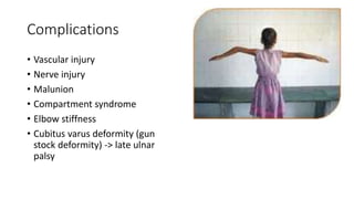


This document discusses paediatric fractures, including physeal injuries, supracondylar fractures of the humerus, and paediatric abuse. Key points include: - Children's bones have a physis/growth plate not present in adults, making physeal injuries common fracture patterns like buckle fractures and greenstick fractures. - Supracondylar fractures frequently occur in the distal humerus and can be posteriorly or anteriorly displaced. Nerve injuries and compartment syndrome are complications. - Paediatric abuse is difficult to diagnose but risk factors include inconsistent history, delay in care, and fractures in non-mobile children. A skeletal survey aids investigation.


