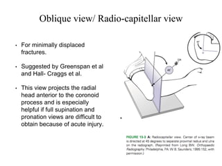
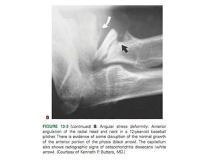
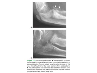
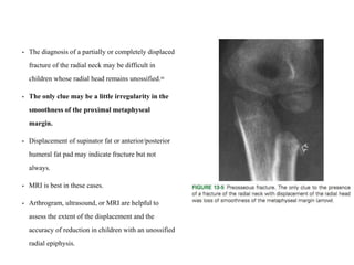
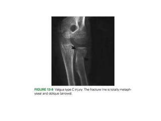
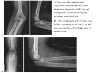
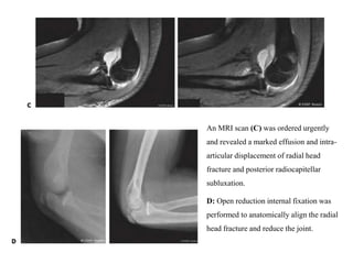
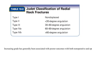
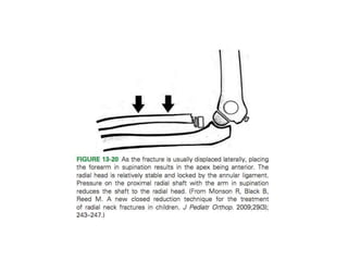
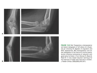
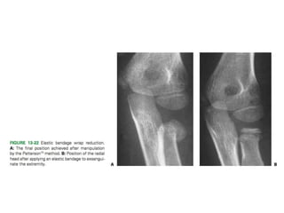
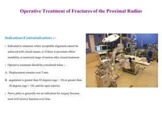
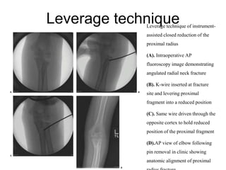
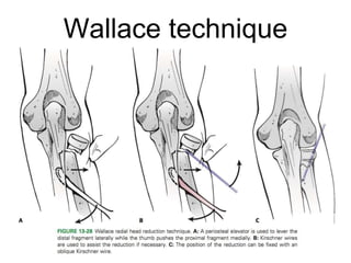
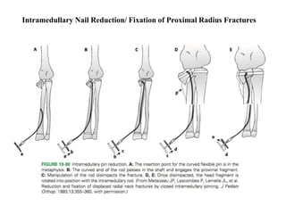
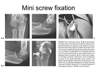
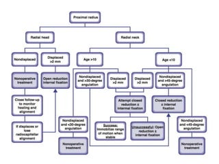
ASSESSMENT OF FRACTURES OF THE
PROXIMAL RADIUS IN CHILDREN
By :-
Dr OPENDER SINGH KAJLA
RNT MEDICAL COLLEGE, UDAIPUR
INTRODUCTION
• Fractures of the proximal radius in skeletally immature patients usually involve the metaphysis
or physis.
• True isolated radial head fractures are rare.
• Fractures of the radial neck account for slightly more than 1% of all children’s fractures.Radial
neck fractures make up approximately 5% of elbow fractures in children.
• Radial head fractures are uncommon, and when they occur usually are Salter–Harris type IV
injuries.
• The median age at injury is 9 to 10 years in the pediatric population.
• There is little difference in the occurrence rates between males and females22,40,68
; however, this injury
seems to occur on an average approximately 2 years earlier in girls than in boys.
MECHANISM OF
INJURY
• Fractures of the proximal radius most commonly occur after a fall on an outstretched arm with
elbow extended and valgus stress at the elbow.
• The cartilaginous head absorbs the force and transmits it to the weaker physis or metaphysis
of the neck.
• These fractures characteristically produce an angular deformity of the head with the neck.
• Vostal showed that in neutral, the pressure is concentrated on the lateral portion of the head
and neck. In supination, the pressure is concentrated anteriorly, and in pronation it is con-
centrated posteriorly.
• Proximal radial fractures also may occur in association with elbow.
FIGURE SHOWS TYPE OF INJURY IN ELBOW
DISLOCATION
Associated Injuries with Fractures
of the Proximal Radius
• Can occur concomitantly with distal humerus, ulna, radial shaft, or distal radius
fractures.
• Fractures in combination with ulnar fractures often are part of the Monteggia fracture
pattern.
• Portends a poor prognosis for patients with proximal radius fractures with higher rates
of persistent stiffness and pain compared to those with isolated proximal radius
fractures.
• The posterior interosseous nerve (PIN) wraps around the proximal radius and
occasionally can be injured in association with proximal radius fractures.
Signs and Symptoms of Fractures
of the Proximal Radius
• The pain is usually increased with forearm supination and pronation
more so than with elbow flexion and extension.
• Displaced fractures frequently result in visible bruising or
ecchymosis on the lateral aspect of the elbow with significant soft
tissue swelling.
• Neurologic examination should in particu- lar evaluate the PIN,
which can be affected by fractures of the proximal radius.
• In a young child, the primary complaint may be wrist pain, and
pressure over the proximal radius may accentuate this referred wrist
pain.2
The wrist pain may be secondary to radial shortening and
subsequent distal radioulnar joint dysfunction.
Imaging and Other Diagnostic Studies for Fractures of the Proximal Radius
• Displaced fractures are easy to evaluate on AP and Lateral views of elbow X-rays.
• Some variants in the ossification process can resemble a fracture. Most of these involve the
radial head, although a step-off also can develop as a normal variant of the metaphysis.
• There may be a persistence of the secondary ossification centers of the epiphysis.
Comparison views of the contralateral elbow are useful for evaluation of unusual ossification
centers after an acute elbow injury.
• If elbow cannot be extended due to pain then one view is taken with the beam perpendicular
to the distal humerus, and the other with the beam perpendicular to the proximal radius.
Oblique view/ Radio-capitellar view
• For minimally displaced
fractures.
• Suggested by Greenspan et al
and Hall- Craggs et al.
• This view projects the radial
head anterior to the coronoid
process and is especially
helpful if full supination and
pronation views are difficult to
obtain because of acute injury.
• The diagnosis of a partially or completely displaced
fracture of the radial neck may be difficult in
children whose radial head remains unossified.88
• The only clue may be a little irregularity in the
smoothness of the proximal metaphyseal
margin.
• Displacement of supinator fat or anterior/posterior
humeral fat pad may indicate fracture but not
always.
• MRI is best in these cases.
• Arthrogram, ultrasound, or MRI are helpful to
assess the extent of the displacement and the
accuracy of reduction in children with an unossified
radial epiphysis.
• In the preossification stage, on the AP radiograph, the edge of the metaphysis of
the proximal radius slopes distally on its lateral border.
• This angulation is normal and not a fracture.
• In the AP view, the lateral angulation varies from 0 to 15 degrees, with the average
being 12.5 degrees.
• In the lateral view, the angulation can vary from 10 degrees anterior to 5 degrees
posterior, with the average being 3.5 degrees anterior .
• Radial head fractures can be associated with ligament injuries so MRI should be
considered if displacement occurs in serial radiographs.
xray showing lateral angulation of
proximal radius Arthrogram of same patient showing
displaced radial head
Chambers Classification of Proximal Radial Fractures
Group I: Primary Displacement of the Radial Head
A. Valgus fractures
1. Type A—Salter–Harris type I and II injuries of the proxi-
mal radial physis
2. Type B—Salter–Harris type IV injuries of the proximal
radial physis
3. Type C—Fractures involving only the proximal radial
metaphysis
B. Fractures associate with elbow dislocation
1. Type D—Reduction injuries
2. 2. Type E—Dislocation injuries
Group II: Primary Displacement of the Radial Neck
A. Angular injuries(Monteggia typeIII variant)
B. Torsional injuries
Group III: Stress Injuries
A: Acute injury films revealing small
displacement of radial head fracture on the
flexed elbow anteroposterior (AP) view and
subtle posterior subluxation not originally
appreciated on the lateral view.
B: Follow-up radiographs at 1 week noted more
difficulty interpreting the AP view in cast, and
more radiocapitellar posterior displacement on
the lateral view.
An MRI scan (C) was ordered urgently
and revealed a marked effusion and intra-
articular displacement of radial head
fracture and posterior radiocapitellar
subluxation.
D: Open reduction internal fixation was
performed to anatomically align the radial
head fracture and reduce the joint.
Angular injuries
Stress Injuries (Group III)
Increasing grade has generally been associated with poorer outcomes with both nonoperative and ope
PATHOANATOMY AND APPLIED ANATOMY RELATING TO FRACTURES OF
THE PROXIMAL RADIUS
• In the embryo, the proximal radius is well defined by 9 weeks of gestation.
• By 4 years of age, the radial head and neck have the same contours as in an
adult.
• Ossification of the proximal radius epiphysis begins at approximately 5 years of
age as a small, flat nucleus.
• This ossific nucleus can originate as a small sphere or it can be bipartite, which
is a normal variation and should not be misinterpreted as a fracture.
• No ligaments attach directly to the radial neck or head. The radial collateral
ligaments attach to the annular ligament, which originates from the radial side
of the ulna.
• The articular capsule attaches to the proximal third of the neck.
• Thus, only a small portion of the neck lies within the articular capsule.
TREATMENT
• NON-OPERATIVE
• OPERATIVE
Nonoperative Treatment of Fractures of the Proximal Radius
Indications/Contraindications :-
• Nonoperative treatment is indicated for the majority of proximal radius fractures.
• Radial neck angulation of 30 to 45 degrees generally remodels and conservative treatment
will lead to good results.
• It is critical to assess forearm rotation, and if a block to full rotation is appreciated
operative treatment should be considered. Intra- articular aspiration of hematoma and
injection of local anesthetic can assist with pain relief and assessment of range of motion.
• Patients not requiring closed reduction should be immobilized for comfort for a short
period of time to allow for comfort and soft tissue healing.
• This is generally 1 to 3 weeks based on extent of injury and age.
• After fracture pain has subsided patients should work on progressively increasing range
Closed Reduction Techniques
• Patterson’s manipulative technique.
• Kaufman et al. technique - elbow is manipulated in the flexed position.
• Neher and Torch reduction technique
• The Israeli technique.
• Esmarch bandage wrap technique - as an adjunct to all techniques. Helps in easy reduction.
• Monson technique - proximal fragment should be held by annular ligament. Radius shaft is
reduced.
• Radial neck angulation should be reduced to less than 45 degrees in children under 10 years
of age and less than 30 degrees in children greater than 10 years of age.
• The radiocapitellar joint should be congruent.
• The elbow joint must be stable to stress.
• Early range of motion should be encouraged once the acute pain has resolved, generally
An assistant uses both thumbs to place a laterally directed force on the proximal radial shaft
while the surgeon applies a varus stress to the elbow. Simultaneously, the surgeon uses his
other thumb to apply a reduction force directly to the radial head
Operative Treatment of Fractures of the Proximal Radius
Indications/Contraindications :-
• Indicated in situations where acceptable alignment cannot be
achieved with closed means, or if there is persistent elbow
instability or restricted range of motion after closed treatment.
• Operative treatment should be considered when :-
A. Displacement remains over 2 mm,
B. angulation is greater than 45 degrees (age < 10) or greater than
30 degrees (age > 10), and for open injuries.
• Nerve palsy is generally not an indication for surgery because
most will recover function over time.
Instrument-Assisted Closed Reduction
Oblique pin insertion
Leverage techniqueLeverage technique of instrument-
assisted closed reduction of the
proximal radius
(A). Intraoperative AP
fluoroscopy image demonstrating
angulated radial neck fracture
(B). K-wire inserted at fracture
site and levering proximal
fragment into a reduced position
(C). Same wire driven through the
opposite cortex to hold reduced
position of the proximal fragment
(D).AP view of elbow following
pin removal in clinic showing
anatomic alignment of proximal
Wallace technique
Intramedullary Nail Reduction/ Fixation of Proximal Radius Fractures
Mini screw fixation
Plate fixation
Open reduction internal fixation
of a proximal radius fracture
(A). AP radiograph of an 11-
year-old female with elbow
dislocation and radial neck
fracture
(B). Lateral radiograph of the
same patient.
C: Lateral radiograph in splint
after closed reduction showing
persistent radiocapitellar sublux-
ation. Examination under
anesthesia demonstrated very
unstable elbow joint and
therefore decision made to
proceed with open reduction
internal fixation
(D). Lateral radiograph after open
predictors of results after treatment. A higher incidence of good
outcomes is found in patients who do not require fracture
manipulation (closed or open) and present with fractures with
minimal angulation and displacement.68,108 For patients having
operative treatment, closed methods generally lead to improved
results compared to open treatments. This is again largely because
of increased severity of fractures requiring open reduction. In
certain cases, however, open treatment is pre- ferred and small
case series demonstrates improved results with open treatment in
appropriately selected patients.
THANKS

Proximal radius fractures in children

  • 1.
    ASSESSMENT OF FRACTURESOF THE PROXIMAL RADIUS IN CHILDREN By :- Dr OPENDER SINGH KAJLA RNT MEDICAL COLLEGE, UDAIPUR
  • 2.
    INTRODUCTION • Fractures ofthe proximal radius in skeletally immature patients usually involve the metaphysis or physis. • True isolated radial head fractures are rare. • Fractures of the radial neck account for slightly more than 1% of all children’s fractures.Radial neck fractures make up approximately 5% of elbow fractures in children. • Radial head fractures are uncommon, and when they occur usually are Salter–Harris type IV injuries. • The median age at injury is 9 to 10 years in the pediatric population. • There is little difference in the occurrence rates between males and females22,40,68 ; however, this injury seems to occur on an average approximately 2 years earlier in girls than in boys.
  • 3.
    MECHANISM OF INJURY • Fracturesof the proximal radius most commonly occur after a fall on an outstretched arm with elbow extended and valgus stress at the elbow. • The cartilaginous head absorbs the force and transmits it to the weaker physis or metaphysis of the neck. • These fractures characteristically produce an angular deformity of the head with the neck. • Vostal showed that in neutral, the pressure is concentrated on the lateral portion of the head and neck. In supination, the pressure is concentrated anteriorly, and in pronation it is con- centrated posteriorly. • Proximal radial fractures also may occur in association with elbow.
  • 4.
    FIGURE SHOWS TYPEOF INJURY IN ELBOW DISLOCATION
  • 5.
    Associated Injuries withFractures of the Proximal Radius • Can occur concomitantly with distal humerus, ulna, radial shaft, or distal radius fractures. • Fractures in combination with ulnar fractures often are part of the Monteggia fracture pattern. • Portends a poor prognosis for patients with proximal radius fractures with higher rates of persistent stiffness and pain compared to those with isolated proximal radius fractures. • The posterior interosseous nerve (PIN) wraps around the proximal radius and occasionally can be injured in association with proximal radius fractures.
  • 6.
    Signs and Symptomsof Fractures of the Proximal Radius • The pain is usually increased with forearm supination and pronation more so than with elbow flexion and extension. • Displaced fractures frequently result in visible bruising or ecchymosis on the lateral aspect of the elbow with significant soft tissue swelling. • Neurologic examination should in particu- lar evaluate the PIN, which can be affected by fractures of the proximal radius. • In a young child, the primary complaint may be wrist pain, and pressure over the proximal radius may accentuate this referred wrist pain.2 The wrist pain may be secondary to radial shortening and subsequent distal radioulnar joint dysfunction.
  • 8.
    Imaging and OtherDiagnostic Studies for Fractures of the Proximal Radius • Displaced fractures are easy to evaluate on AP and Lateral views of elbow X-rays. • Some variants in the ossification process can resemble a fracture. Most of these involve the radial head, although a step-off also can develop as a normal variant of the metaphysis. • There may be a persistence of the secondary ossification centers of the epiphysis. Comparison views of the contralateral elbow are useful for evaluation of unusual ossification centers after an acute elbow injury. • If elbow cannot be extended due to pain then one view is taken with the beam perpendicular to the distal humerus, and the other with the beam perpendicular to the proximal radius.
  • 9.
    Oblique view/ Radio-capitellarview • For minimally displaced fractures. • Suggested by Greenspan et al and Hall- Craggs et al. • This view projects the radial head anterior to the coronoid process and is especially helpful if full supination and pronation views are difficult to obtain because of acute injury.
  • 12.
    • The diagnosisof a partially or completely displaced fracture of the radial neck may be difficult in children whose radial head remains unossified.88 • The only clue may be a little irregularity in the smoothness of the proximal metaphyseal margin. • Displacement of supinator fat or anterior/posterior humeral fat pad may indicate fracture but not always. • MRI is best in these cases. • Arthrogram, ultrasound, or MRI are helpful to assess the extent of the displacement and the accuracy of reduction in children with an unossified radial epiphysis.
  • 13.
    • In thepreossification stage, on the AP radiograph, the edge of the metaphysis of the proximal radius slopes distally on its lateral border. • This angulation is normal and not a fracture. • In the AP view, the lateral angulation varies from 0 to 15 degrees, with the average being 12.5 degrees. • In the lateral view, the angulation can vary from 10 degrees anterior to 5 degrees posterior, with the average being 3.5 degrees anterior . • Radial head fractures can be associated with ligament injuries so MRI should be considered if displacement occurs in serial radiographs.
  • 14.
    xray showing lateralangulation of proximal radius Arthrogram of same patient showing displaced radial head
  • 15.
    Chambers Classification ofProximal Radial Fractures Group I: Primary Displacement of the Radial Head A. Valgus fractures 1. Type A—Salter–Harris type I and II injuries of the proxi- mal radial physis 2. Type B—Salter–Harris type IV injuries of the proximal radial physis 3. Type C—Fractures involving only the proximal radial metaphysis B. Fractures associate with elbow dislocation 1. Type D—Reduction injuries 2. 2. Type E—Dislocation injuries Group II: Primary Displacement of the Radial Neck A. Angular injuries(Monteggia typeIII variant) B. Torsional injuries Group III: Stress Injuries
  • 18.
    A: Acute injuryfilms revealing small displacement of radial head fracture on the flexed elbow anteroposterior (AP) view and subtle posterior subluxation not originally appreciated on the lateral view. B: Follow-up radiographs at 1 week noted more difficulty interpreting the AP view in cast, and more radiocapitellar posterior displacement on the lateral view.
  • 19.
    An MRI scan(C) was ordered urgently and revealed a marked effusion and intra- articular displacement of radial head fracture and posterior radiocapitellar subluxation. D: Open reduction internal fixation was performed to anatomically align the radial head fracture and reduce the joint.
  • 20.
  • 21.
  • 24.
    Increasing grade hasgenerally been associated with poorer outcomes with both nonoperative and ope
  • 25.
    PATHOANATOMY AND APPLIEDANATOMY RELATING TO FRACTURES OF THE PROXIMAL RADIUS • In the embryo, the proximal radius is well defined by 9 weeks of gestation. • By 4 years of age, the radial head and neck have the same contours as in an adult. • Ossification of the proximal radius epiphysis begins at approximately 5 years of age as a small, flat nucleus. • This ossific nucleus can originate as a small sphere or it can be bipartite, which is a normal variation and should not be misinterpreted as a fracture. • No ligaments attach directly to the radial neck or head. The radial collateral ligaments attach to the annular ligament, which originates from the radial side of the ulna. • The articular capsule attaches to the proximal third of the neck. • Thus, only a small portion of the neck lies within the articular capsule.
  • 27.
  • 28.
    Nonoperative Treatment ofFractures of the Proximal Radius Indications/Contraindications :- • Nonoperative treatment is indicated for the majority of proximal radius fractures. • Radial neck angulation of 30 to 45 degrees generally remodels and conservative treatment will lead to good results. • It is critical to assess forearm rotation, and if a block to full rotation is appreciated operative treatment should be considered. Intra- articular aspiration of hematoma and injection of local anesthetic can assist with pain relief and assessment of range of motion. • Patients not requiring closed reduction should be immobilized for comfort for a short period of time to allow for comfort and soft tissue healing. • This is generally 1 to 3 weeks based on extent of injury and age. • After fracture pain has subsided patients should work on progressively increasing range
  • 29.
    Closed Reduction Techniques •Patterson’s manipulative technique. • Kaufman et al. technique - elbow is manipulated in the flexed position. • Neher and Torch reduction technique • The Israeli technique. • Esmarch bandage wrap technique - as an adjunct to all techniques. Helps in easy reduction. • Monson technique - proximal fragment should be held by annular ligament. Radius shaft is reduced. • Radial neck angulation should be reduced to less than 45 degrees in children under 10 years of age and less than 30 degrees in children greater than 10 years of age. • The radiocapitellar joint should be congruent. • The elbow joint must be stable to stress. • Early range of motion should be encouraged once the acute pain has resolved, generally
  • 31.
    An assistant usesboth thumbs to place a laterally directed force on the proximal radial shaft while the surgeon applies a varus stress to the elbow. Simultaneously, the surgeon uses his other thumb to apply a reduction force directly to the radial head
  • 36.
    Operative Treatment ofFractures of the Proximal Radius Indications/Contraindications :- • Indicated in situations where acceptable alignment cannot be achieved with closed means, or if there is persistent elbow instability or restricted range of motion after closed treatment. • Operative treatment should be considered when :- A. Displacement remains over 2 mm, B. angulation is greater than 45 degrees (age < 10) or greater than 30 degrees (age > 10), and for open injuries. • Nerve palsy is generally not an indication for surgery because most will recover function over time.
  • 37.
  • 40.
  • 41.
    Leverage techniqueLeverage techniqueof instrument- assisted closed reduction of the proximal radius (A). Intraoperative AP fluoroscopy image demonstrating angulated radial neck fracture (B). K-wire inserted at fracture site and levering proximal fragment into a reduced position (C). Same wire driven through the opposite cortex to hold reduced position of the proximal fragment (D).AP view of elbow following pin removal in clinic showing anatomic alignment of proximal
  • 42.
  • 43.
    Intramedullary Nail Reduction/Fixation of Proximal Radius Fractures
  • 44.
  • 46.
    Plate fixation Open reductioninternal fixation of a proximal radius fracture (A). AP radiograph of an 11- year-old female with elbow dislocation and radial neck fracture (B). Lateral radiograph of the same patient. C: Lateral radiograph in splint after closed reduction showing persistent radiocapitellar sublux- ation. Examination under anesthesia demonstrated very unstable elbow joint and therefore decision made to proceed with open reduction internal fixation (D). Lateral radiograph after open
  • 48.
    predictors of resultsafter treatment. A higher incidence of good outcomes is found in patients who do not require fracture manipulation (closed or open) and present with fractures with minimal angulation and displacement.68,108 For patients having operative treatment, closed methods generally lead to improved results compared to open treatments. This is again largely because of increased severity of fractures requiring open reduction. In certain cases, however, open treatment is pre- ferred and small case series demonstrates improved results with open treatment in appropriately selected patients.
  • 49.