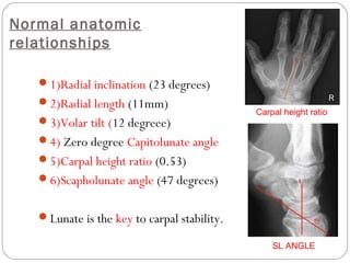
1) The document discusses various carpal dislocations including perilunate dislocations and fractures, scapholunate dissociation, and their anatomy, mechanisms of injury, clinical features, imaging, and management.
2) Perilunate dislocations are high energy injuries that commonly involve disruption of the scapholunate ligament followed by other ligaments and can be classified using the Mayfield classification system. Management involves early closed reduction and surgery including open reduction, ligament repair and fixation.
3) Scapholunate dissociation is an important cause of carpal instability that can lead to DISI deformity if left untreated. The scapholunate ligament maintains carpal stability and its anatomy and biomechan
3. ANATOMY
The wrist is composed of two rows of bones that provide motion and transfer forces.
C, capitate; H, hamate; L, lunate;
S, scaphoid; T, triquetrum; P, pisiform; Td, trapezoid; Tm, trapezium.
4.
5. Gilula arcs outline proximal and distal surfaces of the proximal carpal row and
the proximal cortical margins of capitate and hamate.
6. Normal anatomic
relationships
1)Radial inclination (23 degrees)
2)Radial length (11mm)
3)Volar tilt (12 degreee)
4) Zero degree Capitolunate angle
5)Carpal height ratio (0.53)
6)Scapholunate angle (47 degrees)
Lunate is the key to carpal stability.
Carpal height ratio
SL ANGLE
7. Wrist Ligaments
Extrinsic
Connect radius to carpus & carpus to metacarpals
Intrinsic
Connect carpal to carpal bone.
o Space of Poirier: ligament free area btw
radioscapholunate lig & long radiolunate ligament- at
level of midcarpal joint;an area of potential weakness.
9. Pathomechanics
Classically, the radius, lunate, and capitate have
been described as a central “link” that is colinear in
the sagittal plane.
Scaphoid serves as a connecting strut. Any flexion
moment transmitted across the scaphoid is balanced
by an extension moment at the triquetrum.
10. DISI
When the scaphoid is
destabilized by fracture or
scapholunate ligament
disruption, the lunate and
triquetrum assume a position
of excessive dorsiflexion
(dorsal intercalated segmental
instability [DISI] ) and the
scapholunate angle becomes
abnormally high (>70
degrees).
11. VISI
When the triquetrum is
destabilized (usually by
disruption of the
lunotriquetral ligament
complex), the opposite
pattern (volar intercalated
segmental instability [VISI] )
is seen as the lunate
(intercalated segment) volar
flexes.
12. MECHANISM OF INJURY
FOOSH; axial compressive force
wrist hyperextension,
ulnar deviation, and
intercarpal supination
17. Pathoanatomy
Sequence of events (Mayfield)
scapholunate ligament disrupted -->
disruption of capitolunate articulation -->
disruption of lunotriquetral articulation -->
failure of dorsal radiocarpal ligament -->
lunate rotates and dislocates, usually into carpal tunnel.
18. Dislocation can course through
Greater arc
ligamentous disruptions with associated fractures of
the radius, ulnar, or carpal bones.
Lesser arc
purely ligamentous.
Greater
Lesser
19. Mayfield Classification
STAGE
1 Scapholunate dissociation
2 + Lunocapitate disruption
3 + Lunotriquetral disruption, "perilunate"
4 Lunate dislocated from lunate fossa (usually volar)
•associated with median nerve compression
22. CLINICAL FEATURES
Symptoms
acute wrist swelling and pain
Median nerve symptoms may occur in ~25% of patients
MC in Mayfield stage IV where the lunate dislocates into
the carpal tunnel
23. IMAGING
Radiographs
PA/lateral wrist radiographs
AP
break in Gilula's arc
Lunate and capitate overlap
Lunate appears triangular "piece-of-pie“ sign
Lateral
loss of colinearity of radius, lunate, and capitate
SL angle >70 degrees
MRI
usually not required for diagnosis
24. MANAGEMENT
NON OPERATIVE
Closed reduction and casting
Indications
o no indications when used as definitive
management
Outcomes
o universally poor functional outcomes with non-
operative management
o recurrent dislocation is common
25. Closed Reduction technique of Tavernier
finger traps, elbow at 90 degrees of flexion
hand 5-10 lbs traction for 15 minutes
dorsal dislocations are reduced through wrist extension,
traction, and flexion of wrist.
apply sugar tong splint
follow with surgery.
26. OPERATIVE
1) Emergent closed reduction/splinting followed
by open reduction, ligament repair, fixation, possible
carpal tunnel release.
2) Proximal row carpectomy
3) Total wrist arthrodesis
27. 1) Emergent closed reduction/splinting
followed by open reduction,ligament
repair, fixation,
Indications
all acute injuries <8 weeks old
Outcomes
Emergent closed reduction leads to
Decreased risk of median nerve damage
Decreased risk of cartilage damage
Return to full function unlikely
Decreased grip strength and stiffness are common
28. Approaches – Dorsal, Volar, Combined.
Dorsal approach
longitudinal incision centered at Lister's tubercle
excellent exposure of proximal carpal row and
midcarpal joints
does not allow for carpal tunnel release
Volar approach
extended carpal tunnel incision just proximal to
volar wrist crease
29. Combined dorsal/volar approach
Pros
added exposure
easier reduction
access to distal scaphoid fractures
ability to repair volar ligaments
carpal tunnel decompression
Cons
some believe volar ligament repair not necessary
increased swelling
potential carpal devascularization
difficulty regaining digital flexion and grip
30. Technique
Fix associated fractures
Repair scapholunate ligament
Protect scapholunate ligament repair
Repair of lunotriquetral interosseous ligament
Post-op
Short arm thumb spica splint converted to short arm
cast at first post-op visit
Duration of casting varies, but at least 6 weeks
31. 2) Proximal row carpectomy
Technique
Dorsal and volar incisions if median nerve compression is present
Volar approach allows median nerve decompression with excision
of lunate
Dorsal approach facilitates excision of the scaphoid and
triquetrum
34. INTRODUCTION
Scapholunate ligament is important for carpal stability
chronic scapholunate deficiency DISI
Ligamentous analog of scaphoid #
Acute/ Degenerative injury.
3components: Dorsal, Proximal & Volar
Associated injuries
DISI
Scaphoid flexes palmar and the lunate dorsiflexes
if untreated, progress into a SLAC
35. ANATOMY
Scapholunate interosseous ligament
Location
C-shaped structure connecting the dorsal, proximal and
volar surfaces of the scaphoid and lunate bones
dorsal fiber thickened (2-3mm) compared to volar
Biomechanics
Dorsal component provides the greatest constraint
to translation between the scaphoid and lunate bones
36. CLINICAL EXAMINATION
ASB tenderness
Pain increased with extreme wrist extension and radial deviation
Watson test +ve
When deviating from ulnar to radial, pressure over volar aspect of
scaphoid produces a clunk secondary to dorsal subluxation of the
scaphoid over the dorsal rim of the radius.
37. IMAGING
o Additional radial and ulnar deviation views & clenched fist
Findings
PA radiographs
SL gap > 3mm (Terry Thomas sign)
cortical ring sign (caused by scaphoid malalignment)
scaphoid shortening
Lateral radiographs
dorsal tilt of lunate leads to SL angle > 70°
capitolunate angle > 20°
41. Operative
SURGERY INDICATION
SL Ligament repair acute scapholunate ligament injury
without carpal malalignment
SL reconstruction Acute, SL lig not ammenable to repair
Scaphoid ORIF vs. CRPP SL ligament injury is d/t scaphoid #
Stabilization with wrist fusion
(STT/SLC)
rigid and unreducible DISI deformity