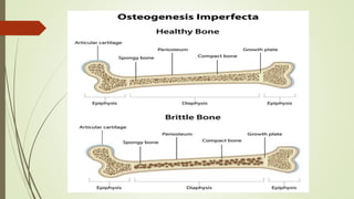
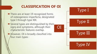
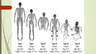
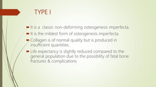
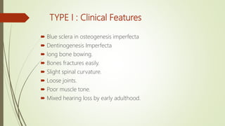
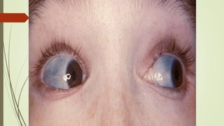
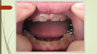
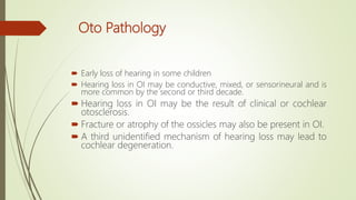
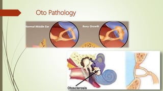
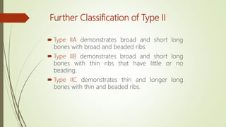
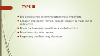
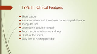
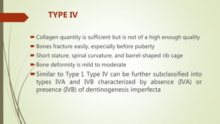
OSTEOGENESIS
IMPERFECTA -By Dr. Rima Jani (PT)
(M.P.T. in Pediatric Science).
OSTEOGENESIS IMPERFECTA
 It is disorder of type I collagen synthesis that affects all connective
tissue in the body.
 Musculoskeletal involvement is diffuse and includes osteoporosis
with excessive fracture even at birth, bowing of long bone, spinal
deformities, muscle weakness and ligamentous laxity.
 Also known as brittle bone disease.
 Incidence is 1 in 15,000 to 1 in 1,00,000
PATHOPHYSIOLOGY
 Osteogenesis imperfecta is caused by defective genes.
 OI is caused by mutations that affect type I collagen
 These genes affect how the body makes collagen, a protein
that helps strengthen bones.
 The condition can be mild, with only a few fractures during a
person's lifetime.
 In more severe cases, it can involve hundreds of fractures
that occur without any apparent cause.
CLINICAL FEATURES
 Blue sclera in eyes
 Dentinogenesis imperfecta
 Hearing loss
 Growth deficiency
 Cardiopulmonary abnormalities
 Easy bruising
 Excessive sweating
 Loose or dislocated joints
OI
Type I
Type II
Type III
Type IV
CLASSIFICATION OF OI
 There are at least 19 recognized forms
of osteogenesis imperfecta, designated
type I through type XIX.
 Several types are distinguished by their
signs and symptoms, although their
characteristic features overlap.
 However, OI is broadly classified into
four main types :
TYPE I
 It is a classic non-deforming osteogenesis imperfecta.
 It is the mildest form of osteogenesis imperfecta.
 Collagen is of normal quality but is produced in
insufficient quantities.
 Life expectancy is slightly reduced compared to the
general population due to the possibility of fatal bone
fractures & complications
TYPE I : Clinical Features
 Blue sclera in osteogenesis imperfecta
 Dentinogenesis Imperfecta
 long bone bowing.
 Bones fractures easily.
 Slight spinal curvature.
 Loose joints.
 Poor muscle tone.
 Mixed hearing loss by early adulthood.
Blue Sclera
 Discoloration of the sclera (whites of the eyes), usually
giving them a blue-gray color.
 The blue-gray color of the sclera is due to the underlying
choroidal veins.
 This is due to the sclera being thinner than normal because
the defective Type I collagen is not forming correctly.
 Slight protrusion of the eyes is also seen.
Dentinogenesis Imperfecta
 It is a genetic disorder of tooth development.
 This condition is a type of dentin dysplasia that causes teeth
to be discolored (most often a blue-gray or yellow-brown
color) and translucent giving teeth an opalescent sheen.
 Teeth are also weaker than normal, making them prone to
rapid wear, breakage, and loss.
 IA and IB are defined to be distinguished by the
absence/presence of dentinogenesis imperfecta
(characterized by opalescent teeth; absent in IA, present in
IB).
Oto Pathology
 Early loss of hearing in some children
 Hearing loss in OI may be conductive, mixed, or sensorineural and is
more common by the second or third decade.
 Hearing loss in OI may be the result of clinical or cochlear
otosclerosis.
 Fracture or atrophy of the ossicles may also be present in OI.
 A third unidentified mechanism of hearing loss may lead to
cochlear degeneration.
Oto Pathology
TYPE II
 It is perinatally lethal osteogenesis imperfecta is the most
severe form.
 Collagen is not of a sufficient quality or quantity.
 Most cases die within the first year of life due to
respiratory failure or intracerebral hemorrhage
TYPE II : Clinical Features
 Severe respiratory problems due to underdeveloped lungs
 Severe bone deformity and small stature.
 Type II can be further sub classified into groups A, B, and C,
which are distinguished by radiographic evaluation of the long
bones and ribs.
Further Classification of Type II
 Type IIA demonstrates broad and short long
bones with broad and beaded ribs.
 Type IIB demonstrates broad and short long
bones with thin ribs that have little or no
beading.
 Type IIC demonstrates thin and longer long
bones with thin and beaded ribs.
 It is progressively deforming osteogenesis imperfecta.
 Collagen improperly formed, enough collagen is made but it
is defective.
 Bones fracture easily, sometimes even before birth.
 Bone deformity, often severe.
 Respiratory problems may also occur.
TYPE III
TYPE III : Clinical Features
 Short stature
 spinal curvature and sometimes barrel-shaped rib cage
 Triangular face
 Loose joints (double-jointed)
 Poor muscle tone in arms and legs
 Bluish of the sclera
 Early loss of hearing possible
 Collagen quantity is sufficient but is not of a high enough quality
 Bones fracture easily, especially before puberty
 Short stature, spinal curvature, and barrel-shaped rib cage
 Bone deformity is mild to moderate
Similar to Type I, Type IV can be further subclassified into
types IVA and IVB characterized by absence (IVA) or
presence (IVB) of dentinogenesis imperfecta
TYPE IV
Diagnosis
 X- rays
 Lab investigations :Collagen testing
 A skin punch biopsy can be performed to determine the
structure and quantity of type I collagen.
 DNA testing
OI type II is often diagnosed by ultrasound during
pregnancy, where already multiple fractures and other
characteristic features may be present.
Treatment
 There is no cure.
 Maintaining a healthy lifestyle by exercising can help to
prevent fractures.
 Treatment may include care of broken bones, pain
medication, physical therapy, braces or wheelchairs, and
surgery.
Medication
 Pharmacologic and vitamin supplements can be given to decrease
the fragility of bones of children
 Calcitonin, fluoride, hormone, and vitamin C and D
 Medication like bisphosphonate group such as pamidronate,
alendronate, zoledronic acid to improve bone density
Surgical Intervention
 A type of surgery that puts metal rods through long bones may be
done to strengthen them.
 Bone infections are treated as and when they occur with the
appropriate antibiotics and antiseptics
 Fractures are managed with a soft splint or fiberglass cast for
immobilization & is kept short to minimize the bone
demineralization, refracture and bony deformity, specifically bowing
of long bones.
Surgical Intervention
 Osteotomy, flexible intramedullary nails, and rod fixation
used to correct bowing deformities or stabilize fractures
 Surgical correction facilitate orthotic use and standing
programs as well as provide support to the bones to
decrease the fracture rate
REHABILITATION
 The goals for the child with severe OI are to:
1. Prevent deformities of the head, spine, and extremities,
2. Avert cardiorespiratory compromise by avoiding constant positioning in
supine,
3. Maximize the child’s ability to move actively.
PHYSIOTHERAPY
 These goals are based on the theory that muscle strengthening and
weight-bearing programs for upper and lower extremities promote
active earlier use of the extremities and may lead to increased bone
mineralization and less severe musculoskeletal deformities
 Instruction in handling of an infant with OI is crucial for all parents
and caregivers.
 The infant should be held with the head and trunk fully supported.
PHYSIOTHERAPY
 Careful positioning of an infant with OI should begin in the first few
days after birth
 Positioning aims to align the infant’s head, trunk, and extremities and
to protect the infant from hitting hard surfaces with activity.
PHYSIOTHERAPY
 Emphasis is on midline orientation of the head and position changes
to prevent the development of a laterally tilted head
 Active movement is encouraged as beginning strengthening
exercises.
PHYSIOTHERAPY
 Careful handling activities can be performed in straight frontal or
sagital plane movements.
 Rotatory movements should be avoided to prevent rotational
Fractures.
 Strengthening activities progress from active movement to playing
with lightweight toys.
PHYSIOTHERAPY
 Active movement can be further encouraged in water either at bath
time or in a swim program with the parent present.
 Standard active- assistive and resistive exercises can be incorporated
as the child becomes a little older.
 Emphasis is also placed on the development of head control in a
variety of positions because children with OI often have a very large
head.
PHYSIOTHERAPY
 Developmental activities such as prone skills and rolling are
encouraged.
 Independent sitting is encouraged when developmentally
appropriate, as is some type of floor mobility.
 Those children who do not have the ability to develop independent
sitting skills should be fitted for a custom- molded seat to promote
head and trunk alignment and afford the child the opportunity to play
in an upright position.
 Throughout the developmental progression, increasing or maintaining
ROM and strength, especially of the pelvic girdle, is incorporated into
activities.
PHYSIOTHERAPY
PHYSIOTHERAPY
 Children should be fitted with orthosis when they have developed
independent sitting skills and balance and are beginning to pull to
stand.
 Those children who cannot sit independently but have developed
head control should be fitted for a standing frame.
 The orthoses recommended in the protocol are containment or
HKAFOs
PHYSIOTHERAPY
 With ambulation and upright positioning, attention must be directed
to the pelvic girdle.
 Hip flexion contractures often develop, and children with OI typically
require ongoing strengthening of their hip extensors and abductors
Osteogenesis Imperfecta

Osteogenesis Imperfecta

  • 1.
    OSTEOGENESIS IMPERFECTA -By Dr.Rima Jani (PT) (M.P.T. in Pediatric Science).
  • 2.
    OSTEOGENESIS IMPERFECTA  Itis disorder of type I collagen synthesis that affects all connective tissue in the body.  Musculoskeletal involvement is diffuse and includes osteoporosis with excessive fracture even at birth, bowing of long bone, spinal deformities, muscle weakness and ligamentous laxity.  Also known as brittle bone disease.  Incidence is 1 in 15,000 to 1 in 1,00,000
  • 4.
    PATHOPHYSIOLOGY  Osteogenesis imperfectais caused by defective genes.  OI is caused by mutations that affect type I collagen  These genes affect how the body makes collagen, a protein that helps strengthen bones.  The condition can be mild, with only a few fractures during a person's lifetime.  In more severe cases, it can involve hundreds of fractures that occur without any apparent cause.
  • 5.
    CLINICAL FEATURES  Bluesclera in eyes  Dentinogenesis imperfecta  Hearing loss  Growth deficiency  Cardiopulmonary abnormalities  Easy bruising  Excessive sweating  Loose or dislocated joints
  • 6.
    OI Type I Type II TypeIII Type IV CLASSIFICATION OF OI  There are at least 19 recognized forms of osteogenesis imperfecta, designated type I through type XIX.  Several types are distinguished by their signs and symptoms, although their characteristic features overlap.  However, OI is broadly classified into four main types :
  • 8.
    TYPE I  Itis a classic non-deforming osteogenesis imperfecta.  It is the mildest form of osteogenesis imperfecta.  Collagen is of normal quality but is produced in insufficient quantities.  Life expectancy is slightly reduced compared to the general population due to the possibility of fatal bone fractures & complications
  • 9.
    TYPE I :Clinical Features  Blue sclera in osteogenesis imperfecta  Dentinogenesis Imperfecta  long bone bowing.  Bones fractures easily.  Slight spinal curvature.  Loose joints.  Poor muscle tone.  Mixed hearing loss by early adulthood.
  • 10.
    Blue Sclera  Discolorationof the sclera (whites of the eyes), usually giving them a blue-gray color.  The blue-gray color of the sclera is due to the underlying choroidal veins.  This is due to the sclera being thinner than normal because the defective Type I collagen is not forming correctly.  Slight protrusion of the eyes is also seen.
  • 12.
    Dentinogenesis Imperfecta  Itis a genetic disorder of tooth development.  This condition is a type of dentin dysplasia that causes teeth to be discolored (most often a blue-gray or yellow-brown color) and translucent giving teeth an opalescent sheen.  Teeth are also weaker than normal, making them prone to rapid wear, breakage, and loss.  IA and IB are defined to be distinguished by the absence/presence of dentinogenesis imperfecta (characterized by opalescent teeth; absent in IA, present in IB).
  • 14.
    Oto Pathology  Earlyloss of hearing in some children  Hearing loss in OI may be conductive, mixed, or sensorineural and is more common by the second or third decade.  Hearing loss in OI may be the result of clinical or cochlear otosclerosis.  Fracture or atrophy of the ossicles may also be present in OI.  A third unidentified mechanism of hearing loss may lead to cochlear degeneration.
  • 15.
  • 16.
    TYPE II  Itis perinatally lethal osteogenesis imperfecta is the most severe form.  Collagen is not of a sufficient quality or quantity.  Most cases die within the first year of life due to respiratory failure or intracerebral hemorrhage
  • 17.
    TYPE II :Clinical Features  Severe respiratory problems due to underdeveloped lungs  Severe bone deformity and small stature.  Type II can be further sub classified into groups A, B, and C, which are distinguished by radiographic evaluation of the long bones and ribs.
  • 18.
    Further Classification ofType II  Type IIA demonstrates broad and short long bones with broad and beaded ribs.  Type IIB demonstrates broad and short long bones with thin ribs that have little or no beading.  Type IIC demonstrates thin and longer long bones with thin and beaded ribs.
  • 19.
     It isprogressively deforming osteogenesis imperfecta.  Collagen improperly formed, enough collagen is made but it is defective.  Bones fracture easily, sometimes even before birth.  Bone deformity, often severe.  Respiratory problems may also occur. TYPE III
  • 20.
    TYPE III :Clinical Features  Short stature  spinal curvature and sometimes barrel-shaped rib cage  Triangular face  Loose joints (double-jointed)  Poor muscle tone in arms and legs  Bluish of the sclera  Early loss of hearing possible
  • 21.
     Collagen quantityis sufficient but is not of a high enough quality  Bones fracture easily, especially before puberty  Short stature, spinal curvature, and barrel-shaped rib cage  Bone deformity is mild to moderate Similar to Type I, Type IV can be further subclassified into types IVA and IVB characterized by absence (IVA) or presence (IVB) of dentinogenesis imperfecta TYPE IV
  • 22.
    Diagnosis  X- rays Lab investigations :Collagen testing  A skin punch biopsy can be performed to determine the structure and quantity of type I collagen.  DNA testing OI type II is often diagnosed by ultrasound during pregnancy, where already multiple fractures and other characteristic features may be present.
  • 23.
    Treatment  There isno cure.  Maintaining a healthy lifestyle by exercising can help to prevent fractures.  Treatment may include care of broken bones, pain medication, physical therapy, braces or wheelchairs, and surgery.
  • 24.
    Medication  Pharmacologic andvitamin supplements can be given to decrease the fragility of bones of children  Calcitonin, fluoride, hormone, and vitamin C and D  Medication like bisphosphonate group such as pamidronate, alendronate, zoledronic acid to improve bone density
  • 25.
    Surgical Intervention  Atype of surgery that puts metal rods through long bones may be done to strengthen them.  Bone infections are treated as and when they occur with the appropriate antibiotics and antiseptics  Fractures are managed with a soft splint or fiberglass cast for immobilization & is kept short to minimize the bone demineralization, refracture and bony deformity, specifically bowing of long bones.
  • 26.
    Surgical Intervention  Osteotomy,flexible intramedullary nails, and rod fixation used to correct bowing deformities or stabilize fractures  Surgical correction facilitate orthotic use and standing programs as well as provide support to the bones to decrease the fracture rate
  • 27.
    REHABILITATION  The goalsfor the child with severe OI are to: 1. Prevent deformities of the head, spine, and extremities, 2. Avert cardiorespiratory compromise by avoiding constant positioning in supine, 3. Maximize the child’s ability to move actively.
  • 28.
    PHYSIOTHERAPY  These goalsare based on the theory that muscle strengthening and weight-bearing programs for upper and lower extremities promote active earlier use of the extremities and may lead to increased bone mineralization and less severe musculoskeletal deformities  Instruction in handling of an infant with OI is crucial for all parents and caregivers.  The infant should be held with the head and trunk fully supported.
  • 29.
    PHYSIOTHERAPY  Careful positioningof an infant with OI should begin in the first few days after birth  Positioning aims to align the infant’s head, trunk, and extremities and to protect the infant from hitting hard surfaces with activity.
  • 30.
    PHYSIOTHERAPY  Emphasis ison midline orientation of the head and position changes to prevent the development of a laterally tilted head  Active movement is encouraged as beginning strengthening exercises.
  • 31.
    PHYSIOTHERAPY  Careful handlingactivities can be performed in straight frontal or sagital plane movements.  Rotatory movements should be avoided to prevent rotational Fractures.  Strengthening activities progress from active movement to playing with lightweight toys.
  • 32.
    PHYSIOTHERAPY  Active movementcan be further encouraged in water either at bath time or in a swim program with the parent present.  Standard active- assistive and resistive exercises can be incorporated as the child becomes a little older.  Emphasis is also placed on the development of head control in a variety of positions because children with OI often have a very large head.
  • 33.
    PHYSIOTHERAPY  Developmental activitiessuch as prone skills and rolling are encouraged.  Independent sitting is encouraged when developmentally appropriate, as is some type of floor mobility.
  • 34.
     Those childrenwho do not have the ability to develop independent sitting skills should be fitted for a custom- molded seat to promote head and trunk alignment and afford the child the opportunity to play in an upright position.  Throughout the developmental progression, increasing or maintaining ROM and strength, especially of the pelvic girdle, is incorporated into activities. PHYSIOTHERAPY
  • 35.
    PHYSIOTHERAPY  Children shouldbe fitted with orthosis when they have developed independent sitting skills and balance and are beginning to pull to stand.  Those children who cannot sit independently but have developed head control should be fitted for a standing frame.  The orthoses recommended in the protocol are containment or HKAFOs
  • 36.
    PHYSIOTHERAPY  With ambulationand upright positioning, attention must be directed to the pelvic girdle.  Hip flexion contractures often develop, and children with OI typically require ongoing strengthening of their hip extensors and abductors

Editor's Notes

  • #15 Otosclerosis is a condition where one or more foci of irregularly laid spongy bone replace part of normally dense enchondral layer of bony otic capsule in the bony labyrinth. This condition affects one of the ossicles (the stapes) resulting in hearing loss, tinnitus, vertigo or a combination of symptoms.