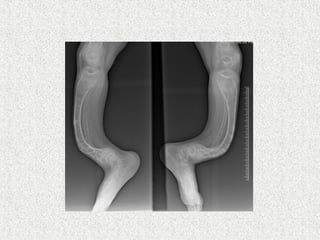
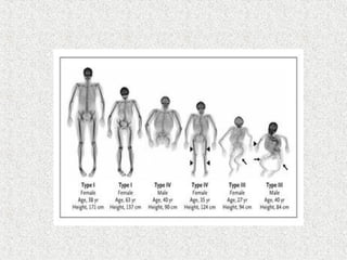
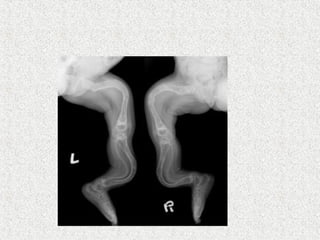
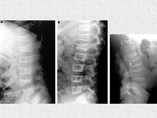
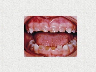
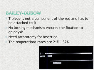
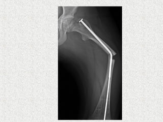
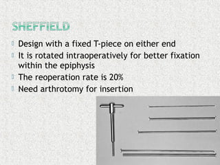
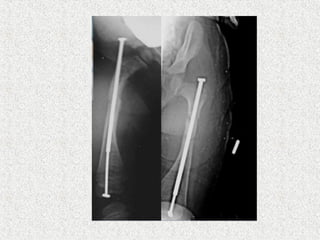
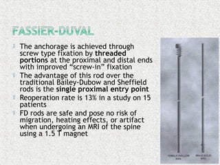
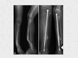
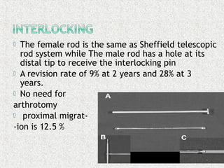
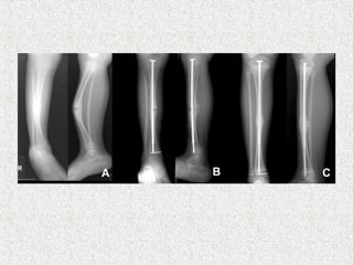
Dr (Major) Parthasarathy S
Pg Resident,MS Orthopaedics
Stanley Medical College,Chennai
Ref :Apley’s system of orthopaedics & fractures 9th
edn
Campbell’s operative orthopaedics 13th
edn
Chapman’s orthopaedic surgery 3rd
edition
Radiographs-Websites
ļ‚ž Brittle bone disease
ļ‚ž Genetic disorder of bone
ļ‚ž 1:20000 incidence
ļ‚ž Abnormal synthesis & structural defect of
type I collagen
ļ‚ž Affected
ļ‚” Bones
ļ‚” Teeth
ļ‚” Ligaments
ļ‚” Sclera
ļ‚” skin
ļ‚ž Considerable variations in
ļ‚” severity of expression
ļ‚” Pattern of inheritance
ļ‚ž Abnormality in gene that codes for alpha
chain type I collagen
ļ‚ž 17q Chromosome
ļ‚ž COL1A1/COL1A2 gene-encodes procollagen
ļ‚ž Type I –quantity
ļ‚ž Other types both quantity & quality
ļ‚ž Alteration in structural integrity
ļ‚ž Reduction in total amount of type I collagen
ļ‚ž Weakening of tissue
ļ‚ž Imperfect ossification of bone
ļ‚” Initiated normally
ļ‚” Progress abnormally
ļ‚” Mixture of lamellar & woven bone
ļ‚” Immature woven bone
ļ‚ž Thinning of dermis
ļ‚ž Laxity of ligaments
ļ‚ž Corneal translucency increased
ļ‚ž Loss of dentin – tooth decay
ļ‚ž Based on
ļ‚” Pattern of inheritance
ļ‚” Age of presentation
ļ‚” Severity of changes in bone & extra-skeletal
tissue
ļ‚ž SILLENCE classification -1981
ļ‚ž Four clinical types-original
ļ‚ž Additional types added later
ļ‚ž Commonest variety
ļ‚ž AD inheritance
ļ‚ž >50%
ļ‚ž # 1-2 yrs of age
ļ‚ž Healing reasonably good
ļ‚ž Deformity not marked
ļ‚ž Deep blue sclera
ļ‚ž Teeth usually normal
ļ‚ž Impaired hearing in adults
ļ‚ž Normal life ecpectancy
ļ‚ž 5-10%
ļ‚ž New dominant mutation/AR
ļ‚ž Intrauterine/neonatal #
ļ‚ž Large skull & wormian bone
ļ‚ž Grey sclera
ļ‚ž Rib # and respiratory difficulty
ļ‚ž Still born/neonatal death
ļ‚ž Sporadic/AR
ļ‚ž Classic but not commonest
ļ‚ž # pesent at birth
ļ‚ž Large skull/wormian bone/pinched looking
face
ļ‚ž Marked deformities/kyphoscoliosis by 6 yrs
ļ‚ž Grey sclera becoming white
ļ‚ž Dentinogenesis imperfect
ļ‚ž Joint laxity marked
ļ‚ž Few survive to adulthood
ļ‚ž Uncommon
ļ‚ž AD
ļ‚ž <5%
ļ‚ž Frequent # during early childhood
ļ‚ž Deformities common
ļ‚ž Pale blue sclera or normal
ļ‚ž Dentinogenesis imperfect
ļ‚ž Survive to adulthood with fairly good
function
ļ‚ž Looser classification
ļ‚” Congenita(Vrolik disease)
ļ‚” Tarda(Ekman-Lobstein disease)
ļ‚ž Vary considerably
ļ‚ž Defining clinincal features
ļ‚” Osteopenia
ļ‚” Liability to #
ļ‚¢ Minor trauma
ļ‚¢ Without much pain & swelling
ļ‚¢ Discovered during infancy
ļ‚¢ Recur frequently throughout childhood
ļ‚¢ Florid callus
ļ‚¢ Lump mistaken for osteosarcoma
ļ‚¢ Abnormal pliable new bone
ļ‚¢ Heal within a normal time interval
ļ‚¢ Minimum time immobilisation
ļ‚¢ Malunion & refracture
ļ‚¢ By 6 yrs severe deformity of long bones & spine
ļ‚¢ After puberty # is infrequent
ļ‚¢ Subclinial forms-recurrent # in adults
ļ‚ž Anterolateral bow or proximal varus
deformity of the femur
ļ‚ž Anterior or anteromedial bow of the tibia
ļ‚ž Spinal deformity---Scoliosis/kyphosis
ļ‚ž Most common type of curve is thoracic
scoliosis.
ļ‚ž Spondylolisthesis, Cervical spinal #
ļ‚ž Adults may be predisposed to rupture of the
patellar ligament or Achilles tendon.
ļ‚ž Acetabular protrusion
ļ‚ž The humerus-- angled laterally or
anterolaterally
ļ‚ž The forearm rotation is often severely
limited
ļ‚ž The elbow joint has cubitus varus with
flexion contracture.
ļ‚ž The faciocranial disproportion--- gives the
face a triangular, elfin shape.
ļ‚ž The ears are displaced downward and
outward. The configuration of the skull ---
soldier's helmet and is called ā€œhelmet head.ā€
ļ‚ž Laxity of ligaments & hypermobile joints
ļ‚ž Blue/grey sclera
ļ‚” Due to uveal pigment showing through
hypertranslucent cornea
ļ‚ž Dentinogenesis imperfecta(crumbling teeth)
ļ‚ž Thin & loose skin
ļ‚ž In severe cases
ļ‚” # present before birth
ļ‚” Stillborn or neonatal death
ļ‚ž Look for evidence of battered baby syndrome
ļ‚ž Motor development delayed in severe form
ļ‚ž Basilar invagination
ļ‚” Basilar stem compression
ļ‚” Macrocephaly
ļ‚” Hydrocephalus
ļ‚” Early death due to respiratory arrest
ļ‚ž OI +AMC
ļ‚ž Alfred Bruck -1897
ļ‚ž Very rare
ļ‚ž Joint contractures + OI features
ļ‚ž Normal sclera/hearing
ļ‚ž Radiograph
ļ‚” Generalised osteopenia
ļ‚” Thinning of long bone
ļ‚” # in various stages of healing
ļ‚” Vertebral compression
ļ‚” Spinal deformity
ļ‚” Skull enlarged
ļ‚” Wormian bones in skull-areas of vicarious
ossification n the calvarium
ļ‚” Widening of metaphysis
ļ‚” Popcorn epiphysis
ļ‚ž Prenatal diagnosis
ļ‚” USG
ļ‚” Chorionic villous sampling
ļ‚ž Preventing #
ļ‚ž Light weight orthoses
ļ‚ž Splinting
ļ‚ž Regular followup
ļ‚ž Plating not recommended
ļ‚ž Skeletal traction not recommended
ļ‚ž Cyclical bisphosphonates
ļ‚ž No optimal dose regimens
ļ‚”zebra stripe sign-- cyclic bisphosphonate
treatment produces sclerotic growth recovery
lines in the long bones
ļ‚ž Sofield & Millar
ļ‚” Multiple osteotomies
ļ‚” Realignment
ļ‚” Medullary nail-solid
ļ‚ž Indication
ļ‚” Deformity correction
ļ‚” Prophylaxis
ļ‚ž 4 main types are developed
ļ‚”Bailey-Dubow
ļ‚”Sheffield
ļ‚”Fassier-Duval
ļ‚”Interlocking telescoping rods
ļ‚ž These rods have a female hollow nail anchored
in the proximal epiphysis of the long bone and a
male solid nail anchored to the distal epiphysis.
They are elongated as the child grow. So, less
revision rates compared to solid
ļ‚ž Rod diameter
ļ‚” Larger diameter gives more stability but causes bone loss
around the rod
ļ‚”Therefore, thinner rods are recommended acting as
internal tutor and not replacing the bone
ļ‚”Leaving 2 mm around the rod in any plan is a safe
method
ļ‚ž Rod length
ļ‚” Pre-operative templates are essential taking into account the
osteotomies needed to straighten the bone
ļ‚”Measure the length (L) between greater trochanter
and distal growth plate
ļ‚”Female rod length is (L) - 7 mm
ļ‚”Male rod length is (L) + (10-15) mm
ļ‚ž T piece is not a component of the rod and has to
be attached to it
ļ‚ž No locking mechanism ensures the fixation to
epiphysis
ļ‚ž Need arthrotomy for insertion
ļ‚ž The reoperations rates are 21% - 32%
Complications of Bailey and Dubow:
ļ‚ž Proximal rod migration
ļ‚ž Disengagement of the
epiphyseal T-piece
ļ‚ž Bending
ļ‚ž Infection
ļ‚ž Design with a fixed T-piece on either end
ļ‚ž It is rotated intraoperatively for better fixation
within the epiphysis
ļ‚ž The reoperation rate is 20%
ļ‚ž Need arthrotomy for insertion
ļ‚ž The anchorage is achieved through
screw type fixation by threaded
portions at the proximal and distal ends
with improved ā€œscrew-inā€ fixation
ļ‚ž The advantage of this rod over the
traditional Bailey-Dubow and Sheffield
rods is the single proximal entry point
ļ‚ž Reoperation rate is 13% in a study on 15
patients
ļ‚ž FD rods are safe and pose no risk of
migration, heating effects, or artifact
when undergoing an MRI of the spine
using a 1.5 T magnet
ļ‚ž The female rod is the same as Sheffield telescopic
rod system while The male rod has a hole at its
distal tip to receive the interlocking pin
ļ‚ž A revision rate of 9% at 2 years and 28% at 3
years.
ļ‚ž No need for
arthrotomy
ļ‚ž proximal migrat-
-ion is 12.5 %
ļ‚ž Regular nail
ļ‚” In older patient
Osteogenesis imperfecta

Osteogenesis imperfecta

  • 1.
    Dr (Major) ParthasarathyS Pg Resident,MS Orthopaedics Stanley Medical College,Chennai Ref :Apley’s system of orthopaedics & fractures 9th edn Campbell’s operative orthopaedics 13th edn Chapman’s orthopaedic surgery 3rd edition Radiographs-Websites
  • 2.
    ļ‚ž Brittle bonedisease ļ‚ž Genetic disorder of bone ļ‚ž 1:20000 incidence ļ‚ž Abnormal synthesis & structural defect of type I collagen ļ‚ž Affected ļ‚” Bones ļ‚” Teeth ļ‚” Ligaments ļ‚” Sclera ļ‚” skin
  • 3.
    ļ‚ž Considerable variationsin ļ‚” severity of expression ļ‚” Pattern of inheritance
  • 4.
    ļ‚ž Abnormality ingene that codes for alpha chain type I collagen ļ‚ž 17q Chromosome ļ‚ž COL1A1/COL1A2 gene-encodes procollagen ļ‚ž Type I –quantity ļ‚ž Other types both quantity & quality
  • 5.
    ļ‚ž Alteration instructural integrity ļ‚ž Reduction in total amount of type I collagen ļ‚ž Weakening of tissue ļ‚ž Imperfect ossification of bone ļ‚” Initiated normally ļ‚” Progress abnormally ļ‚” Mixture of lamellar & woven bone ļ‚” Immature woven bone
  • 9.
    ļ‚ž Thinning ofdermis ļ‚ž Laxity of ligaments ļ‚ž Corneal translucency increased ļ‚ž Loss of dentin – tooth decay
  • 10.
    ļ‚ž Based on ļ‚”Pattern of inheritance ļ‚” Age of presentation ļ‚” Severity of changes in bone & extra-skeletal tissue ļ‚ž SILLENCE classification -1981 ļ‚ž Four clinical types-original ļ‚ž Additional types added later
  • 12.
    ļ‚ž Commonest variety ļ‚žAD inheritance ļ‚ž >50% ļ‚ž # 1-2 yrs of age ļ‚ž Healing reasonably good ļ‚ž Deformity not marked ļ‚ž Deep blue sclera ļ‚ž Teeth usually normal ļ‚ž Impaired hearing in adults ļ‚ž Normal life ecpectancy
  • 13.
    ļ‚ž 5-10% ļ‚ž Newdominant mutation/AR ļ‚ž Intrauterine/neonatal # ļ‚ž Large skull & wormian bone ļ‚ž Grey sclera ļ‚ž Rib # and respiratory difficulty ļ‚ž Still born/neonatal death
  • 15.
    ļ‚ž Sporadic/AR ļ‚ž Classicbut not commonest ļ‚ž # pesent at birth ļ‚ž Large skull/wormian bone/pinched looking face ļ‚ž Marked deformities/kyphoscoliosis by 6 yrs ļ‚ž Grey sclera becoming white ļ‚ž Dentinogenesis imperfect ļ‚ž Joint laxity marked ļ‚ž Few survive to adulthood
  • 16.
    ļ‚ž Uncommon ļ‚ž AD ļ‚ž<5% ļ‚ž Frequent # during early childhood ļ‚ž Deformities common ļ‚ž Pale blue sclera or normal ļ‚ž Dentinogenesis imperfect ļ‚ž Survive to adulthood with fairly good function
  • 18.
    ļ‚ž Looser classification ļ‚”Congenita(Vrolik disease) ļ‚” Tarda(Ekman-Lobstein disease)
  • 19.
    ļ‚ž Vary considerably ļ‚žDefining clinincal features ļ‚” Osteopenia ļ‚” Liability to # ļ‚¢ Minor trauma ļ‚¢ Without much pain & swelling ļ‚¢ Discovered during infancy ļ‚¢ Recur frequently throughout childhood ļ‚¢ Florid callus
  • 20.
    ļ‚¢ Lump mistakenfor osteosarcoma ļ‚¢ Abnormal pliable new bone ļ‚¢ Heal within a normal time interval ļ‚¢ Minimum time immobilisation ļ‚¢ Malunion & refracture ļ‚¢ By 6 yrs severe deformity of long bones & spine ļ‚¢ After puberty # is infrequent ļ‚¢ Subclinial forms-recurrent # in adults
  • 21.
    ļ‚ž Anterolateral bowor proximal varus deformity of the femur ļ‚ž Anterior or anteromedial bow of the tibia ļ‚ž Spinal deformity---Scoliosis/kyphosis ļ‚ž Most common type of curve is thoracic scoliosis. ļ‚ž Spondylolisthesis, Cervical spinal # ļ‚ž Adults may be predisposed to rupture of the patellar ligament or Achilles tendon.
  • 24.
    ļ‚ž Acetabular protrusion ļ‚žThe humerus-- angled laterally or anterolaterally ļ‚ž The forearm rotation is often severely limited ļ‚ž The elbow joint has cubitus varus with flexion contracture. ļ‚ž The faciocranial disproportion--- gives the face a triangular, elfin shape. ļ‚ž The ears are displaced downward and outward. The configuration of the skull --- soldier's helmet and is called ā€œhelmet head.ā€
  • 29.
    ļ‚ž Laxity ofligaments & hypermobile joints ļ‚ž Blue/grey sclera ļ‚” Due to uveal pigment showing through hypertranslucent cornea ļ‚ž Dentinogenesis imperfecta(crumbling teeth) ļ‚ž Thin & loose skin ļ‚ž In severe cases ļ‚” # present before birth ļ‚” Stillborn or neonatal death ļ‚ž Look for evidence of battered baby syndrome ļ‚ž Motor development delayed in severe form
  • 33.
    ļ‚ž Basilar invagination ļ‚”Basilar stem compression ļ‚” Macrocephaly ļ‚” Hydrocephalus ļ‚” Early death due to respiratory arrest
  • 34.
    ļ‚ž OI +AMC ļ‚žAlfred Bruck -1897 ļ‚ž Very rare ļ‚ž Joint contractures + OI features ļ‚ž Normal sclera/hearing
  • 35.
    ļ‚ž Radiograph ļ‚” Generalisedosteopenia ļ‚” Thinning of long bone ļ‚” # in various stages of healing ļ‚” Vertebral compression ļ‚” Spinal deformity ļ‚” Skull enlarged ļ‚” Wormian bones in skull-areas of vicarious ossification n the calvarium ļ‚” Widening of metaphysis ļ‚” Popcorn epiphysis
  • 36.
    ļ‚ž Prenatal diagnosis ļ‚”USG ļ‚” Chorionic villous sampling
  • 37.
    ļ‚ž Preventing # ļ‚žLight weight orthoses ļ‚ž Splinting ļ‚ž Regular followup ļ‚ž Plating not recommended ļ‚ž Skeletal traction not recommended
  • 38.
    ļ‚ž Cyclical bisphosphonates ļ‚žNo optimal dose regimens ļ‚”zebra stripe sign-- cyclic bisphosphonate treatment produces sclerotic growth recovery lines in the long bones
  • 39.
    ļ‚ž Sofield &Millar ļ‚” Multiple osteotomies ļ‚” Realignment ļ‚” Medullary nail-solid ļ‚ž Indication ļ‚” Deformity correction ļ‚” Prophylaxis
  • 43.
    ļ‚ž 4 maintypes are developed ļ‚”Bailey-Dubow ļ‚”Sheffield ļ‚”Fassier-Duval ļ‚”Interlocking telescoping rods ļ‚ž These rods have a female hollow nail anchored in the proximal epiphysis of the long bone and a male solid nail anchored to the distal epiphysis. They are elongated as the child grow. So, less revision rates compared to solid
  • 44.
    ļ‚ž Rod diameter ļ‚”Larger diameter gives more stability but causes bone loss around the rod ļ‚”Therefore, thinner rods are recommended acting as internal tutor and not replacing the bone ļ‚”Leaving 2 mm around the rod in any plan is a safe method ļ‚ž Rod length ļ‚” Pre-operative templates are essential taking into account the osteotomies needed to straighten the bone ļ‚”Measure the length (L) between greater trochanter and distal growth plate ļ‚”Female rod length is (L) - 7 mm ļ‚”Male rod length is (L) + (10-15) mm
  • 45.
    ļ‚ž T pieceis not a component of the rod and has to be attached to it ļ‚ž No locking mechanism ensures the fixation to epiphysis ļ‚ž Need arthrotomy for insertion ļ‚ž The reoperations rates are 21% - 32%
  • 47.
    Complications of Baileyand Dubow: ļ‚ž Proximal rod migration ļ‚ž Disengagement of the epiphyseal T-piece ļ‚ž Bending ļ‚ž Infection
  • 49.
    ļ‚ž Design witha fixed T-piece on either end ļ‚ž It is rotated intraoperatively for better fixation within the epiphysis ļ‚ž The reoperation rate is 20% ļ‚ž Need arthrotomy for insertion
  • 51.
    ļ‚ž The anchorageis achieved through screw type fixation by threaded portions at the proximal and distal ends with improved ā€œscrew-inā€ fixation ļ‚ž The advantage of this rod over the traditional Bailey-Dubow and Sheffield rods is the single proximal entry point ļ‚ž Reoperation rate is 13% in a study on 15 patients ļ‚ž FD rods are safe and pose no risk of migration, heating effects, or artifact when undergoing an MRI of the spine using a 1.5 T magnet
  • 54.
    ļ‚ž The femalerod is the same as Sheffield telescopic rod system while The male rod has a hole at its distal tip to receive the interlocking pin ļ‚ž A revision rate of 9% at 2 years and 28% at 3 years. ļ‚ž No need for arthrotomy ļ‚ž proximal migrat- -ion is 12.5 %
  • 56.