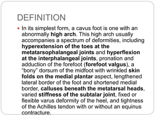
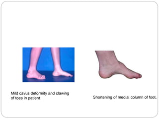
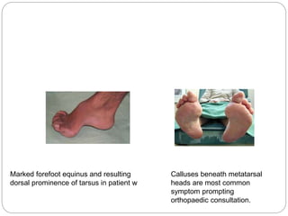
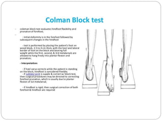
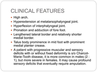
DR PRATIK DHABALIA
CAVUS FOOT
DEFINITION
 In its simplest form, a cavus foot is one with an
abnormally high arch. This high arch usually
accompanies a spectrum of deformities, including
hyperextension of the toes at the
metatarsophalangeal joints and hyperflexion
at the interphalangeal joints, pronation and
adduction of the forefoot (forefoot valgus), a
“bony” dorsum of the midfoot with wrinkled skin
folds on the medial plantar aspect, lengthened
lateral border of the foot and shortened medial
border, calluses beneath the metatarsal heads,
varied stiffness of the subtalar joint, fixed or
flexible varus deformity of the heel, and tightness
of the Achilles tendon with or without an equinus
contracture.
Mild cavus deformity and clawing
of toes in patient Shortening of medial column of foot.
Marked forefoot equinus and resulting
dorsal prominence of tarsus in patient w
Calluses beneath metatarsal
heads are most common
symptom prompting
orthopaedic consultation.
ETIOLOGY
 NEUROMUSCULAR CAUSES
 The most common neuromuscular diseases
causing pes cavus in skeletally mature feet are
Charcot-Marie-Tooth disease and poliomyelitis.
 Patients with spinal dysraphism.
 cerebral palsy.
 primary cerebellar disease.
 arthrogryposis.
 severe clubfeet may develop cavus deformity,
but these conditions usually are recognized and
treated before skeletal maturity.
 Traumatic cavus deformity can be caused by
deep posterior compartment syndrome after
fracture of the tibia or fibula or by malunion of
midfoot fractures or fracture dislocations.
 In patients with neuromuscular diseases and
patients with idiopathic deformities, the underlying
pathologic mechanism of the cavus deformity is
believed to be an imbalance of the extrinsic-
intrinsic muscles.
 The principle that the hindfoot deformity follows
forefoot equinus is supported by the observations
of Paulos et al.: the rigid plantarflexed first ray
forces the heel into varus, and eventually the
deformity becomes fixed.
 The Coleman and Chestnut block test is an
excellent method of determining the hindfoot-
forefoot relationship in pes cavus and determining
whether the hindfoot component is flexible. Other
than the cause and possibly the age of the
patient, the flexibility of any or all of the anatomic
components of a cavus foot is the most important
factor for determining persistence of symptoms
and appropriate treatment
CLINICAL FEATURES
 High arch.
 Hyperextension at metatarsophalyngeal joint.
 Hyperflexion of interphalyngeal joint.
 Pronation and adduction of fore foot.
 Lengthened lateral border and relatively shorter
medial border.
 Talus body prominence in mid foot with prominent
medial planter crease.
 A patient with progressive muscular and sensory
deficits with or without fixed deformity is s/o Charcot-
Marie-Tooth disease, it is more common in males (2 :
1), but more severe in females. It may cause profound
sensory deficits that eventually require amputation.
HOW TO DIFFERENTIATE
 A patient presenting with spasticity, even of the
mildest degree, or with a preulcerative or ulcerative
lesion of the plantar surface of the foot must be
offered nonoperative treatment with shoe or ankle-
foot orthoses. Surgery in the presence of decreased
plantar sensation is fraught with complications that
are most difficult to overcome.
 The postpoliomyelitis cavus foot has anterior and
posterior (or forefoot and hindfoot) components to the
deformity, in contrast to patients with Charcot-Marie-
Tooth disease, who usually have no fixed hindfoot
calcaneal deformity.
 Because of intact sensation and the nonprogressive
nature of the deformities, patients with
postpoliomyelitis cavus feet have a better, or at least
more predictable, prognosis than patients with
Charcot-Marie-Tooth disease, with or without
treatment.
 Posttraumatic cavovarus results from injury to the
extrinsic foot musculature, creating an imbalance
with the intrinsic musculature. Affecting the deep
posterior leg compartment, compartment
syndrome and subsequent Volkmann contracture
can lead to a cavus foot with claw toe deformities.
Soft-tissue injuries from crush mechanisms or
severe burns can lead to muscle imbalance that
results in cavus deformity
RADIOGRAPHIC FINDING
 A standing lateral view allows assessment of
ankle joint position, calcaneal pitch, and midfoot
and forefoot position, especially the degree of
plantarflexion of the first ray.This information is
invaluable in preoperative planning.
 The standing lateral radiograph also allows
estimation of the contribution of the hindfoot
(talus and calcaneus), midfoot (navicular and
cuboid-cuneiform), and forefoot (Lisfranc) to the
cavus deformity. The extension deformity of the
phalanges on the metatarsal heads during weight
bearing helps determine the severity of the fixed
deformity
A line is drawn from planter most surface of calcaneum to the inferior
border of distal articular surface.
Normal 17-32 degree
Usually normal in CMT with forefoot equinus, increased in idiopathic
cases and in poliomyelitis
 Standing anteroposterior views with the hindfoot
in as neutral a position as possible help
corroborate any metatarsus adductus component
suspected clinically The talocalcaneal angle (Kite
angle) is determined on this view. The closer the
talocalcaneal angle approaches zero, the more
parallel the talus is in relation to the calcaneus,
indicating hindfoot varus.
 Other radiographic findings that may be helpful
include (1) degenerative changes (2) rotation of
the talus in the ankle mortise (3) dystrophic
ossification in soft tissue suggesting tendon or
ligament injury on the oblique view
The talocalcaneal angle, also
known as the kite angle, refers to
the angle between lines drawn
down the axis of
the talus and calcaneus measured
on a weightbearing DP foot
radiograph.
The mid-talar line should pass
through (or just medial to) the base
of the 1st metatarsal and the mid-
calcaneal line should pass through
the base of the 4th metatarsal.
The talocalcaneal angle should
measure between 25 and 40
degrees.
TREATMENT
CLAW TOES
 In patients with traumatic pes cavus, only the claw toe
deformities and possibly tight plantar fascia may require
surgical treatment, leaving the bony midfoot deformity to
appropriate shoe and orthotic management.
 For fixed contractures at the metatarsophalangeal and
interphalangeal joints, the following are recommended:
 1. Lengthening of the extensor hallucis longus and
extensor digitorum longus.
 2. Tenotomy of the extensor digitorum brevis and the
extensor hallucis brevis.
 3. Dorsal capsulotomy of the metatarsophalangeal joints.
 4. Resection of the head and neck of the proximal
phalanges.
 5. Release of the plantar fascia, if indicated.
 6. Arthrodesis of the interphalangeal joint of the
hallux or plantar plate release and sectioning of
the collateral ligaments at the interphalangeal
joint of the hallux with temporary Kirschner wire
fixation
1.PLANTAR FASCIA RELEASE
 Make a longitudinal incision along the medial side of the
calcaneus and carry it distally to a point 4 cm anterior to
the medial tubercle
 Separate the superficial and deep surfaces of the plantar
fascia from the muscle and fat and free it throughout its
breadth.
 Incise the fascia transversely close to where it blends into
the plantar surface of the calcaneus.
 Place a periosteal elevator or retractor on the deep surface
of the fascia as it is released.
 If the plantar fascia still feels tight, incise the medial band
again through a separate incision 2 cm proximal to the first
metatarsal head. Protect the flexor hallucis longus while
releasing the medial band of the plantar fascia down to,
but not through, the flexor hallucis brevis, and dorsiflex the
first metatarsal by pushing up on the metatarsal head.
 Secure hemostasis and close the wound with
nonabsorbable sutures in adult patients.
Correction of clawing of great and
second toe
 Surgical technique for clawing of great and second
toes.
 A- Incision.
 B- Extensor hallucis longus is lengthened in coronal
or sagittal plane, and extensor hallucis brevis is
tenotomized.
 C- Dorsal capsulotomy and collateral ligament
release.
 D- Approach to interphalangeal joint through separate
dorsal incision.
 E-Corrected position on lateral view; arthrodesis of
interphalangeal joint of great toe with longitudinal wire
down to base of proximal phalanx.
 F, Correction of second toe by excision of head and
neck of proximal phalanx, dorsal capsulotomy at
metatarsophalangeal joint, lengthening of extensor
digitorum longus, and tenotomy of extensor digitorum
brevis.
 G, Correction at metatarsophalangeal and proximal
interphalangeal joints.
2.TENDON SUSPENSION OF THE FIRST
METATARSAL AND INTERPHALANGEAL JOINT
ARTHRODESIS
 The Jones procedure, which is basically a tendon
suspension of the first metatarsal combined with
arthrodesis of the interphalangeal joint, has
proved valuable over many decades. Instead of
Z-lengthening of the extensor hallucis longus, the
proximal end is placed through a hole in the first
metatarsal neck.
3.Proximal First Metatarsal Osteotomy and Plantar
Fasciotomy.
 This combined procedure is applicable in
hereditary motor sensory neuropathy, after
trauma and peripheral nerve injury, although
today it is probably used more often in Charcot-
Marie-Tooth disease to correct a predominantly
forefoot (first ray) driven cavus deformity.
In Charcot-Marie-Tooth disease, the correction of the forefoot
equinus should proceed in an orderly fashion
Plantar fascia relese and transfer of
peroneus longus to peroneus brevis
Basilar close wedge osteotomy of the
first metatarsal is done
Transfer of EHL to the neck of 1st
metatarsal and in skeletally mature
patient arthrodesis of interphalengeal
joint of halux.
4.COMBINED PROXIMAL FIRST METATARSAL
OSTEOTOMY, PLANTAR FASCIOTOMY, AND
TRANSFER OF THE ANTERIOR TIBIAL TENDON
 In treatment for cavus deformity secondary to
Charcot-Marie-Tooth, also included transfer of the
anterior tibial tendon to the lateral cuneiform if
grade 4 or 5 strength was present. Later in the
study, this transfer was done for a tenodesis to
negate any residual deforming force that the
anterior tibial muscle may have on the varus
component of the cavovarus deformity.
5.PLANTAR FASCIOTOMIES AND CLOSING WEDGE
OSTEOTOMY
 DOUBLE PLANTAR
FASCIOTOMIES (IF
REQUIRED— MOST OFTEN IN
ADULTS AND AFTER
TRAUMA) BY GOULD TECH.
 CLOSING WEDGE
GREENSTICK DORSAL
PROXIMAL METATARSAL
OSTEOTOMIES
5.EXTENSOR TENDON TRANSFER
 (HIBBS) ■ Make a curved
incision 7.5 to 10 cm long
on the dorsum of the foot
lateral to the midline and
expose the common
extensor tendons ■ Divide
the tendons as far distally
as feasible, draw their
proximal ends through a
tunnel in the third
cuneiform, and fix them
with a nonabsorbable
suture ■ As an alternative,
use a plantar button and
felt with a Bunnell pull-out
stitch. ■ Close the wounds
and apply a plaster boot
cast with the foot in the
corrected position
TARSOMETATARSAL TRUNCATEDWEDGE
ARTHRODESIS
 Another procedure for forefoot equinus advocated
by Jahss is arthrodesis of all tarsometatarsal
joints
 Indications :
 1. Equinus deformity of the forefoot with
persistent painful metatarsalgia and associated
plantar keratoses, unrelieved by conservative
management. Such patients usually have more
than 10 degrees of equinus angulation.
 2. Pes cavus with normal muscle balance and
without advanced metatarsal fat pad atrophy
 3. Equinovarus or equinoadductovarus deformity
of the forefoot with the heel in neutral or almost
neutral position. This group includes the residual
clubfoot deformities and compartment
syndromes. If the lesion is caused by
neuromuscular disease, such as poliomyelitis, the
foot should be stable.
 4. Normal vascular status and normal skin
coverage.
 5. The procedure preferably is done at an early
stage. If the associated hammertoes are still
flexible, the toes straighten as the dorsal wedge
is closed and do not require separate surgery.
 Contraindications :
 1. When skin coverage of the forefoot is poor
from previous surgery, or vascularity of the skin is
questionable, no surgery should be performed.
 2. Surgery should not be done before skeletal
maturity.
MIDFOOT CAVUS
 Cavus deformity can occur at the midtarsal joints
(talonavicular-calcaneocuboid) or
naviculocuneiform joints, but is most common at
the former. Depending on the rigidity of the
deformity, plantar fascia release combined with
calcaneal or metatarsal osteotomy may correct
the deformity sufficiently to achieve a plantigrade
foot. For mild-to-moderate fixed cavus deformity
at the midfoot, the following osteotomies have
been described.
 Any of these midfoot osteotomies may produce a
short, wide, unattractive foot, depending on how
much bone is removed.
1.ANTERIOR TARSAL WEDGE OSTEOTOMY
2.V-OSTEOTOMY OF THE TARSUS
 The disadvantage of the anterior tarsal wedge
osteotomy is the foot is shortened, widened, and
thickened. Japas described a technique to produce a
more normal-appearing foot. It consists of a V-
osteotomy in which the apex of the V is proximal and
at the highest point of the cavus, usually within the
navicular. One limb of the V extends laterally and the
other medially through the first cuneiform to the
medial border.
 No bone is excised; instead, the proximal border of
the distal fragment of the osteotomy is depressed
plantarward while the metatarsal heads are elevated,
correcting the deformity and lengthening the plantar
surface of the foot. The technique is recommended
for moderate deformity in children 6 years old or older.
Deformities of the hindfoot or midtarsal joint are not
corrected by this osteotomy and may require later
correction by triple arthrodesis or the Dwyer
COMBINED CAVUS (CALCANEOCAVUS
DEFORMITY)
 1. OSTEOTOMY OF THE CALCANEUS
(DWYER’S)
2. CRESCENTIC CALCANEAL OSTEOTOMY
 Samilson recommended
crescentic calcaneal
osteotomy for
ambulatory patients with
symptomatic
calcaneocavus feet. On
a lateral radiograph, the
calcaneus must be
relatively vertical, and
the apex of the cavus
must be posterior to the
midtarsus. The
operation does not
correct midtarsal or
forefoot cavus but does
correct hindfoot cavus
(calcaneocavus).
CALCANEOCAVOVARUS AND CAVOVARUS
DEFORMITY
 Seen in patients with Charcot-Marie-Tooth disease,
but occasionally this deformity occurs after
poliomyelitis or malunion of displaced fractures of the
talus subtalar arthrodesis (removing more bone
laterally) combined with lateral closing wedge (7 to 10
mm) osteotomy of the anterior aspect of the
calcaneus 1 cm proximal to the calcaneocuboid
joint (reverse Evans osteotomy) may correct the
deformities, while allowing some degree of midtarsal
motion. If the talonavicular or calcaneocuboid joints
show arthritic changes, triple arthrodesis is
indicated. The wedges of bone must be planned
carefully to correct the multiplane deformity. In
neuromuscular cavovarus or calcaneocavovarus
deformities, one of the following methods is
recommended.
1. TRIPLANAR OSTEOTOMY AND LATERAL
LIGAMENT RECONSTRUCTION
 For unstable ankle joints without significant
degenerative changes associated with
calcaneocavovarus deformity, Saxby and
Myerson recommended performing a lateral
ligament reconstruction at the time of calcaneal
triplanar osteotomy.
 If degenerative changes are noted: triple
arthrodesis with lateral ligament reconstruction.
 Saxby and Myerson emphasized that in
hereditary sensorimotor neuropathy (Charcot-
Marie-Tooth disease), the peroneus brevis is not
functioning and tendon can be used to help
stabilize the ankle joint that tilts into varus.
2. Z-SHAPED CALCANEAL OSTEOTOMY
 Knupp et al. described a procedure using a step-
cut (scarf) osteotomy that allows correction of the
heel in frontal and transverse planes.
 Indications: 1.rigid varus hindfoot combined with
forefoot valgus and an excessively plantarflexed
first ray. Osteotomy of the calcaneus is necessary
if the varus deformity is caused by metatarsus
primus flexus without a rigid hindfoot deformity.
Likewise, additional tendon transfers may be
required in an excessively pronated forefoot.
 Degenerative disease and coalitions are
contraindications to this procedure. A neurologic
assessment should be done, especially if
Charcot-Marie-Tooth disease is suspected.
3.PERONEUS BREVIS TENODESIS
 When the ankle joint is unstable in varus from chronic
weakness of the peroneal tendons, Myerson
recommended a tenodesis using the peroneus brevis
tendon because it has no active function.
 Realignment of the hindfoot with an osteotomy or
arthrodesis may correct the tibiotalar tilt by shifting the
weight-bearing axis of the leg laterally.
 In patients with hindfoot varus and ankle instability not
associated with any neuromuscular imbalance, the
peroneus brevis tendon is split and reconstruction is
performed (Chrisman and Snook). If the talar tilt is
fixed and rigid and articular pain is present, a pantalar
arthrodesis should be performed.
CALCANEOCAVOVARUS AND CAVOVARUS
DEFORMITY ASSOCIATED WITH ARTHRITIC
CHANGES OF THE SUBTALAR AND MIDTARSAL
JOINTS
 1.TRIPLE ARTHRODESIS
 Siffert, Forster, and Nachamie triple arthrodesis
DUNN TECHNIQUE
 An alternative method is . Occasionally, the
deformity is so severe that the entire navicular is
removed :Dunn technique
LAMBRINUDI TRIPLE ARTHRODESIS
THANK YOU

Pes cavus

  • 1.
  • 2.
    DEFINITION  In itssimplest form, a cavus foot is one with an abnormally high arch. This high arch usually accompanies a spectrum of deformities, including hyperextension of the toes at the metatarsophalangeal joints and hyperflexion at the interphalangeal joints, pronation and adduction of the forefoot (forefoot valgus), a “bony” dorsum of the midfoot with wrinkled skin folds on the medial plantar aspect, lengthened lateral border of the foot and shortened medial border, calluses beneath the metatarsal heads, varied stiffness of the subtalar joint, fixed or flexible varus deformity of the heel, and tightness of the Achilles tendon with or without an equinus contracture.
  • 3.
    Mild cavus deformityand clawing of toes in patient Shortening of medial column of foot.
  • 4.
    Marked forefoot equinusand resulting dorsal prominence of tarsus in patient w Calluses beneath metatarsal heads are most common symptom prompting orthopaedic consultation.
  • 5.
    ETIOLOGY  NEUROMUSCULAR CAUSES The most common neuromuscular diseases causing pes cavus in skeletally mature feet are Charcot-Marie-Tooth disease and poliomyelitis.  Patients with spinal dysraphism.  cerebral palsy.  primary cerebellar disease.  arthrogryposis.  severe clubfeet may develop cavus deformity, but these conditions usually are recognized and treated before skeletal maturity.
  • 6.
     Traumatic cavusdeformity can be caused by deep posterior compartment syndrome after fracture of the tibia or fibula or by malunion of midfoot fractures or fracture dislocations.  In patients with neuromuscular diseases and patients with idiopathic deformities, the underlying pathologic mechanism of the cavus deformity is believed to be an imbalance of the extrinsic- intrinsic muscles.
  • 8.
     The principlethat the hindfoot deformity follows forefoot equinus is supported by the observations of Paulos et al.: the rigid plantarflexed first ray forces the heel into varus, and eventually the deformity becomes fixed.
  • 9.
     The Colemanand Chestnut block test is an excellent method of determining the hindfoot- forefoot relationship in pes cavus and determining whether the hindfoot component is flexible. Other than the cause and possibly the age of the patient, the flexibility of any or all of the anatomic components of a cavus foot is the most important factor for determining persistence of symptoms and appropriate treatment
  • 11.
    CLINICAL FEATURES  Higharch.  Hyperextension at metatarsophalyngeal joint.  Hyperflexion of interphalyngeal joint.  Pronation and adduction of fore foot.  Lengthened lateral border and relatively shorter medial border.  Talus body prominence in mid foot with prominent medial planter crease.  A patient with progressive muscular and sensory deficits with or without fixed deformity is s/o Charcot- Marie-Tooth disease, it is more common in males (2 : 1), but more severe in females. It may cause profound sensory deficits that eventually require amputation.
  • 12.
    HOW TO DIFFERENTIATE A patient presenting with spasticity, even of the mildest degree, or with a preulcerative or ulcerative lesion of the plantar surface of the foot must be offered nonoperative treatment with shoe or ankle- foot orthoses. Surgery in the presence of decreased plantar sensation is fraught with complications that are most difficult to overcome.  The postpoliomyelitis cavus foot has anterior and posterior (or forefoot and hindfoot) components to the deformity, in contrast to patients with Charcot-Marie- Tooth disease, who usually have no fixed hindfoot calcaneal deformity.  Because of intact sensation and the nonprogressive nature of the deformities, patients with postpoliomyelitis cavus feet have a better, or at least more predictable, prognosis than patients with Charcot-Marie-Tooth disease, with or without treatment.
  • 13.
     Posttraumatic cavovarusresults from injury to the extrinsic foot musculature, creating an imbalance with the intrinsic musculature. Affecting the deep posterior leg compartment, compartment syndrome and subsequent Volkmann contracture can lead to a cavus foot with claw toe deformities. Soft-tissue injuries from crush mechanisms or severe burns can lead to muscle imbalance that results in cavus deformity
  • 14.
    RADIOGRAPHIC FINDING  Astanding lateral view allows assessment of ankle joint position, calcaneal pitch, and midfoot and forefoot position, especially the degree of plantarflexion of the first ray.This information is invaluable in preoperative planning.  The standing lateral radiograph also allows estimation of the contribution of the hindfoot (talus and calcaneus), midfoot (navicular and cuboid-cuneiform), and forefoot (Lisfranc) to the cavus deformity. The extension deformity of the phalanges on the metatarsal heads during weight bearing helps determine the severity of the fixed deformity
  • 15.
    A line isdrawn from planter most surface of calcaneum to the inferior border of distal articular surface. Normal 17-32 degree Usually normal in CMT with forefoot equinus, increased in idiopathic cases and in poliomyelitis
  • 16.
     Standing anteroposteriorviews with the hindfoot in as neutral a position as possible help corroborate any metatarsus adductus component suspected clinically The talocalcaneal angle (Kite angle) is determined on this view. The closer the talocalcaneal angle approaches zero, the more parallel the talus is in relation to the calcaneus, indicating hindfoot varus.  Other radiographic findings that may be helpful include (1) degenerative changes (2) rotation of the talus in the ankle mortise (3) dystrophic ossification in soft tissue suggesting tendon or ligament injury on the oblique view
  • 17.
    The talocalcaneal angle,also known as the kite angle, refers to the angle between lines drawn down the axis of the talus and calcaneus measured on a weightbearing DP foot radiograph. The mid-talar line should pass through (or just medial to) the base of the 1st metatarsal and the mid- calcaneal line should pass through the base of the 4th metatarsal. The talocalcaneal angle should measure between 25 and 40 degrees.
  • 18.
    TREATMENT CLAW TOES  Inpatients with traumatic pes cavus, only the claw toe deformities and possibly tight plantar fascia may require surgical treatment, leaving the bony midfoot deformity to appropriate shoe and orthotic management.  For fixed contractures at the metatarsophalangeal and interphalangeal joints, the following are recommended:  1. Lengthening of the extensor hallucis longus and extensor digitorum longus.  2. Tenotomy of the extensor digitorum brevis and the extensor hallucis brevis.  3. Dorsal capsulotomy of the metatarsophalangeal joints.  4. Resection of the head and neck of the proximal phalanges.  5. Release of the plantar fascia, if indicated.
  • 19.
     6. Arthrodesisof the interphalangeal joint of the hallux or plantar plate release and sectioning of the collateral ligaments at the interphalangeal joint of the hallux with temporary Kirschner wire fixation
  • 20.
    1.PLANTAR FASCIA RELEASE Make a longitudinal incision along the medial side of the calcaneus and carry it distally to a point 4 cm anterior to the medial tubercle  Separate the superficial and deep surfaces of the plantar fascia from the muscle and fat and free it throughout its breadth.  Incise the fascia transversely close to where it blends into the plantar surface of the calcaneus.  Place a periosteal elevator or retractor on the deep surface of the fascia as it is released.  If the plantar fascia still feels tight, incise the medial band again through a separate incision 2 cm proximal to the first metatarsal head. Protect the flexor hallucis longus while releasing the medial band of the plantar fascia down to, but not through, the flexor hallucis brevis, and dorsiflex the first metatarsal by pushing up on the metatarsal head.  Secure hemostasis and close the wound with nonabsorbable sutures in adult patients.
  • 21.
    Correction of clawingof great and second toe
  • 22.
     Surgical techniquefor clawing of great and second toes.  A- Incision.  B- Extensor hallucis longus is lengthened in coronal or sagittal plane, and extensor hallucis brevis is tenotomized.  C- Dorsal capsulotomy and collateral ligament release.  D- Approach to interphalangeal joint through separate dorsal incision.  E-Corrected position on lateral view; arthrodesis of interphalangeal joint of great toe with longitudinal wire down to base of proximal phalanx.  F, Correction of second toe by excision of head and neck of proximal phalanx, dorsal capsulotomy at metatarsophalangeal joint, lengthening of extensor digitorum longus, and tenotomy of extensor digitorum brevis.  G, Correction at metatarsophalangeal and proximal interphalangeal joints.
  • 23.
    2.TENDON SUSPENSION OFTHE FIRST METATARSAL AND INTERPHALANGEAL JOINT ARTHRODESIS  The Jones procedure, which is basically a tendon suspension of the first metatarsal combined with arthrodesis of the interphalangeal joint, has proved valuable over many decades. Instead of Z-lengthening of the extensor hallucis longus, the proximal end is placed through a hole in the first metatarsal neck.
  • 25.
    3.Proximal First MetatarsalOsteotomy and Plantar Fasciotomy.  This combined procedure is applicable in hereditary motor sensory neuropathy, after trauma and peripheral nerve injury, although today it is probably used more often in Charcot- Marie-Tooth disease to correct a predominantly forefoot (first ray) driven cavus deformity.
  • 26.
    In Charcot-Marie-Tooth disease,the correction of the forefoot equinus should proceed in an orderly fashion Plantar fascia relese and transfer of peroneus longus to peroneus brevis Basilar close wedge osteotomy of the first metatarsal is done Transfer of EHL to the neck of 1st metatarsal and in skeletally mature patient arthrodesis of interphalengeal joint of halux.
  • 27.
    4.COMBINED PROXIMAL FIRSTMETATARSAL OSTEOTOMY, PLANTAR FASCIOTOMY, AND TRANSFER OF THE ANTERIOR TIBIAL TENDON  In treatment for cavus deformity secondary to Charcot-Marie-Tooth, also included transfer of the anterior tibial tendon to the lateral cuneiform if grade 4 or 5 strength was present. Later in the study, this transfer was done for a tenodesis to negate any residual deforming force that the anterior tibial muscle may have on the varus component of the cavovarus deformity.
  • 28.
    5.PLANTAR FASCIOTOMIES ANDCLOSING WEDGE OSTEOTOMY  DOUBLE PLANTAR FASCIOTOMIES (IF REQUIRED— MOST OFTEN IN ADULTS AND AFTER TRAUMA) BY GOULD TECH.  CLOSING WEDGE GREENSTICK DORSAL PROXIMAL METATARSAL OSTEOTOMIES
  • 29.
    5.EXTENSOR TENDON TRANSFER (HIBBS) ■ Make a curved incision 7.5 to 10 cm long on the dorsum of the foot lateral to the midline and expose the common extensor tendons ■ Divide the tendons as far distally as feasible, draw their proximal ends through a tunnel in the third cuneiform, and fix them with a nonabsorbable suture ■ As an alternative, use a plantar button and felt with a Bunnell pull-out stitch. ■ Close the wounds and apply a plaster boot cast with the foot in the corrected position
  • 30.
    TARSOMETATARSAL TRUNCATEDWEDGE ARTHRODESIS  Anotherprocedure for forefoot equinus advocated by Jahss is arthrodesis of all tarsometatarsal joints  Indications :  1. Equinus deformity of the forefoot with persistent painful metatarsalgia and associated plantar keratoses, unrelieved by conservative management. Such patients usually have more than 10 degrees of equinus angulation.  2. Pes cavus with normal muscle balance and without advanced metatarsal fat pad atrophy
  • 31.
     3. Equinovarusor equinoadductovarus deformity of the forefoot with the heel in neutral or almost neutral position. This group includes the residual clubfoot deformities and compartment syndromes. If the lesion is caused by neuromuscular disease, such as poliomyelitis, the foot should be stable.  4. Normal vascular status and normal skin coverage.  5. The procedure preferably is done at an early stage. If the associated hammertoes are still flexible, the toes straighten as the dorsal wedge is closed and do not require separate surgery.
  • 32.
     Contraindications : 1. When skin coverage of the forefoot is poor from previous surgery, or vascularity of the skin is questionable, no surgery should be performed.  2. Surgery should not be done before skeletal maturity.
  • 34.
    MIDFOOT CAVUS  Cavusdeformity can occur at the midtarsal joints (talonavicular-calcaneocuboid) or naviculocuneiform joints, but is most common at the former. Depending on the rigidity of the deformity, plantar fascia release combined with calcaneal or metatarsal osteotomy may correct the deformity sufficiently to achieve a plantigrade foot. For mild-to-moderate fixed cavus deformity at the midfoot, the following osteotomies have been described.  Any of these midfoot osteotomies may produce a short, wide, unattractive foot, depending on how much bone is removed.
  • 35.
  • 36.
    2.V-OSTEOTOMY OF THETARSUS  The disadvantage of the anterior tarsal wedge osteotomy is the foot is shortened, widened, and thickened. Japas described a technique to produce a more normal-appearing foot. It consists of a V- osteotomy in which the apex of the V is proximal and at the highest point of the cavus, usually within the navicular. One limb of the V extends laterally and the other medially through the first cuneiform to the medial border.  No bone is excised; instead, the proximal border of the distal fragment of the osteotomy is depressed plantarward while the metatarsal heads are elevated, correcting the deformity and lengthening the plantar surface of the foot. The technique is recommended for moderate deformity in children 6 years old or older. Deformities of the hindfoot or midtarsal joint are not corrected by this osteotomy and may require later correction by triple arthrodesis or the Dwyer
  • 38.
    COMBINED CAVUS (CALCANEOCAVUS DEFORMITY) 1. OSTEOTOMY OF THE CALCANEUS (DWYER’S)
  • 39.
    2. CRESCENTIC CALCANEALOSTEOTOMY  Samilson recommended crescentic calcaneal osteotomy for ambulatory patients with symptomatic calcaneocavus feet. On a lateral radiograph, the calcaneus must be relatively vertical, and the apex of the cavus must be posterior to the midtarsus. The operation does not correct midtarsal or forefoot cavus but does correct hindfoot cavus (calcaneocavus).
  • 40.
    CALCANEOCAVOVARUS AND CAVOVARUS DEFORMITY Seen in patients with Charcot-Marie-Tooth disease, but occasionally this deformity occurs after poliomyelitis or malunion of displaced fractures of the talus subtalar arthrodesis (removing more bone laterally) combined with lateral closing wedge (7 to 10 mm) osteotomy of the anterior aspect of the calcaneus 1 cm proximal to the calcaneocuboid joint (reverse Evans osteotomy) may correct the deformities, while allowing some degree of midtarsal motion. If the talonavicular or calcaneocuboid joints show arthritic changes, triple arthrodesis is indicated. The wedges of bone must be planned carefully to correct the multiplane deformity. In neuromuscular cavovarus or calcaneocavovarus deformities, one of the following methods is recommended.
  • 41.
    1. TRIPLANAR OSTEOTOMYAND LATERAL LIGAMENT RECONSTRUCTION  For unstable ankle joints without significant degenerative changes associated with calcaneocavovarus deformity, Saxby and Myerson recommended performing a lateral ligament reconstruction at the time of calcaneal triplanar osteotomy.  If degenerative changes are noted: triple arthrodesis with lateral ligament reconstruction.  Saxby and Myerson emphasized that in hereditary sensorimotor neuropathy (Charcot- Marie-Tooth disease), the peroneus brevis is not functioning and tendon can be used to help stabilize the ankle joint that tilts into varus.
  • 43.
    2. Z-SHAPED CALCANEALOSTEOTOMY  Knupp et al. described a procedure using a step- cut (scarf) osteotomy that allows correction of the heel in frontal and transverse planes.  Indications: 1.rigid varus hindfoot combined with forefoot valgus and an excessively plantarflexed first ray. Osteotomy of the calcaneus is necessary if the varus deformity is caused by metatarsus primus flexus without a rigid hindfoot deformity. Likewise, additional tendon transfers may be required in an excessively pronated forefoot.  Degenerative disease and coalitions are contraindications to this procedure. A neurologic assessment should be done, especially if Charcot-Marie-Tooth disease is suspected.
  • 46.
    3.PERONEUS BREVIS TENODESIS When the ankle joint is unstable in varus from chronic weakness of the peroneal tendons, Myerson recommended a tenodesis using the peroneus brevis tendon because it has no active function.  Realignment of the hindfoot with an osteotomy or arthrodesis may correct the tibiotalar tilt by shifting the weight-bearing axis of the leg laterally.  In patients with hindfoot varus and ankle instability not associated with any neuromuscular imbalance, the peroneus brevis tendon is split and reconstruction is performed (Chrisman and Snook). If the talar tilt is fixed and rigid and articular pain is present, a pantalar arthrodesis should be performed.
  • 48.
    CALCANEOCAVOVARUS AND CAVOVARUS DEFORMITYASSOCIATED WITH ARTHRITIC CHANGES OF THE SUBTALAR AND MIDTARSAL JOINTS  1.TRIPLE ARTHRODESIS  Siffert, Forster, and Nachamie triple arthrodesis
  • 49.
    DUNN TECHNIQUE  Analternative method is . Occasionally, the deformity is so severe that the entire navicular is removed :Dunn technique
  • 50.
  • 51.

Editor's Notes

  • #12 Cmt is of 4 types type 1 m/c, inheritance AD, In 10% x linked, type 4 is AR.
  • #31 Jahss did not recommend plantar fascial release as a separate procedure; rather than internal fixation, he used the shortened plantar fascia to add stability to the osteotomies when the forefoot was dorsiflexed.
  • #45  A, Exposure of lateral wall of calcaneus. B and C, First horizontal cut. D, Two vertical cuts. E, Completion of osteotomy with chisel. F, After wedge removal and before closing of the osteotomy and lateralization of the tuberosity.
  • #46 Knupp et al. Z-shaped calcaneal osteotomy. A, Lateralization and valgus placement of tuberosity after Z-osteotomy and translation of calcaneus. B, Site of osteotomy and removal of bone wedge. C, Lateral translation of calcaneal tuberosity from posterior and from proximal
  • #49 Joints fused are: • Subtalar joint • Calcaneo cuboid joint • Talo navicular joint