Multiple atraumatic osteoporotic vertebral fractures: Unusual cause of pain i...
Osteogenesis Imperfecta (OI)
1. Osteogenesis Imperfecta (OI)
By: Tasha Taylor
Introduction
Osteogenesis Imperfecta (OI), also known as “Brittle Bone
Disease”, is a genetic disorder based on the mutation of type 1
collagen fiber, increased bone fragility, and decreased bone mass.
There is no significant difference in the likelihood of having OI
when it comes to men,women,or race. Each year there is
approximately 1 in 20,000 born with OI, with 25,000 to 50,000
diagnosed in the US.There are four major types of OI (type I-
mildest, type II-fatal, type III-severe, and type IV-mild) and two
which are classified as “Silenced”(type V and type VI). Type IV will
be the primary focus when going into histologic detail (see figure
1).
Diagnosis
● Clinical Diagnosis
● Physician or geneticist diagnoses types of OI based on clinical
features and bone fractures
● Blood and urine tests
● Prenatal diagnosis by ultrasound
● First indication is broken bones from little or no trauma as an
infant or young child.
● Milder cases of OI, such as type I, are not diagnosed til early
teen or young adult years.
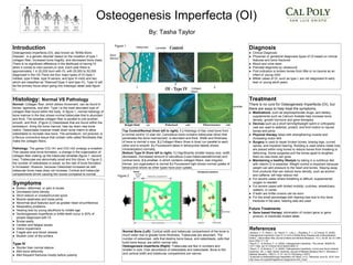
Histology: Normal VS Pathology
Normal: Collagen fiber, which allows movement, can be found in
bones, ligaments, and skin. Type I is the most abundant type of
collagen fiber found within the body. In figure 1, normal histology of
bone marrow in the iliac shows normal trabeculae that is abundant
and thick. The lamellae collagen fiber is parallel to one another,
smooth, and thick. (Figure 2) Osteoblasts that are found within the
endosteum, lining the bone marrow, help lay down new bone
matrix. Osteoclasts however break down bone matrix to allow
osteoblasts to recreate new bone. The periosteum, not pictured, is
a fibrous connective tissue that contain cells called fibroblasts that
make the collagen fibers.
Pathology: The genes COL1A1 and COL1A2 undergo a mutation.
This causes slow bone formation, a change in the organization of
collagen fiber making up the trabecular lamellae (figure 1: second
row). Trabeculae are abnormally small and thin (blue). In Figure 2,
the number of osteoblasts is raised, so the rate of bone formation
is increased. However, because Osteoclast increased as well,
trabecular bone mass does not increase. Cortical and trabecular
compartments shrink causing thin bones compared to normal.
Symptoms
● Broken, deformed, or pain in bones
● Decreased bone density
● Short stature or crooked/curved spine
● Muscle weakness and loose joints
● Abnormal skull features such as greater head circumference
● Respiratory problems
● Hearing loss by young adulthood to middle age
● Dentinogenesis imperfecta or brittle teeth occur in 50% of
people diagnosed with OI
● Bruise easily
● Cardiac and fatigue issues
● Vision impairment
● Fragile skin and blood vessels
● Blueish color of the schlera
Type IV
● Shorter than normal stature
● Mild bone deformity
● Mild frequent fractures mostly before puberty
Treatment
There is no cure for Osteogenesis Imperfecta (OI), but
there are ways to help treat the symptoms.
● Medications, such as bisphosphonate drugs, and dietary
supplements such as Calcium Acetate help increase bone
density: growth hormone and gene therapies
● Devices such as a short term/lightweight splint or orthopedic
cast are used to stabilize, protect, and limit motion to injured
bones and joints
● Physical therapy helps with strengthening muscle and
increasing motor skill
● Surgery is used to repair broken bones, bowing legs, curved
spines, and impaired hearing. Rodding is used where metal rods
are placed within long bones to reduce bones from breaking or
deforming. Some surgeries pull the bones apart to lengthen
them so new bone can grow
● Maintaining a healthy lifestyle by taking in a nutritious diet
with vitamin D is essential. Weight control is important because
weight can add pressure to the heart and bones. Staying away
from products that can reduce bone density, such as alcohol
and caffeine, will help reduce risk.
● For severe cases where breathing is difficult, supplemental
oxygen is needed
● For severe cases with limited mobility: crutches, wheelchairs,
walkers, or canes
● If teeth are brittle crowns can be worn
● For the small percentage with hearing loss due to tiny bone
fractures in the ears, hearing aids are used
Future Treatments
● Gene based therapy: elimination of mutant gene or gene
product, or inactivate mutant allele
References
Glorieux, F. H., Ward, L. M., Rauch, F., Lalic, L., Roughley, P. J., & Travers, R. (2002).
Osteogenesis Imperfecta Type VI: A Form of Brittle Bone Disease with a Mineralization
Defect. J Bone Miner Res Journal of Bone and Mineral Research, 17(1), 30-38. doi:10.1359
/jbmr.2002.17.1.30
Rauch, F., & Glorieux, F. H. (2004). Osteogenesis imperfecta. The Lancet, 363(9418),
1377-1385. doi:10.1016/s0140-6736(04)16051-0
Rauch, F., & Glorieux, F. H. (2005). Osteogenesis imperfecta, current and future medical
treatment. Am. J. Med. Genet. American Journal of Medical Genetics Part C: Seminars in
Medical Genetics, 139C(1), 31-37. doi:10.1002/ajmg.c.30072
Subscribe to theBreakthrough Newsletter orE-News. (n.d.). Retrieved June 02, 2016, from
http://www.oif.org/site/PageServer?pagename=AOI_Facts
Normal Bone (Left): Cortical width and trabecular compartment of the bone is
much wider due to greater bone thickness. Trabeculae are abundant. The
number of osteoclast, cells that destroy bone tissue, and osteoblasts, cells that
build bone tissue, are within normal ratio.
Osteogenesis Imperfecta (Right): Trabeculae are few in numbers and
smaller in size. Over abundance of osteoblasts and osteoclasts. Bone is thin
and cortical width and trabecular compartment are narrow.
Top Control/Normal (from left to right): 1.) Histology of iliac crest bone from
a normal control 12 year old. Cancellous bone contains trabeculae (blue) that
penetrates the bone marrow(red): is abundant and thick. Width and thickness
of bone is normal in size. 2.) Collagen fibers (lines in blue) run parallel to each
other and is smooth. 3.) Fluorescent takes in tetracycline labels( shows
mineralization) normally.
Bottom Type IV (from left to right): 1.) Significantly smaller biopsy size: width
decreased. Decreased amount of cancellous (Less trabeculae/abnormal) and
cortical bone. 2.)Lamellae, in which contains collagen fibers, was irregular,
thinner, but organization is normal 3.) Fluorescent light shows normal uptake of
tetracycline where as other types have poor uptake.
Figure 1
1
2
3
Figure 2
trabeculae Lamellae
Bone
Marrow
Lamellae
Collagen
fibers