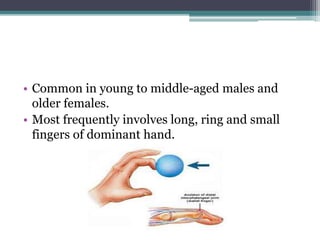
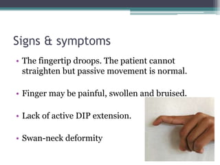
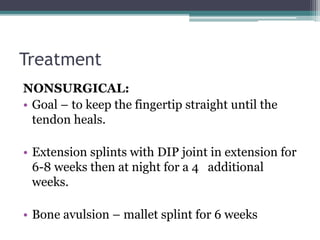
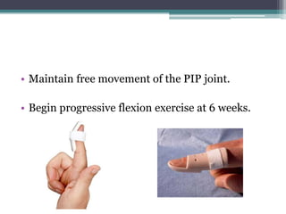
Mallet finger
Dr. Santosh Batajoo
Introduction
• Deformity of the finger caused by damaged
extensor tendon distal to DIP.
• Also called baseball finger or hammer finger or
drop finger.
• Disruption may be bony or tendinous.
• Injury at zone 1.
• Common in young to middle-aged males and
older females.
• Most frequently involves long, ring and small
fingers of dominant hand.
Mechanism
• Ball or the object strikes the tip of the finger or
thumb and forcibly bends it.
• A minor force such as tucking in a bed sheet.
• Force tears the tendon or even pull away a piece
of bone.
• Less common mechanism is a sharp or crushing-
type laceration to the dorsal DIP joint.
• Less commonly, a forceful hyperextension injury
of the DIP joint.
Classification (Doyle’s)
Type I Closed injury with or without small dorsal avulsion fracture
Type II Open injury (laceration)
Type III Open injury ( deep soft tissue abrasion involving loss skin and
tendon substance)
Type IV Mallet fracture
A- distal phalanx physeal injury (pediatrics)
B- fracture fragment involving 20% to 50% of articular surface
(adult)
C- fracture fragment >50% of articular surface (adult)
Signs & symptoms
• The fingertip droops. The patient cannot
straighten but passive movement is normal.
• Finger may be painful, swollen and bruised.
• Lack of active DIP extension.
• Swan-neck deformity
Diagnosis
• X-ray : Avulsion of distal phalanx
May be a ligamentous injury with normal
bony anatomy.
Treatment
NONSURGICAL:
• Goal – to keep the fingertip straight until the
tendon heals.
• Extension splints with DIP joint in extension for
6-8 weeks then at night for a 4 additional
weeks.
• Bone avulsion – mallet splint for 6 weeks
• Maintain free movement of the PIP joint.
• Begin progressive flexion exercise at 6 weeks.
Surgical :
Indications-
• volar subluxation of distal phalanx,
• large bone fragments with >50% articular suface
involved,
• non-surgical treatment was not successful.
• >2mm articular gap
• CRPP or ORIF – pin fixation or dorsal/extension
blocking pin.
• ORIF with a pull-out wire.
• Surgical reconstruction of terminal tendon in
chronic injury > 12 weeks with healthy joint –
direct repair, tenodermodesis or retinacular
ligament reconstruction
• Central slip release (Fowler)
• Type 1 and type 2 can be treated closed.
• Type 3 require soft tissue coverage and pinning
of the DIP joint.
• DIP arthrodesis – painful, stiff, arthritic DIP
joint
• Swan neck deformity – lateral band tenodesis,
FDS tenodisis, Fowler central slip tenotomy
Complications
• Extensor lag
• Non union
• Swan neck deformities due to – attenuation of
volar plate and transverse retinacular ligament
at PIP joint, dorsal subluxation of lateral bands,
hyperextension of PIP, contracture of
traiangular ligament.
Thank you…

Mallet finger

  • 1.
  • 2.
    Introduction • Deformity ofthe finger caused by damaged extensor tendon distal to DIP. • Also called baseball finger or hammer finger or drop finger. • Disruption may be bony or tendinous. • Injury at zone 1.
  • 3.
    • Common inyoung to middle-aged males and older females. • Most frequently involves long, ring and small fingers of dominant hand.
  • 4.
    Mechanism • Ball orthe object strikes the tip of the finger or thumb and forcibly bends it. • A minor force such as tucking in a bed sheet. • Force tears the tendon or even pull away a piece of bone.
  • 5.
    • Less commonmechanism is a sharp or crushing- type laceration to the dorsal DIP joint. • Less commonly, a forceful hyperextension injury of the DIP joint.
  • 6.
    Classification (Doyle’s) Type IClosed injury with or without small dorsal avulsion fracture Type II Open injury (laceration) Type III Open injury ( deep soft tissue abrasion involving loss skin and tendon substance) Type IV Mallet fracture A- distal phalanx physeal injury (pediatrics) B- fracture fragment involving 20% to 50% of articular surface (adult) C- fracture fragment >50% of articular surface (adult)
  • 7.
    Signs & symptoms •The fingertip droops. The patient cannot straighten but passive movement is normal. • Finger may be painful, swollen and bruised. • Lack of active DIP extension. • Swan-neck deformity
  • 8.
    Diagnosis • X-ray :Avulsion of distal phalanx May be a ligamentous injury with normal bony anatomy.
  • 9.
    Treatment NONSURGICAL: • Goal –to keep the fingertip straight until the tendon heals. • Extension splints with DIP joint in extension for 6-8 weeks then at night for a 4 additional weeks. • Bone avulsion – mallet splint for 6 weeks
  • 10.
    • Maintain freemovement of the PIP joint. • Begin progressive flexion exercise at 6 weeks.
  • 11.
    Surgical : Indications- • volarsubluxation of distal phalanx, • large bone fragments with >50% articular suface involved, • non-surgical treatment was not successful. • >2mm articular gap
  • 12.
    • CRPP orORIF – pin fixation or dorsal/extension blocking pin. • ORIF with a pull-out wire. • Surgical reconstruction of terminal tendon in chronic injury > 12 weeks with healthy joint – direct repair, tenodermodesis or retinacular ligament reconstruction • Central slip release (Fowler)
  • 13.
    • Type 1and type 2 can be treated closed. • Type 3 require soft tissue coverage and pinning of the DIP joint. • DIP arthrodesis – painful, stiff, arthritic DIP joint • Swan neck deformity – lateral band tenodesis, FDS tenodisis, Fowler central slip tenotomy
  • 14.
    Complications • Extensor lag •Non union • Swan neck deformities due to – attenuation of volar plate and transverse retinacular ligament at PIP joint, dorsal subluxation of lateral bands, hyperextension of PIP, contracture of traiangular ligament.
  • 15.