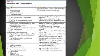
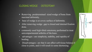
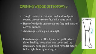
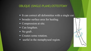
Osteotomy
Dr. Dibyendunarayan Bid, PhD
Osteotomy
 The major objective of this procedure on the bones is to
improve the functional status of the limb.
 It is achieved either by surgical correction of the bony
deformity or by providing stability to the joint.
 It is done by dividing or cutting the bone wedge and
realigning or reconstructing the joint.
 Basically, it converts the shearing force into a compressive
force by changing the line of weight bearing.
 It is performed in three distinct stages.
Stage I: A stage of division of the bone
 The bone is cut or divided close to the deformity by:
 (a) single transverse cut, or
 (b) by removing a piece of bone (wedge),
 (c) by reconstructing the joint following division, the two
ends of the bone are brought together to correct the
anatomical alignment, or reconstructed to provide
stability to the joint, or by altering shearing forces at the
fracture site into compressive forces by changing the line
of weight bearing (Fig. 24-24).
Stage II: A stage of immobilization
 After achieving the objective of correct anatomical
alignment or stability to the joint, immobilization is done
in this position either by POP cast, internal fixation or by
external fixation as feasible.
Stage III: A stage of achieving the optimal
functional restoration
 Once the proper consolidation of osteotomy is ensured,
vigorous physiotherapy is required to restore the optimal
function of the concerned joint or limb.
 Osteotomy is indicated in several bony and joint
dysfunctions or bony deformities for various purposes
(Tables 24-6–24-8).
Types of osteotomies:
 A: Opening wedge.
 B: Closing wedge.
 C: Displacement.
 D: Stepcut.
 E: Crescentic (dome).
 F: Transverse derotation.
 G: Oblique (single plane).
TRANSVERSE OSTEOTOMY
 Ideal for correcting rotation alone.
 Diaphysis or metaphysis plane of cut is
transverse to long axis of bone to avoid frontal or
sagittal deformity.
 Simple to perform but it is relatively unstable and
is not ideally suited for interfragmentary
compression.
 Resists axial load but weak to torsion or bending
loads.
 Angular corrections difficult to control .
CLOSING WEDGE OSTEOTOMY
 Removing predetermined sized wedge of bone from
maximal deformity.
 base of wedge is at covex surface of deformity.
 After removing wedge gap is closed and internal fixation
done.
 commonly used high tibial osteotomy performed to treat
unicompartmental arthritis of the knee.
 advantages :-are simplicity, stability, and rapidityof
healing.
 Disadvantages:- are that it can affect soft-tissue balance if
close to joints, and it will result in some shortening.
OPENING WEDGE OSTEOTOMY :-
 Single transverse cut was used and wedge is
opened on concave surface with bone graft.
 Base of wedge is on concave surface and apex on
convex surface.
 Advantage - some gain in length.
 Disadvantages :- filled by a bone graft, which
slows healing, nonunions can occur, and the
intercalary bone graft used must remodel before
full weight bearing can begin.
OBLIQUE (SINGLE-PLANE)OSTEOTOMY
 It can correct all deformities with a single cut.
 broader surface area for healing.
 Compression at site.
 Can lengthen.
 No graft .
 Creates some rotation.
 useful in the metaphyseal region.
CRESCENTIC (DOME) OSTEOTOMY
 Used in metaphyseal or epiphyseal cancellous
bone, where irregular nature of bone and cut
provide good inherent stability, and broad surface
area and cancellous bone lead to rapid healing .
 Dome shaped – one side shallow and another side
deep cut was done done and deformity was
corrected .
 Bone saving procedure.
 It is ideal for correcting deformity near joints that
are in a single plane, preferably the frontal plane.
DISPLACEMENT OSTEOTOMY
 Described by wagner is useful to address
a major juxtaarticular deformity .
 Transverse metaphyseal osteotomy in which
periarticular fragment is rotated, impacting one
corner of the metaphysis into medullary canal of
other fragment.
 This transforms bending loads into compressive
loads while preserving length and improving joint
alignment.
STEPCUT OSTEOTOMY:-
 In rare cases, such as one-stage diaphyseal
lengthening.
 Rotational and angular corrections are limited .
Physiotherapeutic management following
osteotomy
 Objective: To improve both ROM and muscle functions.
Pre-operative education
 The related musculoskeletal structure of the joint to be operated
should be given special sessions of exercise and movement training.
It may include compensatory trick movements, postural
adaptations, orthotic aids, etc. to perform functional tasks (e.g.,
substitution of shoulder abduction by shoulder girdle elevation, trunk
lateral flexion to the opposite side – a planned shoulder joint
arthrodesis).
 If not, restore full ROM with improvement of strength, endurance
and flexibility of the related joints of the joint to be arthrodesed.
 During immobilization
 Measures to reduce pain and swelling, placing a limb in comfortable
relaxed position.
 Keeping a watch on the expected postsurgical complications as related to
the procedure of surgery and the site.
 Ensure the stability of the operated joint.
 Initiate repetitive isometrics in repetitive small bouts to the muscle groups
under immobilization.
 Strong active movements to the joints free of immobilization and the
functional muscle groups.
 Initiate preliminary functional activities with guidance and assistance (e.g.,
shifting in the bed, sitting up, supported standing progressing to non-
weight bearing (NWB) ambulation or using operated upper extremity as an
assisting arm).
 During mobilization
 Improving all the functions of the muscle groups, especially
around the operated joint and concentrated mobility exercise
training to improve the ROM should be initiated immediately.
Objective training on tricky movements.
 Isometrics and self-resistive type of exercise training to
improve strength and endurance and relaxed free repetitive
movements are the mainstay as soon as the immobilization is
discontinued.
 Adjunctive procedures of thermotherapy like hydropack around
the operated area (excluding skin incision) are ideal to induce
relaxation and comfort before mobilization.
 Initially well-supported objective functional re-education
should be begun as early as possible in a graded manner and
guidance.
 Manual therapy procedures and PNF techniques play an
important role in achieving early optimal functional
restoration.
 Note: Longer period of immobilization is always associated with limitation
of joint movement and disuse atrophy of the muscle groups under
immobilization needs extra efforts and early attention.
COMPLICATIONS OF OSTEOTOMY AND
DEFORMITY CORRECTION:-
 General : thrombo-embolism and infections.
 Undercorrection and overcorrection.
 Nerve tension: acute long-bone corrections > 20
degrees should be avoided and if there is a known risk
of nerve injury it should be limited to 10 degrees.
example is peroneal nerve palsy
 Compartment syndrome: Osteotomy of the tibia
or forearm bones.
 Non-union: may occur if fixation is inadequate or
if soft tissues are damaged by excessive stripping
during surgical exposure.
Osteotomy and physiotherapy

Osteotomy and physiotherapy

  • 1.
  • 2.
    Osteotomy  The majorobjective of this procedure on the bones is to improve the functional status of the limb.  It is achieved either by surgical correction of the bony deformity or by providing stability to the joint.  It is done by dividing or cutting the bone wedge and realigning or reconstructing the joint.  Basically, it converts the shearing force into a compressive force by changing the line of weight bearing.  It is performed in three distinct stages.
  • 3.
    Stage I: Astage of division of the bone  The bone is cut or divided close to the deformity by:  (a) single transverse cut, or  (b) by removing a piece of bone (wedge),  (c) by reconstructing the joint following division, the two ends of the bone are brought together to correct the anatomical alignment, or reconstructed to provide stability to the joint, or by altering shearing forces at the fracture site into compressive forces by changing the line of weight bearing (Fig. 24-24).
  • 5.
    Stage II: Astage of immobilization  After achieving the objective of correct anatomical alignment or stability to the joint, immobilization is done in this position either by POP cast, internal fixation or by external fixation as feasible.
  • 6.
    Stage III: Astage of achieving the optimal functional restoration  Once the proper consolidation of osteotomy is ensured, vigorous physiotherapy is required to restore the optimal function of the concerned joint or limb.  Osteotomy is indicated in several bony and joint dysfunctions or bony deformities for various purposes (Tables 24-6–24-8).
  • 10.
    Types of osteotomies: A: Opening wedge.  B: Closing wedge.  C: Displacement.  D: Stepcut.  E: Crescentic (dome).  F: Transverse derotation.  G: Oblique (single plane).
  • 11.
    TRANSVERSE OSTEOTOMY  Idealfor correcting rotation alone.  Diaphysis or metaphysis plane of cut is transverse to long axis of bone to avoid frontal or sagittal deformity.  Simple to perform but it is relatively unstable and is not ideally suited for interfragmentary compression.  Resists axial load but weak to torsion or bending loads.  Angular corrections difficult to control .
  • 12.
    CLOSING WEDGE OSTEOTOMY Removing predetermined sized wedge of bone from maximal deformity.  base of wedge is at covex surface of deformity.  After removing wedge gap is closed and internal fixation done.  commonly used high tibial osteotomy performed to treat unicompartmental arthritis of the knee.  advantages :-are simplicity, stability, and rapidityof healing.  Disadvantages:- are that it can affect soft-tissue balance if close to joints, and it will result in some shortening.
  • 13.
    OPENING WEDGE OSTEOTOMY:-  Single transverse cut was used and wedge is opened on concave surface with bone graft.  Base of wedge is on concave surface and apex on convex surface.  Advantage - some gain in length.  Disadvantages :- filled by a bone graft, which slows healing, nonunions can occur, and the intercalary bone graft used must remodel before full weight bearing can begin.
  • 14.
    OBLIQUE (SINGLE-PLANE)OSTEOTOMY  Itcan correct all deformities with a single cut.  broader surface area for healing.  Compression at site.  Can lengthen.  No graft .  Creates some rotation.  useful in the metaphyseal region.
  • 16.
    CRESCENTIC (DOME) OSTEOTOMY Used in metaphyseal or epiphyseal cancellous bone, where irregular nature of bone and cut provide good inherent stability, and broad surface area and cancellous bone lead to rapid healing .  Dome shaped – one side shallow and another side deep cut was done done and deformity was corrected .  Bone saving procedure.  It is ideal for correcting deformity near joints that are in a single plane, preferably the frontal plane.
  • 18.
    DISPLACEMENT OSTEOTOMY  Describedby wagner is useful to address a major juxtaarticular deformity .  Transverse metaphyseal osteotomy in which periarticular fragment is rotated, impacting one corner of the metaphysis into medullary canal of other fragment.  This transforms bending loads into compressive loads while preserving length and improving joint alignment.
  • 19.
    STEPCUT OSTEOTOMY:-  Inrare cases, such as one-stage diaphyseal lengthening.  Rotational and angular corrections are limited .
  • 20.
    Physiotherapeutic management following osteotomy Objective: To improve both ROM and muscle functions. Pre-operative education  The related musculoskeletal structure of the joint to be operated should be given special sessions of exercise and movement training. It may include compensatory trick movements, postural adaptations, orthotic aids, etc. to perform functional tasks (e.g., substitution of shoulder abduction by shoulder girdle elevation, trunk lateral flexion to the opposite side – a planned shoulder joint arthrodesis).  If not, restore full ROM with improvement of strength, endurance and flexibility of the related joints of the joint to be arthrodesed.
  • 21.
     During immobilization Measures to reduce pain and swelling, placing a limb in comfortable relaxed position.  Keeping a watch on the expected postsurgical complications as related to the procedure of surgery and the site.  Ensure the stability of the operated joint.  Initiate repetitive isometrics in repetitive small bouts to the muscle groups under immobilization.  Strong active movements to the joints free of immobilization and the functional muscle groups.  Initiate preliminary functional activities with guidance and assistance (e.g., shifting in the bed, sitting up, supported standing progressing to non- weight bearing (NWB) ambulation or using operated upper extremity as an assisting arm).
  • 22.
     During mobilization Improving all the functions of the muscle groups, especially around the operated joint and concentrated mobility exercise training to improve the ROM should be initiated immediately. Objective training on tricky movements.  Isometrics and self-resistive type of exercise training to improve strength and endurance and relaxed free repetitive movements are the mainstay as soon as the immobilization is discontinued.
  • 23.
     Adjunctive proceduresof thermotherapy like hydropack around the operated area (excluding skin incision) are ideal to induce relaxation and comfort before mobilization.  Initially well-supported objective functional re-education should be begun as early as possible in a graded manner and guidance.  Manual therapy procedures and PNF techniques play an important role in achieving early optimal functional restoration.  Note: Longer period of immobilization is always associated with limitation of joint movement and disuse atrophy of the muscle groups under immobilization needs extra efforts and early attention.
  • 24.
    COMPLICATIONS OF OSTEOTOMYAND DEFORMITY CORRECTION:-  General : thrombo-embolism and infections.  Undercorrection and overcorrection.  Nerve tension: acute long-bone corrections > 20 degrees should be avoided and if there is a known risk of nerve injury it should be limited to 10 degrees. example is peroneal nerve palsy
  • 25.
     Compartment syndrome:Osteotomy of the tibia or forearm bones.  Non-union: may occur if fixation is inadequate or if soft tissues are damaged by excessive stripping during surgical exposure.