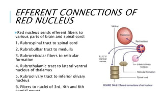
The midbrain connects the brainstem to the forebrain and cerebellum. It consists of the tectum and cerebral peduncles. The tectum contains the superior and inferior colliculi, which are involved in visual and auditory reflexes. The cerebral peduncles contain the substantia nigra and red nucleus. The red nucleus receives input from the motor cortex and dentate nucleus, and sends outputs to control muscle tone, complex movements, righting reflexes, and eye movements.
BRAINSTEM
The Brainstem lies at the base of the brain and the top of the spinal cord.
The brainstem is located in the posterior cranial fossa.
The brainstem is the structure that connects the cerebrum of the brain to the spinal cord and cerebellum.
Provides a pathway for tracts running between higher and lower neural centers.
Divided into 3 major divisions:
midbrain,
pons, and
medulla oblongata.
It is responsible for many vital functions of life, such as breathing, consciousness, blood pressure, heart rate, and sleep.
It contains many critical collections of white and grey matter.
The grey matter within the brainstem consists of nerve cell bodies and form many important brainstem nuclei. Ten of the twelve cranial nerves arise from their cranial nerve nuclei in the brainstem.
The white matter tracts of the brainstem include axons of nerves traversing their course to different structures. These tracts travel both to the brain (afferent) and from the brain (efferent) such as the somatosensory pathways and the corticospinal tracts, respectively.
Mid Brain
The midbrain is continuous with the cerebral hemisphere.
The upper posterior (i.e. rear) portion of the midbrain is called the tectum, which means "roof."
The surface of the tectum is covered with four bumps representing two paired structures: the superior and inferior colliculi.
The superior colliculi are involved in eye movements and visual processing, while the inferior colliculi are involved in auditory processing.
Another important nucleus, the substantia nigra, is located here.
The substantia nigra is rich in dopamine neurons and is considered part of the basal ganglia.
Pons
An important pathway for tracts that run from the cerebrum down to the medulla and spinal cord, as well as for tracts that travel up into the brain.
It also forms important connections with the cerebellum via fibre bundles known as the cerebellar peduncles.
Posteriorly, the pons and medulla are separated from the cerebellum by the fourth ventricle.
Home to several nuclei for cranial nerves.
Medulla
The point where the brainstem connects to the spinal cord.
Contains a nucleus called the nucleus of the solitary tract that is crucial for our survival (receives information about blood flow, along with information about levels of oxygen and carbon dioxide in the blood, from the heart and major blood vessels).
When this information suggests a discordance with bodily needs (e.g. blood pressure is too low), there are reflexive actions initiated in the nucleus of the solitary tract to bring things back to within the desired range.
Blood Supply
The brain stem receives its blood supply exclusively from the posterior circulation, including the vertebrae and basilar artery.
The medulla receives its blood supply from the vertebral via medial and lateral perforating arteries.
The pons and midbrain receive their blood from the basilar via the medial and lateral perforating arteries.
gross Anatomy of Mid Brain.location an relation of midbrain. external an internal features of mid brain. cross section at the level of superior and inferior colliculus. Anterior and posterior view of midbrain.
clinical correlation of midbrain.
Thalamus-Anatomy,Physiology,Applied aspectsRanadhi Das
Thalamus is a very important relay station.
All general and special sensory impulses (except smell) & afferent impulses from RAS are integrated here.
Thalamus however is the center of pain and protopathic sensations.
It has other non sensory functions as well, like motor control, sleep, wakefulness.
It is the largest structure deriving from the embryonic diencephalon, the posterior part of the forebrain situated between the midbrain and the cerebrum.
The thalamus is part of a nuclear complex structured of 4 parts, the hypothalamus, epithalamus, prethalamus (formerly called ventral thalamus) and dorsal thalamus.
Largest part of hind brain.
Called “ silent area/Little Brain ”
Weight- 150 gms.
Cerebellar cortex is a large folded sheet, each fold is called Folium.
Connected to brain stem by 3 pairs of peduncles- Superior (Brachium conjunctiva), Middle (Brachium Pontis) & Inferior (Restiform body) peduncle.
understanding spinal cord, its bransches, lesions, functions and anatomy.
hope to give you better knowledge of spinal cord by the end of it.
plese review ans comment for my future updates and corrections that iw ill be needing in this.
The reference material used to make video is: Principles of Anatomy and Physiology Gerard J. Tortora, Bryan H. Derrickson.
Pons is part of brain stem, present superior to medulla, inferior to mid brain and anterior to cerebellum.
Pons means a bridge. As the name denotes, it connects other areas of brain.
Neurons extending from cerebral cortex to pons makes corticopontine tract.
Pons is connected to cerebellum by middle cerebral peduncle.
Pons has vestibular nuclei, which is part of equilibrium pathways from inner ear to brain.
Pons has also respiratory nuclei. Along with rhythmicity area of medulla, pons controls basal respiratory rhythm.
Pons also contains nuclei for cranial nerve number V, VI,VII, and VIII.
For the video, Kindly visit my you tube channel.
https://www.youtube.com/channel/UC7xXKrK7pDObSfXvk9aOgDA.
Here is anatomy and physiology of brain stem. Where we will discuss all three parts of brain stem. Starting from medulla, second is pons and third is mid brain. In this video I am presenting anatomy and physiology of medulla. Anatomy of medulla: Medulla Oblongata or more simply medulla is part of brain stem which forms base of the brain stem. Location of medulla oblongata is superior to spinal cord and inferior to Pons. It contains pyramid, olive and above pyramidal structure, there is decussation of pyramids which explains why each part of brain controls opposite part of body. Adding to that medulla also has several nuclei which controls activity of cardiovascular system and respiratory system. Medulla also has nuclei for controlling reflexes of vomiting, swallowing, hiccuping, coughing and sneezing. It has also nuclei for test, hearing and balance. Medulla also contains nuclei of cranial nerve number VIII, IX, X, XI and XII. Functions of medulla or what dose medulla do? So medulla controls blood pressure, diameter of wall of arteries, heart rate, basal respiration rate and also vomiting, swallowing, hiccuping, coughing and sneezing.
In this video, we explain you about anatomy and physiology of Pons. The reference material used to make video is: Principles of Anatomy and Physiology Gerard J. Tortora, Bryan H. Derrickson. Pons is part of brain stem, present superior to medulla, inferior to mid brain and anterior to cerebellum. Pons means a bridge. As the name denotes, it connects other areas of brain. Neurons extending from cerebral cortex to pons makes corticopontine tract. Pons is connected to cerebellum by middle cerebral peduncle. Pons has vestibular nuclei, which is part of equilibrium pathways from inner ear to brain. Pons has also respiratory nuclei. Along with rhythmicity area of medulla, pons controls basal respiratory rhythm. Pons also contains nuclei for cranial nerve number V, VI,VII, and VIII.
You can watch the video on my you tube channel: https://youtu.be/I0FaX-iQfa0
Medulla oblongata or more simply medulla is part of brain stem which forms base of the brain stem. It contains pyramid, olive and above pyramidal structure, there is decussation of pyramids which explains why each part of brain controls opposite part of body. Adding to that medulla also has several nuclei which controls activity of cardiovascular system and respiratory system. Medulla also has nuclei for controlling reflexes of vomiting, swallowing, hiccuping, coughing and sneezing. It has also nuclei for test, hearing and balance. Medulla also contains nuclei of cranial nerve number VIII, IX, X, XI and XII.
Nutrition in New born and Kids
Calorie requirement of newborn and growing kids
Protein energy malnutrition
Vitamin deficiency disorders in kids
Ricketts
Scurvy
Kwashiorkor
Marasmus
The limbic system, also known as the paleomammalian cortex, is a set of brain structures located on both sides of the thalamus, immediately beneath the medial temporal lobe of the cerebrum primarily in the forebrain.[1]
It supports a variety of functions including emotion, behavior, motivation, long-term memory, and olfaction.[2] Emotional life is largely housed in the limbic system, and it critically aids the formation of memories.
With a primordial structure, the limbic system is involved in lower order emotional processing of input from sensory systems
This PPT describes neurological gait deviations.
It describes Hemiplegic/circumductory gait, Spastic Diplegic gait, Parkinson gait, Myopathic & Ataxic gait in detail along with its causes and management in with Physiotherapy treatment. detail
This PPT is made to explain basic techniques of therapeutic massage in detail.
It includes : Stroking, Pressure Manipulation, Tapotement & Vibration
Physiological effects, Therapeutic uses, Caution & Contraindications.
Description : Osteogenesis Imperfecta/
Brittle bone disease :
It is disorder of type I collagen synthesis that affects all connective tissue in the body.
Musculoskeletal involvement is diffuse and includes osteoporosis with excessive fracture even at birth, bowing of long bone, spinal deformities, muscle weakness and ligamentous laxity.
Key words :
Osteogenesis Imperfecta, Brittle bone disease, Genetic disorder, Pathophysiology, Types of OI, Denetinogenesis Imperfecta, Bluish sclera, Frequent fractures, fractures, Hearing loss, Management, orthopedic, Rehabilitation
Physiotherapy, pediatrics, physiotherapist, pediatric orthopedic surgery.
Arthrogryposis multiplex congenita is a disorder that affects the early development of body joints in a fetus, most commonly the large joints in the arms and legs. An infant who is born with the condition typically has limited mobility and obvious physical deformities in one or more joints.
1. Scar Tissue
2.What makes a scar
3. Characteristics of Scar
4. Scar & Adhesion
5. Classification as External and Internal scars
6 Detriments/Disadvantages
7. How scar tissue forms in muscle
8. Phases of scar healing
9.Phases of wound healing
10. Release of scar and adhesion
11. Massage
12. Massage techniques
13. Conditions : Burns & Skin grafting
14. Cautions
15. Procedure
16. When to start massage after surgery
17. How to massage scar tissue
18. Effleurage, Kneading, Skin rolling and Friction
19. Caution
20. Keloid
21. Warning.
What is oedema?
types of oedema
1. soft and mobile
2. consolidated/endurated
Conditions in which oedema is treated with massage
a) Radical mastectomy
b) Venous ulcer
c) gravitational/paralytic oedema
Technique of massage to reduce oedema
Kneading
Effleurage
Picking Up &
Friction
Instructions to patient
Position of Patient
Aim
Procedure &
Steps of Massage Application
Diabetes Mellitus
Introduction
Pathophysiology
Types of Diabetes Mellitus
Type 1, 2 and
gestational diabetes
rescent research in Type 1 diabetes
Risk factors and causes
Complications short term and long term of diabetes
Management
Treatment with Insulin
Diabetic drugs
Healthy Diet
Exercises prescription
aerobic exercises,
resistance exercises and
flexibility
Report Back from SGO 2024: What’s the Latest in Cervical Cancer?bkling
Are you curious about what’s new in cervical cancer research or unsure what the findings mean? Join Dr. Emily Ko, a gynecologic oncologist at Penn Medicine, to learn about the latest updates from the Society of Gynecologic Oncology (SGO) 2024 Annual Meeting on Women’s Cancer. Dr. Ko will discuss what the research presented at the conference means for you and answer your questions about the new developments.
Ozempic: Preoperative Management of Patients on GLP-1 Receptor Agonists Saeid Safari
Preoperative Management of Patients on GLP-1 Receptor Agonists like Ozempic and Semiglutide
ASA GUIDELINE
NYSORA Guideline
2 Case Reports of Gastric Ultrasound
Lung Cancer: Artificial Intelligence, Synergetics, Complex System Analysis, S...Oleg Kshivets
RESULTS: Overall life span (LS) was 2252.1±1742.5 days and cumulative 5-year survival (5YS) reached 73.2%, 10 years – 64.8%, 20 years – 42.5%. 513 LCP lived more than 5 years (LS=3124.6±1525.6 days), 148 LCP – more than 10 years (LS=5054.4±1504.1 days).199 LCP died because of LC (LS=562.7±374.5 days). 5YS of LCP after bi/lobectomies was significantly superior in comparison with LCP after pneumonectomies (78.1% vs.63.7%, P=0.00001 by log-rank test). AT significantly improved 5YS (66.3% vs. 34.8%) (P=0.00000 by log-rank test) only for LCP with N1-2. Cox modeling displayed that 5YS of LCP significantly depended on: phase transition (PT) early-invasive LC in terms of synergetics, PT N0—N12, cell ratio factors (ratio between cancer cells- CC and blood cells subpopulations), G1-3, histology, glucose, AT, blood cell circuit, prothrombin index, heparin tolerance, recalcification time (P=0.000-0.038). Neural networks, genetic algorithm selection and bootstrap simulation revealed relationships between 5YS and PT early-invasive LC (rank=1), PT N0—N12 (rank=2), thrombocytes/CC (3), erythrocytes/CC (4), eosinophils/CC (5), healthy cells/CC (6), lymphocytes/CC (7), segmented neutrophils/CC (8), stick neutrophils/CC (9), monocytes/CC (10); leucocytes/CC (11). Correct prediction of 5YS was 100% by neural networks computing (area under ROC curve=1.0; error=0.0).
CONCLUSIONS: 5YS of LCP after radical procedures significantly depended on: 1) PT early-invasive cancer; 2) PT N0--N12; 3) cell ratio factors; 4) blood cell circuit; 5) biochemical factors; 6) hemostasis system; 7) AT; 8) LC characteristics; 9) LC cell dynamics; 10) surgery type: lobectomy/pneumonectomy; 11) anthropometric data. Optimal diagnosis and treatment strategies for LC are: 1) screening and early detection of LC; 2) availability of experienced thoracic surgeons because of complexity of radical procedures; 3) aggressive en block surgery and adequate lymph node dissection for completeness; 4) precise prediction; 5) adjuvant chemoimmunoradiotherapy for LCP with unfavorable prognosis.
Title: Sense of Taste
Presenter: Dr. Faiza, Assistant Professor of Physiology
Qualifications:
MBBS (Best Graduate, AIMC Lahore)
FCPS Physiology
ICMT, CHPE, DHPE (STMU)
MPH (GC University, Faisalabad)
MBA (Virtual University of Pakistan)
Learning Objectives:
Describe the structure and function of taste buds.
Describe the relationship between the taste threshold and taste index of common substances.
Explain the chemical basis and signal transduction of taste perception for each type of primary taste sensation.
Recognize different abnormalities of taste perception and their causes.
Key Topics:
Significance of Taste Sensation:
Differentiation between pleasant and harmful food
Influence on behavior
Selection of food based on metabolic needs
Receptors of Taste:
Taste buds on the tongue
Influence of sense of smell, texture of food, and pain stimulation (e.g., by pepper)
Primary and Secondary Taste Sensations:
Primary taste sensations: Sweet, Sour, Salty, Bitter, Umami
Chemical basis and signal transduction mechanisms for each taste
Taste Threshold and Index:
Taste threshold values for Sweet (sucrose), Salty (NaCl), Sour (HCl), and Bitter (Quinine)
Taste index relationship: Inversely proportional to taste threshold
Taste Blindness:
Inability to taste certain substances, particularly thiourea compounds
Example: Phenylthiocarbamide
Structure and Function of Taste Buds:
Composition: Epithelial cells, Sustentacular/Supporting cells, Taste cells, Basal cells
Features: Taste pores, Taste hairs/microvilli, and Taste nerve fibers
Location of Taste Buds:
Found in papillae of the tongue (Fungiform, Circumvallate, Foliate)
Also present on the palate, tonsillar pillars, epiglottis, and proximal esophagus
Mechanism of Taste Stimulation:
Interaction of taste substances with receptors on microvilli
Signal transduction pathways for Umami, Sweet, Bitter, Sour, and Salty tastes
Taste Sensitivity and Adaptation:
Decrease in sensitivity with age
Rapid adaptation of taste sensation
Role of Saliva in Taste:
Dissolution of tastants to reach receptors
Washing away the stimulus
Taste Preferences and Aversions:
Mechanisms behind taste preference and aversion
Influence of receptors and neural pathways
Impact of Sensory Nerve Damage:
Degeneration of taste buds if the sensory nerve fiber is cut
Abnormalities of Taste Detection:
Conditions: Ageusia, Hypogeusia, Dysgeusia (parageusia)
Causes: Nerve damage, neurological disorders, infections, poor oral hygiene, adverse drug effects, deficiencies, aging, tobacco use, altered neurotransmitter levels
Neurotransmitters and Taste Threshold:
Effects of serotonin (5-HT) and norepinephrine (NE) on taste sensitivity
Supertasters:
25% of the population with heightened sensitivity to taste, especially bitterness
Increased number of fungiform papillae
Ethanol (CH3CH2OH), or beverage alcohol, is a two-carbon alcohol
that is rapidly distributed in the body and brain. Ethanol alters many
neurochemical systems and has rewarding and addictive properties. It
is the oldest recreational drug and likely contributes to more morbidity,
mortality, and public health costs than all illicit drugs combined. The
5th edition of the Diagnostic and Statistical Manual of Mental Disorders
(DSM-5) integrates alcohol abuse and alcohol dependence into a single
disorder called alcohol use disorder (AUD), with mild, moderate,
and severe subclassifications (American Psychiatric Association, 2013).
In the DSM-5, all types of substance abuse and dependence have been
combined into a single substance use disorder (SUD) on a continuum
from mild to severe. A diagnosis of AUD requires that at least two of
the 11 DSM-5 behaviors be present within a 12-month period (mild
AUD: 2–3 criteria; moderate AUD: 4–5 criteria; severe AUD: 6–11 criteria).
The four main behavioral effects of AUD are impaired control over
drinking, negative social consequences, risky use, and altered physiological
effects (tolerance, withdrawal). This chapter presents an overview
of the prevalence and harmful consequences of AUD in the U.S.,
the systemic nature of the disease, neurocircuitry and stages of AUD,
comorbidities, fetal alcohol spectrum disorders, genetic risk factors, and
pharmacotherapies for AUD.
ARTIFICIAL INTELLIGENCE IN HEALTHCARE.pdfAnujkumaranit
Artificial intelligence (AI) refers to the simulation of human intelligence processes by machines, especially computer systems. It encompasses tasks such as learning, reasoning, problem-solving, perception, and language understanding. AI technologies are revolutionizing various fields, from healthcare to finance, by enabling machines to perform tasks that typically require human intelligence.
Anti ulcer drugs and their Advance pharmacology ||
Anti-ulcer drugs are medications used to prevent and treat ulcers in the stomach and upper part of the small intestine (duodenal ulcers). These ulcers are often caused by an imbalance between stomach acid and the mucosal lining, which protects the stomach lining.
||Scope: Overview of various classes of anti-ulcer drugs, their mechanisms of action, indications, side effects, and clinical considerations.
Explore natural remedies for syphilis treatment in Singapore. Discover alternative therapies, herbal remedies, and lifestyle changes that may complement conventional treatments. Learn about holistic approaches to managing syphilis symptoms and supporting overall health.
MANAGEMENT OF ATRIOVENTRICULAR CONDUCTION BLOCK.pdfJim Jacob Roy
Cardiac conduction defects can occur due to various causes.
Atrioventricular conduction blocks ( AV blocks ) are classified into 3 types.
This document describes the acute management of AV block.
Factory Supply Best Quality Pmk Oil CAS 28578–16–7 PMK Powder in Stockrebeccabio
Factory Supply Best Quality Pmk Oil CAS 28578–16–7 PMK Powder in Stock
Telegram: bmksupplier
signal: +85264872720
threema: TUD4A6YC
You can contact me on Telegram or Threema
Communicate promptly and reply
Free of customs clearance, Double Clearance 100% pass delivery to USA, Canada, Spain, Germany, Netherland, Poland, Italy, Sweden, UK, Czech Republic, Australia, Mexico, Russia, Ukraine, Kazakhstan.Door to door service
Hot Selling Organic intermediates
3. SO, WHAT’S THE BRAIN STEM ?
Brain stem is the area which is located in the lower part of
the brain, gives connection and serves as bridge between the
lower part and the upper part of the CNS.
Brain stem is composed of three structures from cranial to
caudal
Midbrain
Pons
Medulla Oblongata
4. CONT…
Brain stem connects to the cerebellum via 6 peduncles, one
pair arising from each brain stem component. So ,
Superior peduncle arises from Mid brain
Middle peduncle arises from pons
Inferior peduncle arises from Medulla Oblongata
The ventral layer of brain stem is motor in function , while
the middle layer is sensory in function
5.
6. FUNCTIONS OF THE BRAIN STEM
1.Conduct functions.
All information related from the body to the cerebrum and cerebellum
and vice versa, must traverse the brain stem. Including:
a. The ascending sensory pathways
b. Descending tracts
2.Provides the origin of the cranial nerves (CN III-XII).
3.Conjugate eye movement.
4. Integrative functions
7. INTEGRATIVE FUNCTIONS
It controls consciousness & sleep cycle (alertness and arousal)
through reticular formation.
Centers for cardiovascular, respiratory & autonomic nervous system.
It has centers for cough, gag, swallow, and vomit.
Sense of body balance (Vestibular functions) CN VIII
Pain sensitivity control: Periaqueductal grey matter of mesencephalon
is an area which is rich in endogenous opioid and is important in
modulation of painful stimuli.
8.
9.
10.
11. Medulla oblongata or medulla is the lowermost part of
brain.
It is situated below pons and is continued downwards as
spinal cord.
Medulla forms the main pathway for ascending and
descending tracts of the spinal cord.
It also has many important centers which
control the vital functions.
MEDULLA OBLONGATA
12. 1. Respiratory Centers
Dorsal and ventral group of neurons form the medullary
respiratory centers, which maintain normal rhythmic respiration.
2. Vasomotor Center
Vasomotor center controls blood pressure and heart rate.
3. Deglutition Center
Deglutition center regulates the pharyngeal and esophageal
stages of deglutition.
MEDULLA OBLONGATA
13. 4. Vomiting Center
Vomiting center induces vomiting during irritation or
inflammation of gastrointestinal (GI) tract.
5. Superior and Inferior Salivatory Nuclei
Salivatory nuclei control the secretion of saliva.
MEDULLA OBLONGATA
14. 6. Cranial Nerve Nuclei
Nuclei of 12th, 11th, 10th and some nuclei of 8th and 5th
cranial nerves are located in the medulla oblongata.
12th cranial (hypoglossal) nerve controls the movements
of tongue.
11th cranial (accessory) nerve controls the movements of
shoulder and
10th cranial (vagus) nerve controls almost all the vital
functions in the body, viz. cardiovascular system,
respiratory system, GI system, etc.
MEDULLA OBLONGATA
15. 7. Vestibular Nuclei
Vestibular nuclei contain the second order neurons of
vestibular nerve.
There are four vestibular nuclei, situated in the rostral part
of medulla and caudal part of pons:
superior, medial, lateral and inferior vestibular nuclei.
Medial and inferior vestibular nuclei extend into medulla.
MEDULLA OBLONGATA
19. All the medullary centers and nuclei of
cranial nerves are controlled by higher
centers, situated in cerebral cortex and
hypothalamus.
MEDULLA OBLONGATA
21. PONS
Pons forms a bridge between medulla and midbrain.
Functions of Pons
1. Axons of pontine nuclei join to form the middle cerebellar peduncle
or the brachium pontis. Pons forms the pathway that connects
cerebellum with cerebral cortex.
2. Pyramidal tracts pass through the pons.
3. Medial lemniscus is joined by the fibers of 10th, 9th, 7th & 5th
cranial nerves in pons.
4. Nuclei of 8th, 7th, 6th and 5th cranial nerves are located in pons.
5. Pons contains the pneumotaxic and apneustic centers for regulation
of respiration.
25. MIDBRAIN
Midbrain lies between pons
and diencephalon.
It consists of two parts:
A. Tectum
B. Cerebral peduncles.
TECTUM
Tectum is formed by two
structures:
1. Superior colliculus
2. Inferior colliculus.
26. MIDBRAIN : TECTUM
1. Superior Colliculus
Superior colliculus is a small structure and is an
important center for reflexes.
Through tectospinal tract, superior colliculus controls
the movements of the eyes, head, trunk and limbs, in
response to visual impulses.
Efferent fibers from superior colliculus going to the
nucleus of III cranial (oculomotor) nerve cause
constriction of pupil during light reflex.
Thus, it forms the center for light reflex. Superior
colliculus also receives afferents from optic tract,
which helps in the integration of optical and postural
reflexes.
28. MIDBRAIN : TECTUM
2. Inferior Colliculus
Inferior colliculus consists of single layer of neurons to which the
lateral lemniscus (auditory fibers) synapses.
Inferior colliculus is the center for auditory reflexes.
Stimulation of this also produces reflex vocalization.
30. MIDBRAIN : CEREBRAL PEDUNCLES
Cerebral peduncles
include:
1. Basis pedunculi
2. Substantia nigra
3. Tegmentum, which
includes red nucleus.
31. 1. Basis Pedunculus
Basis pedunculus consists of pyramidal tract
fibers
in the middle, temporopontine fibers laterally and
frontopontine fibers medially.
As a whole, the cerebral peduncles assist in
refining motor movements, learning of new
motor skills, and converting
proprioceptive information into balance and
posture maintenance
MIDBRAIN : CEREBRAL PEDUNCLES
32. 2. Substantia Nigra
Substantia nigra is situated
below the red nucleus.
Substantia nigra is considered
as one of the components of
basal ganglia
MIDBRAIN : CEREBRAL PEDUNCLES
33. 3. Tegmentum
Tegmentum lies dorsal to substantia
nigra and is actually the upward
continuation of the reticular
formation in pons.
Tegmentum comprises three
decussations and red nucleus.
MIDBRAIN : CEREBRAL PEDUNCLES
34. DECUSSATIONS IN
TEGMENTUM
i. Superior cerebellar peduncle, which is formed by fibers between
cerebellum and other parts of CNS.
These fibers are predominantly efferent fibers from dentate
nucleus of cerebellum; few
fibers are from other cerebellar nuclei such as nucleus globosus
and nucleus emboliformis.
ii. Forel decussation, which is due to the crossing of rubrospinal
tracts from either side
iii. Meynert decussation, which is due to the crossing of medial
longitudinal bundle that is formed by efferent fibers of 3rd , 4th
and 6th cranial nerves.
35.
36. RED NUCLEUS
Red nucleus is a large oval
or round mass of gray
matter, extending between
the superior colliculus and
hypothalamus.
37. PARTS OF RED NUCLEUS
Red nucleus has two parts:
1. Nucleus magnocellularis, which is formed by large cells. Fibers
from this form the rubrospinal and rubrobulbar tracts.
2. Nucleus parvocellularis, which is formed by smaller cells. Fibers
from this form mainly the rubroreticular tract.
38. AFFERENT CONNECTIONS OF
RED NUCLEUS
Red nucleus receives fibers from:
1. Nucleus parvocellularis, which receives
fibers from
motor cortex (area 6) – corticorubral fibers
2. Nucleus magnocellularis, which receives
fibers
from motor cortex (area 6) – pallidorubral
fibers
3. Nucleus magnocellularis, which receives
fibers from
dentate nucleus (of opposite side) –
39. EFFERENT CONNECTIONS OF
RED NUCLEUS
Red nucleus sends efferent fibers to
various parts of brain and spinal cord:
1. Rubrospinal tract to spinal cord
2. Rubrobulbar tract to medulla
3. Rubroreticular fibers to reticular
formation
4. Rubrothalamic tract to lateral ventral
nucleus of thalamus
5. Rubroolivary tract to inferior olivary
nucleus
6. Fibers to nuclei of 3rd, 4th and 6th
40. FUNCTIONS OF RED NUCLEUS
1. Control of muscle tone: Because of its connections with cerebellum,
vestibular apparatus and skeletal muscle,
the red nucleus plays an important role in facilitating the muscle tone.
2. Control of complex muscular movements: Red nucleus controls the
complex muscular movements.
It plays an important role in the integration of various impulses received
from many important areas of brain.
3. Control of righting reflexes: Red nucleus is the center for all righting
reflexes except optical righting reflexes
41.
42.
43. FUNCTIONS OF RED NUCLEUS
4. Control of movements of eyeball: Through its efferent connections
with nuclei of 3rd, 4th and 6th cranial nerves, red nucleus plays an
important role in the control of ocular movements .
5. Control of skilled movements: Red nucleus plays an important role in
controlling the skilled muscular movements by its connections with
spinal cord and cerebral cortex.