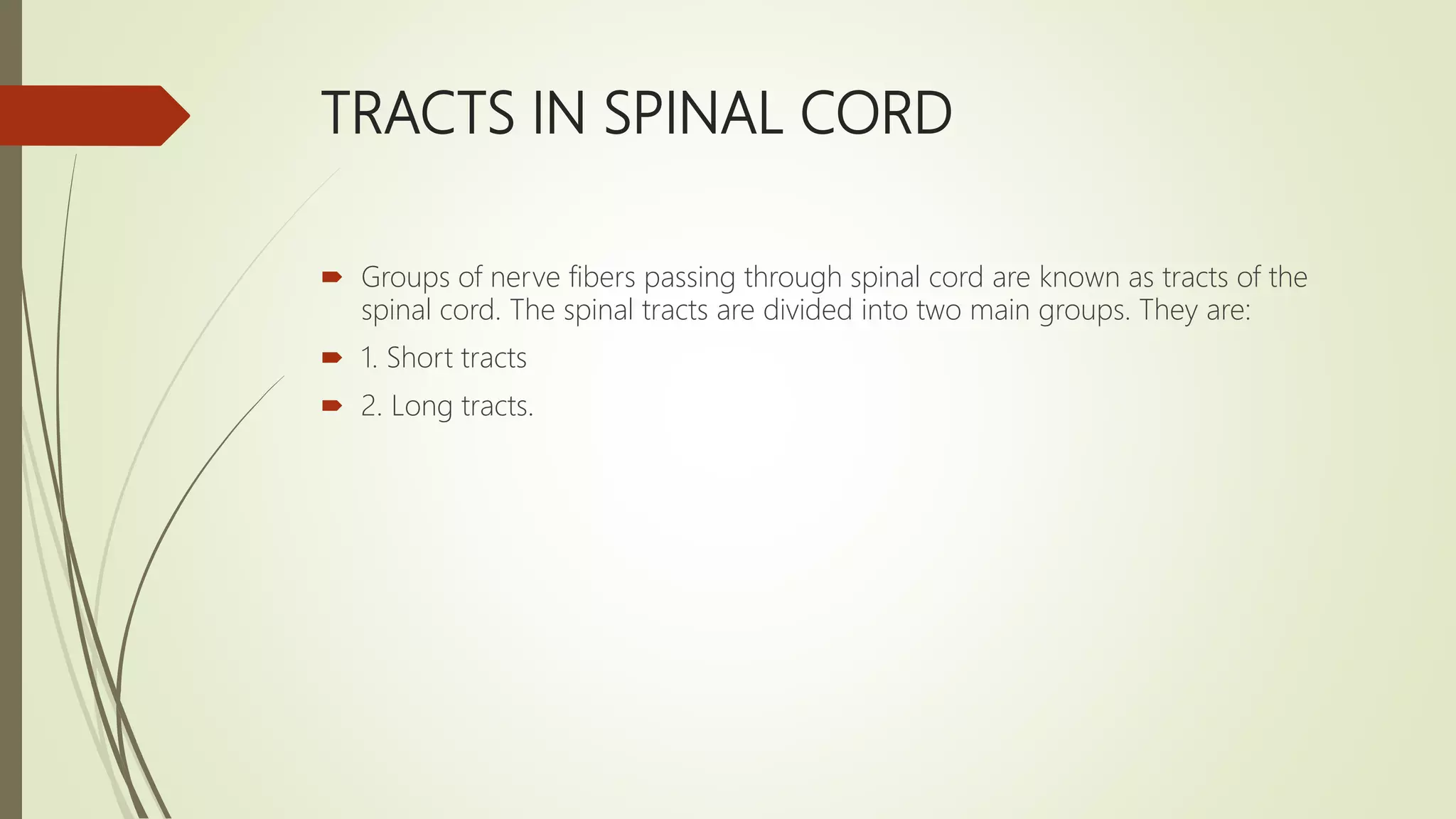
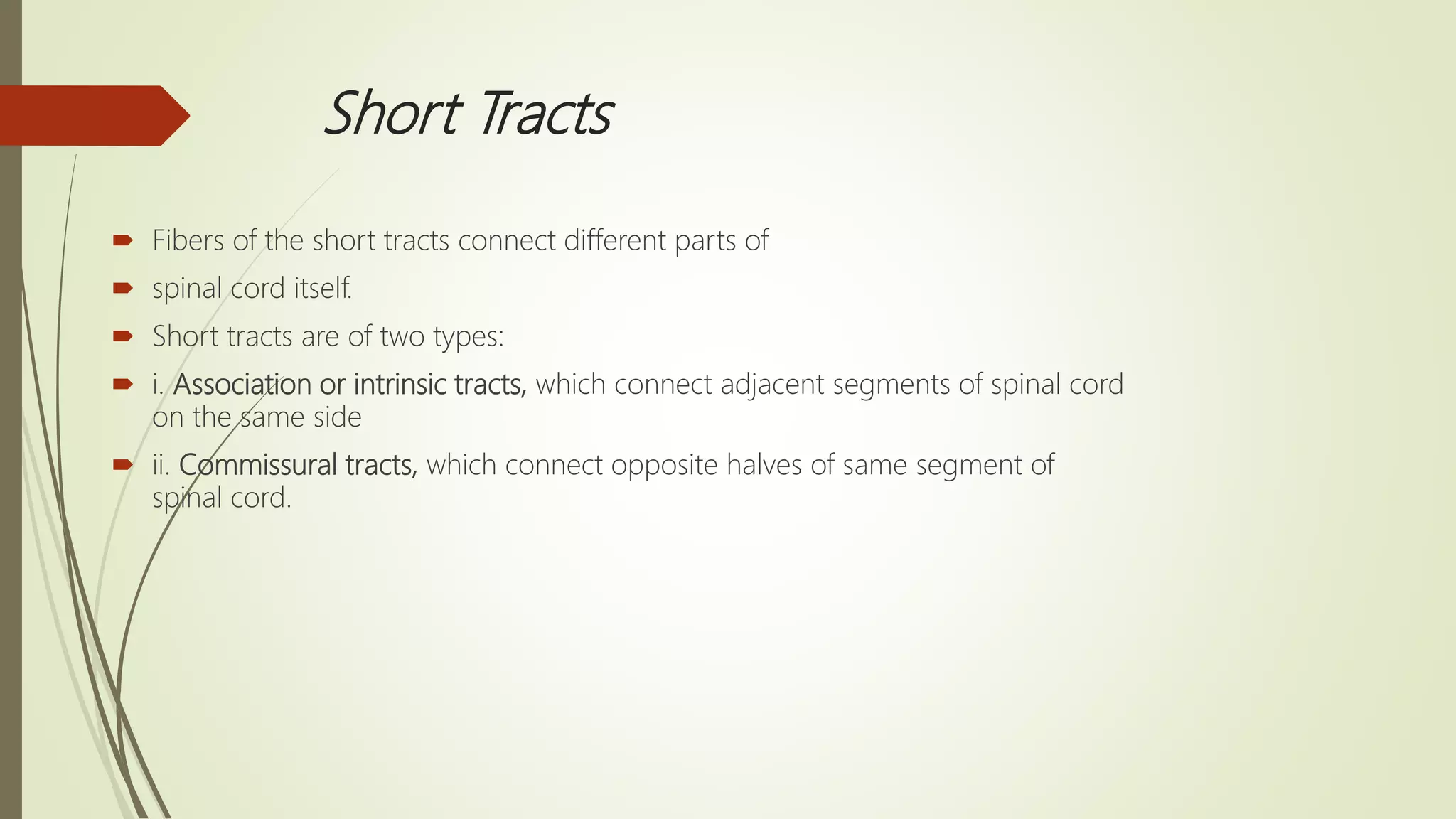
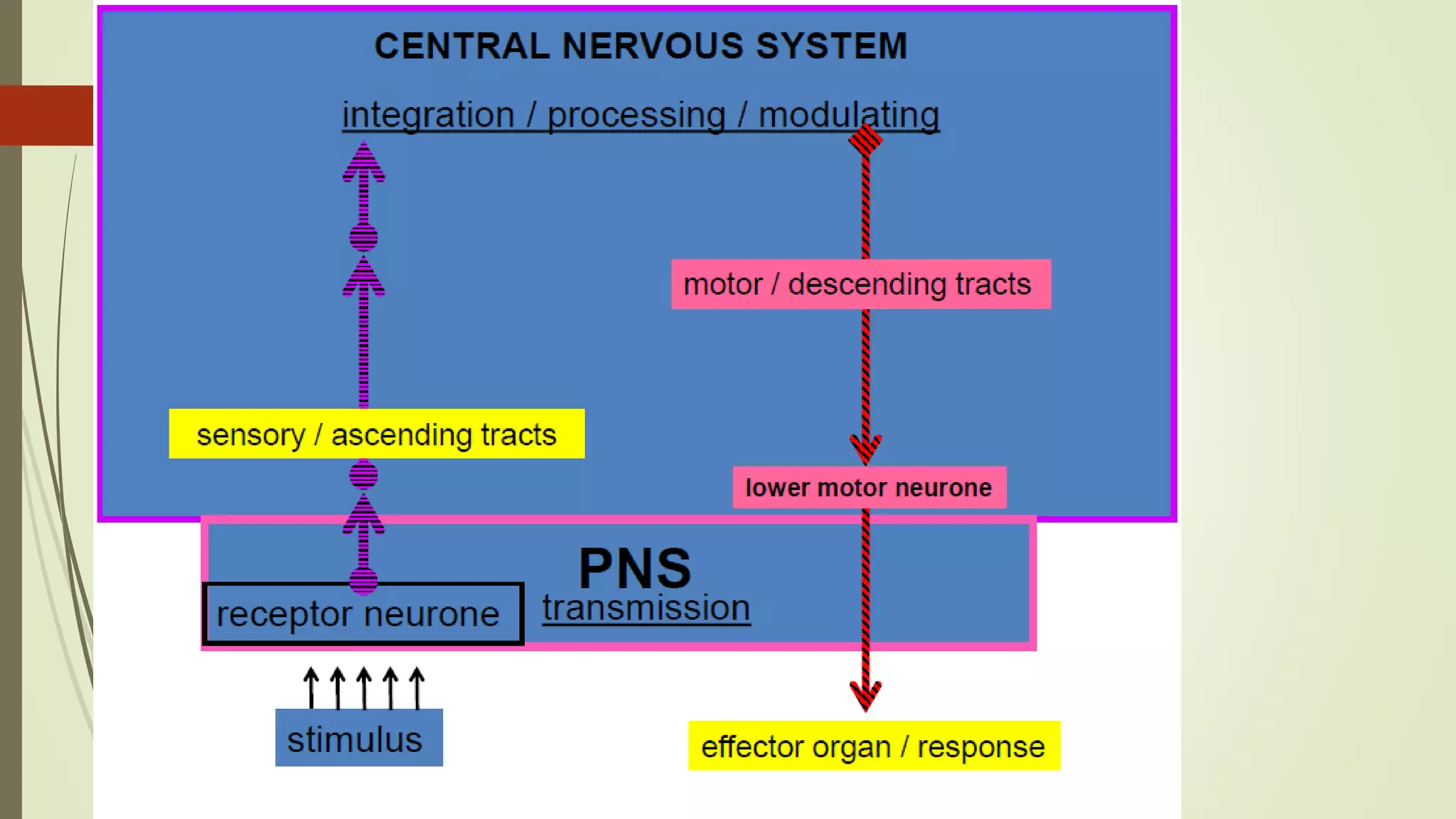
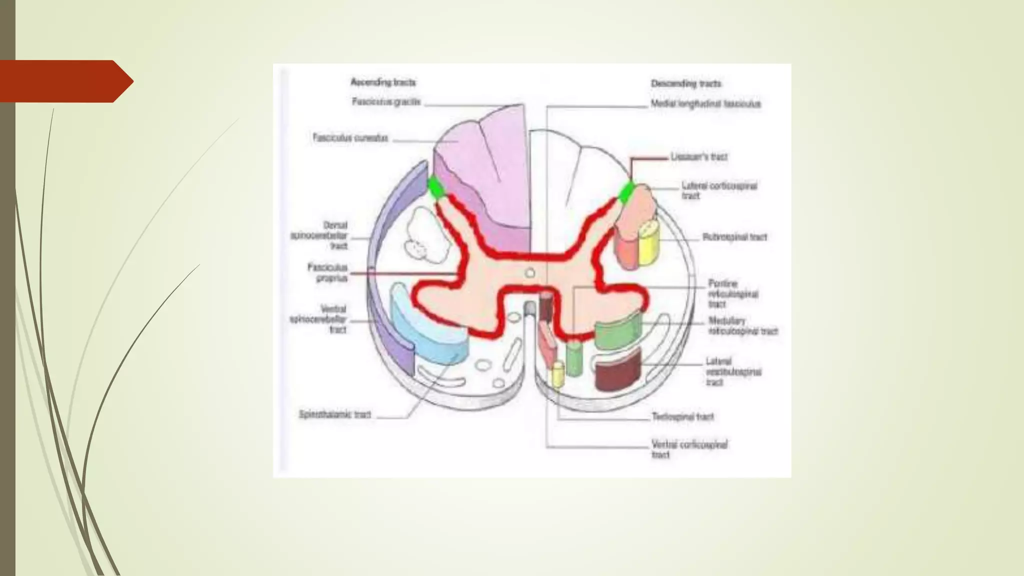
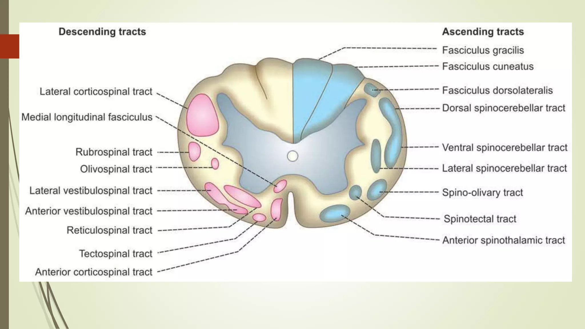
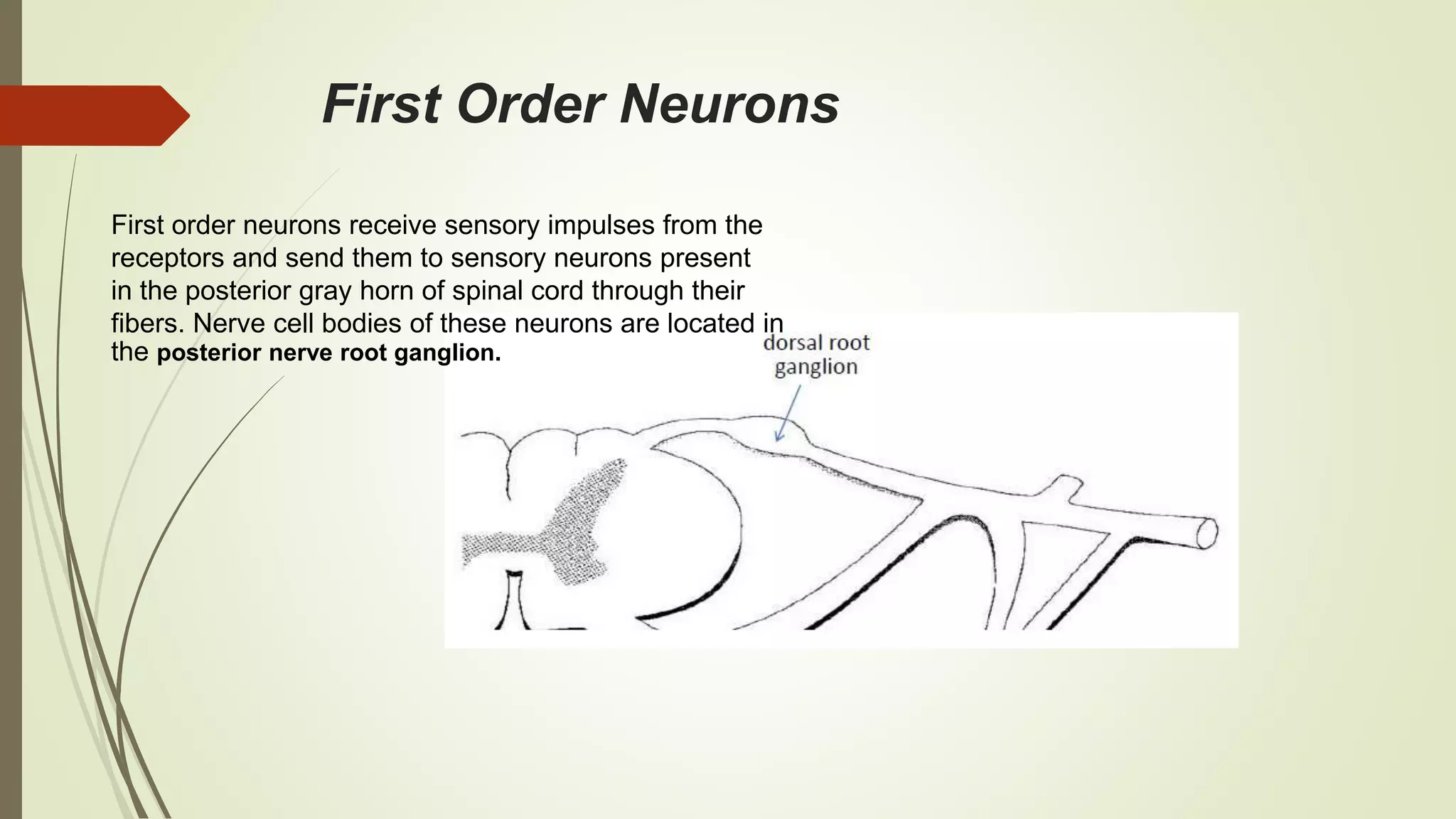
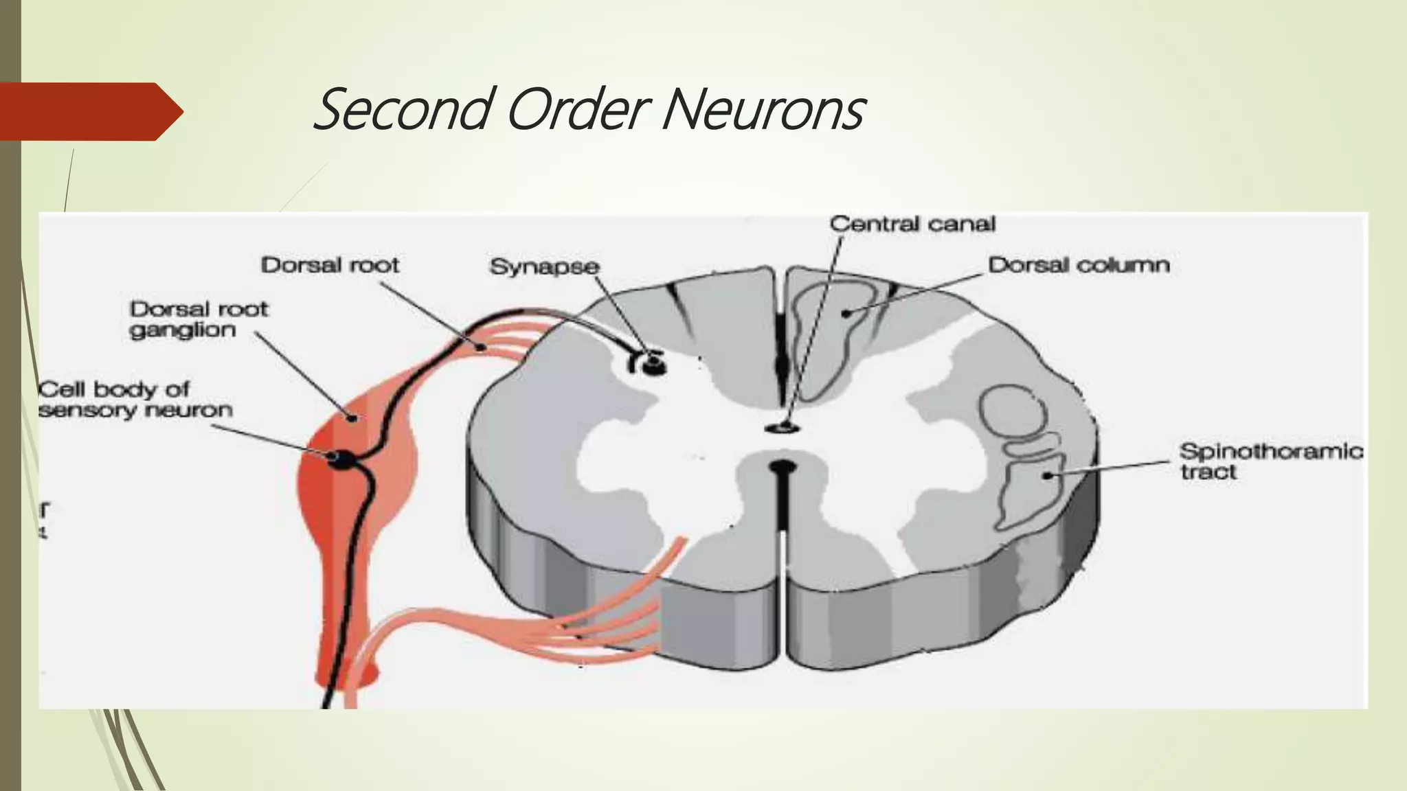
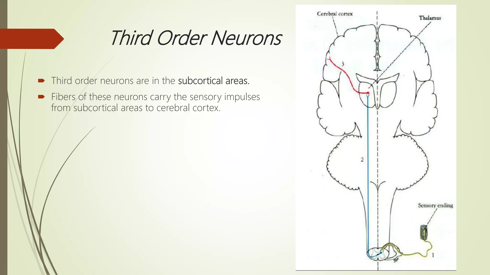
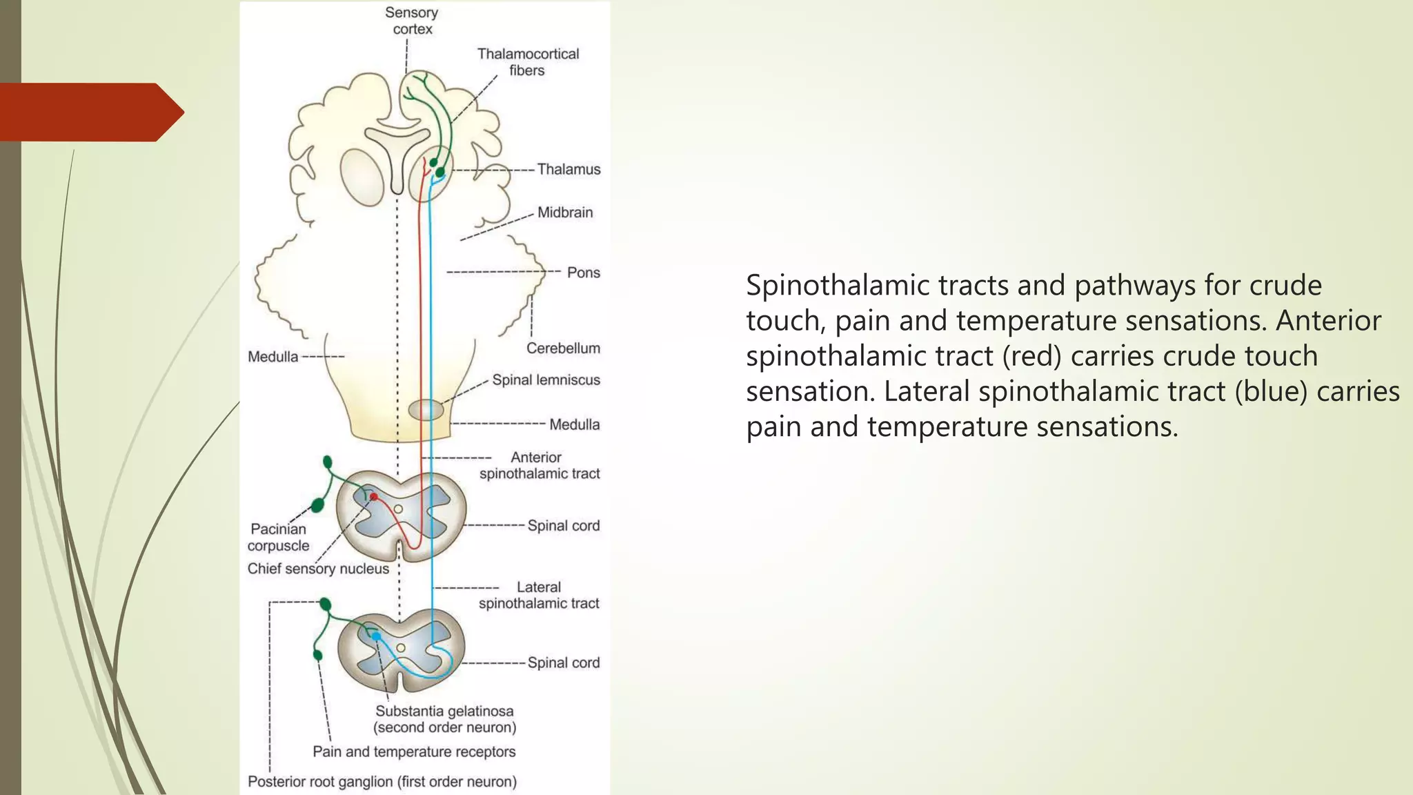
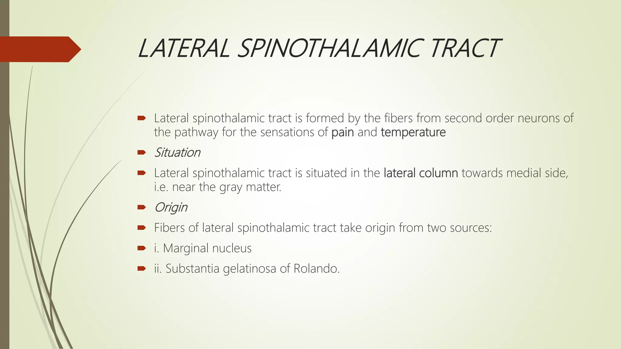
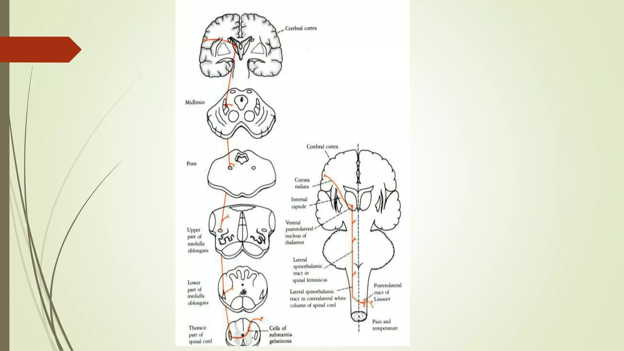
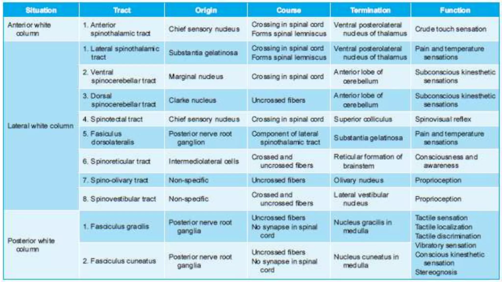
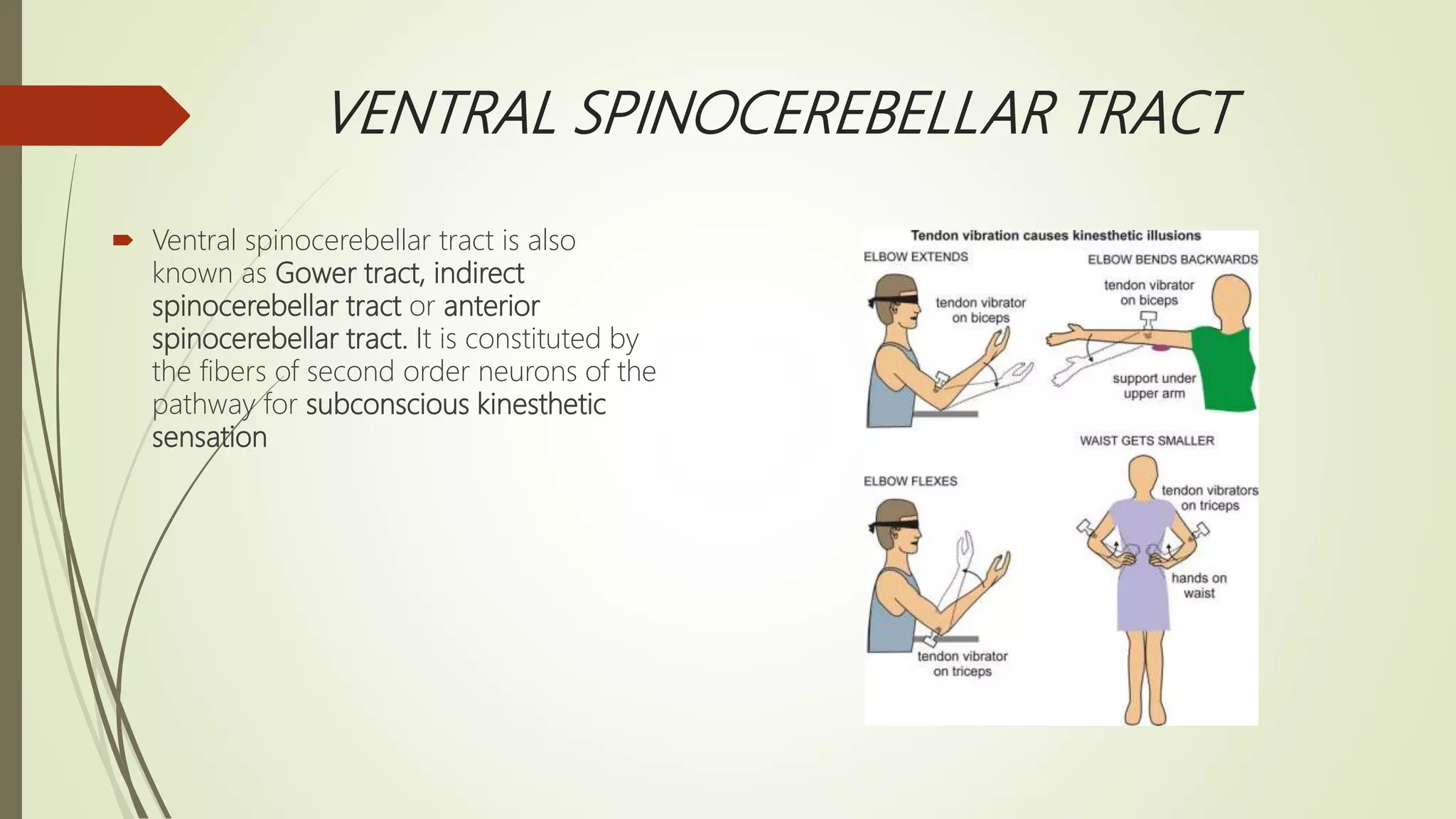
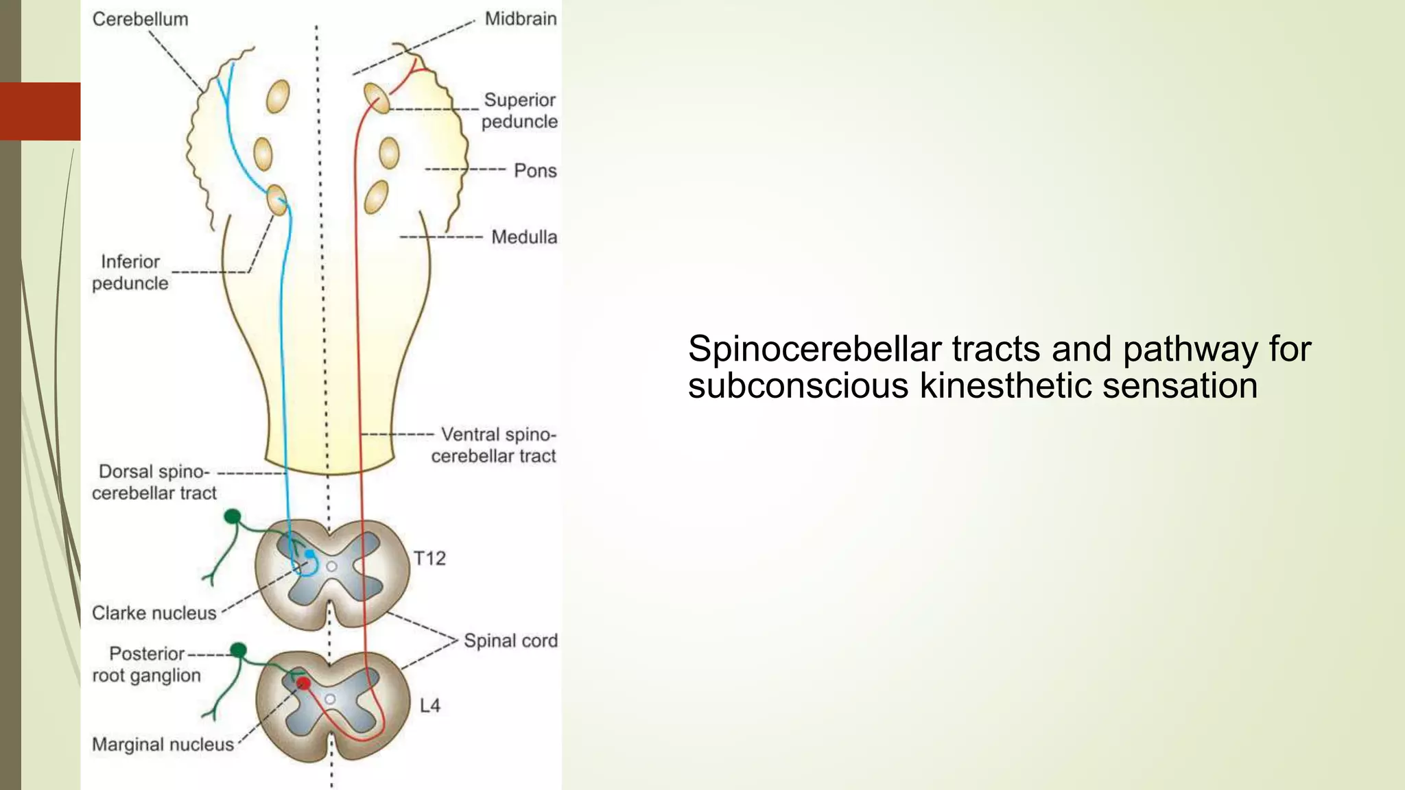
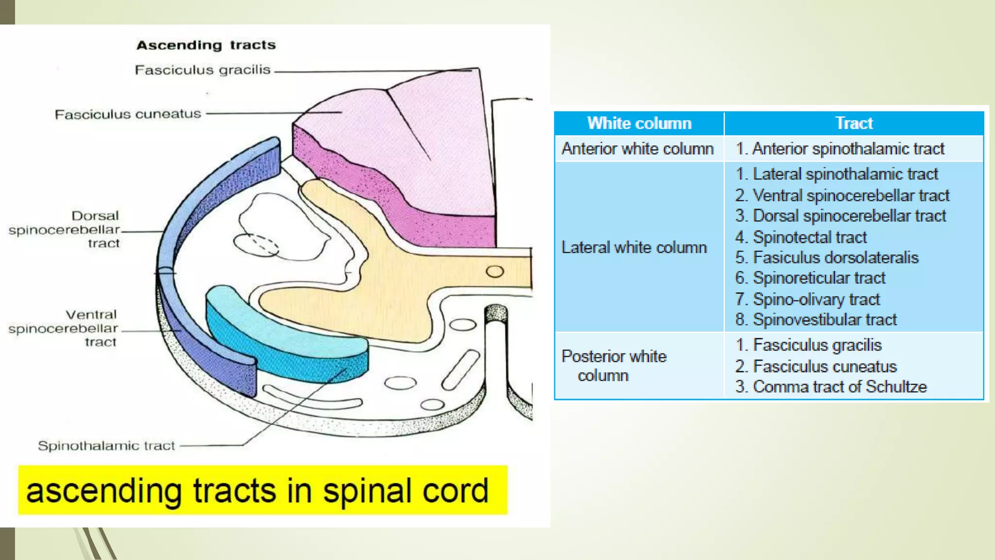
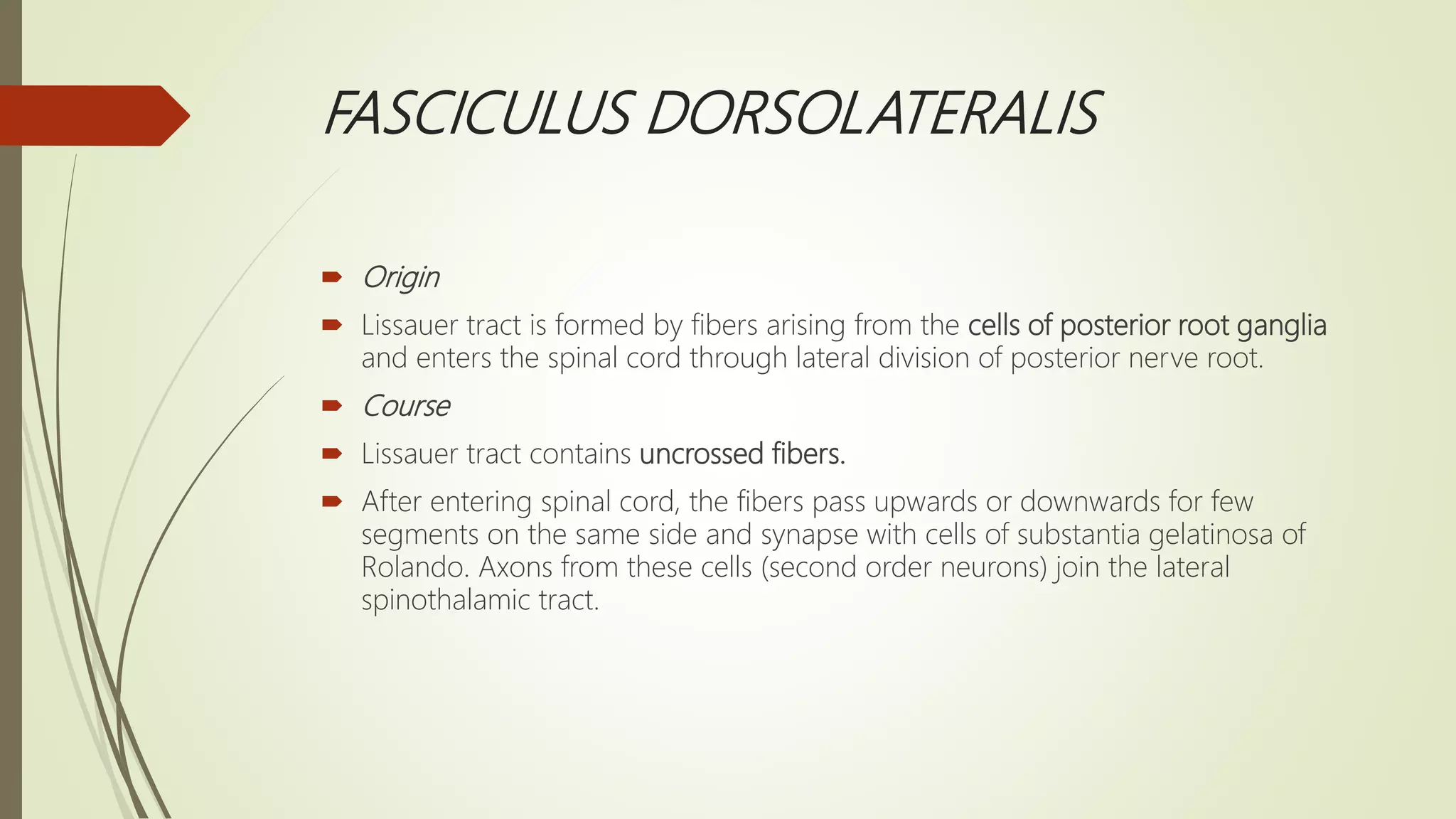
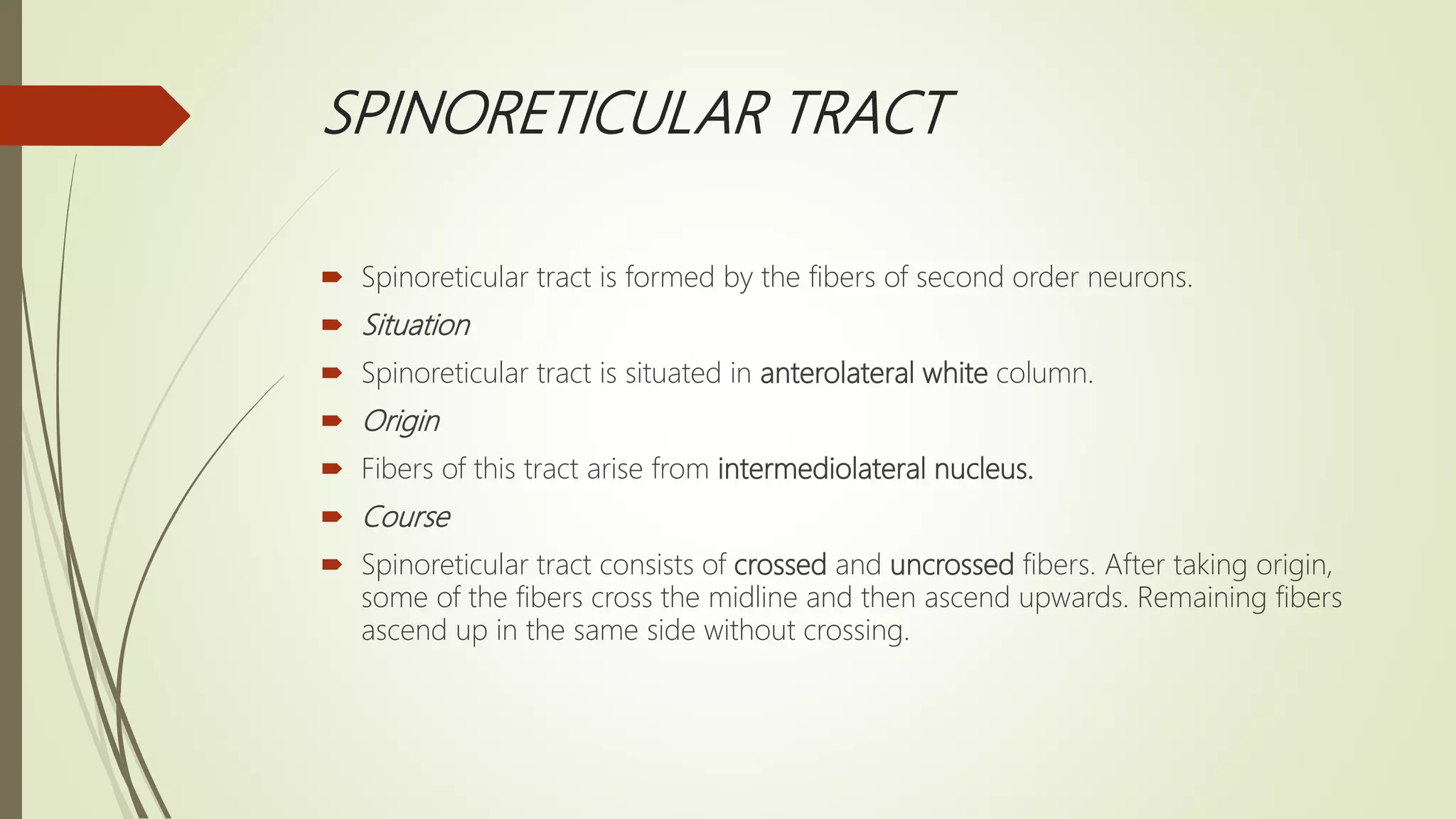
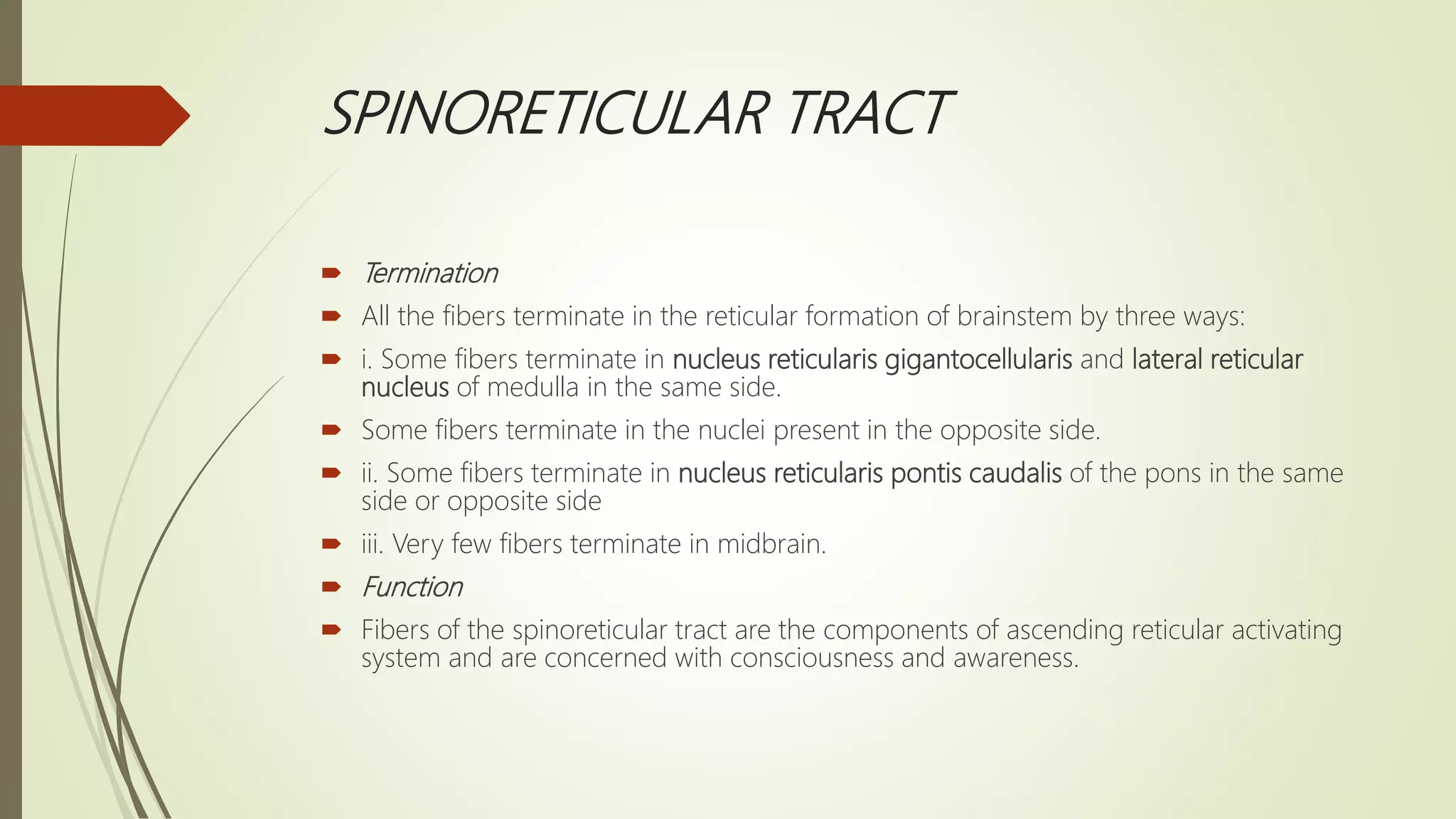
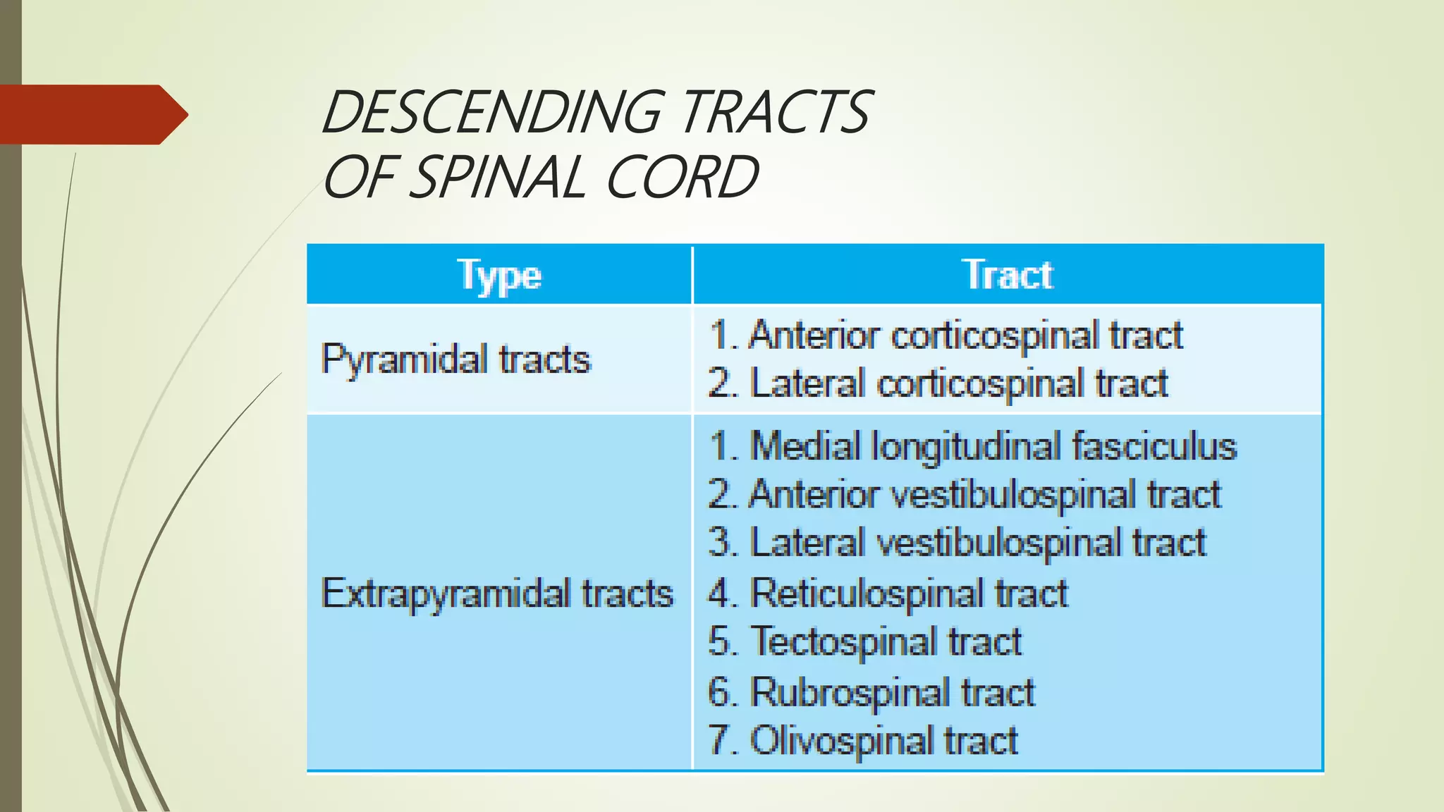
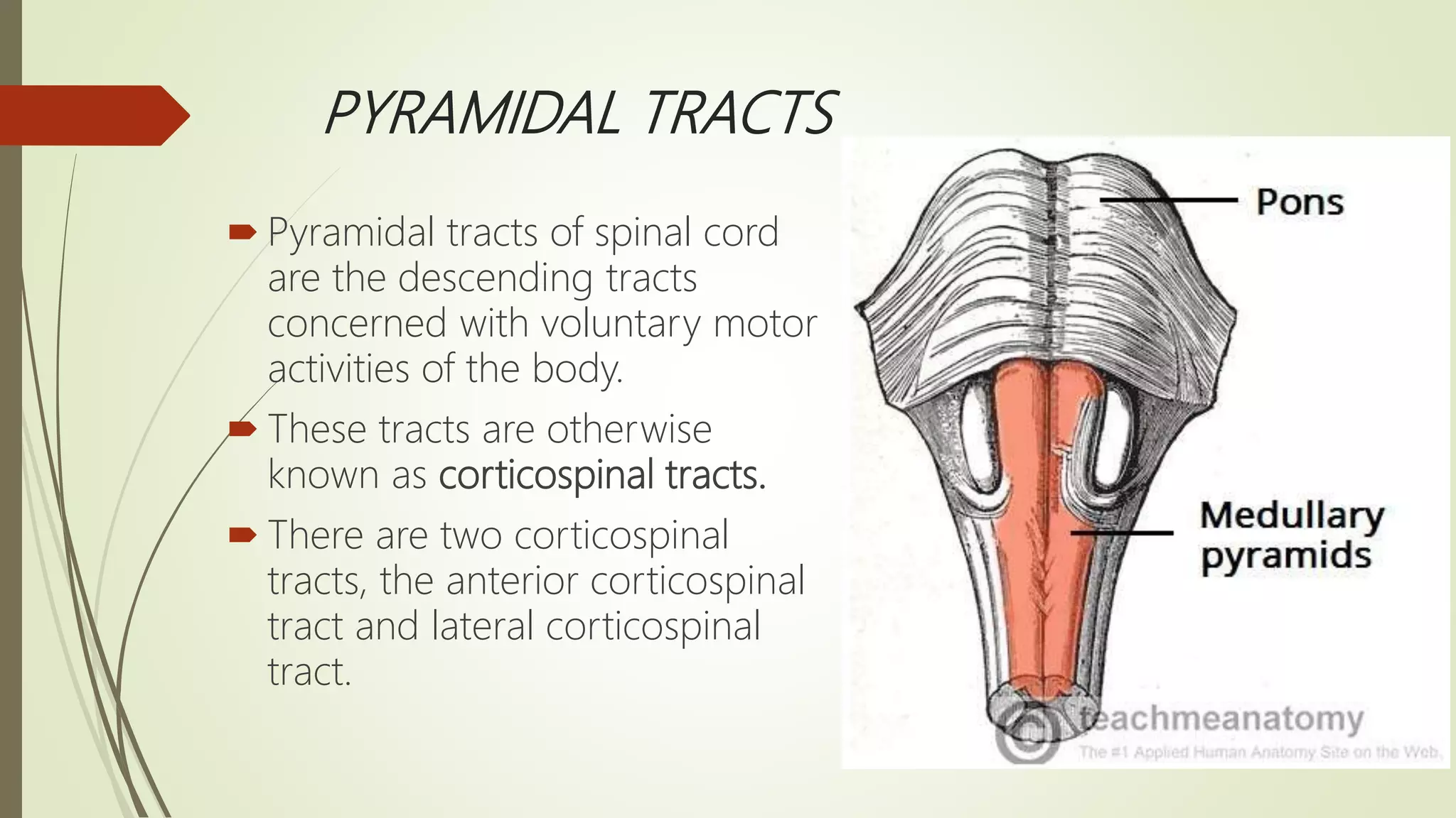
The document discusses the ascending and descending spinal pathways. It describes the short and long tracts of the spinal cord. The long tracts include ascending tracts that carry sensory impulses from the spinal cord to the brain, and descending tracts that carry motor impulses from the brain to the spinal cord. It provides details on three major ascending tracts - the anterior spinothalamic tract, lateral spinothalamic tract, and ventral spinocerebellar tract. It explains the origin, course, termination and functions of these tracts. The document also describes the dorsal spinocerebellar tract and its role in proprioception.
spinal cord, ascending tracts of the the spinal cord, spinocortical tracts, gray matter of spinal cord, white mater of spinal cord, organization of neuron, first order second order and third order neuron, anterolateral spinal tract anteroposterior spinal tract, spinolivary tract, visceral sensory tract, dorsal column tract, spino cerebellar tract , spinorectal pathway, spino olivary pathway, cerebellar peduncles,
Pyramidal tract by Sunita.M.Tiwale,Prof. Dept of physiology,D.Y.Patil Medical...Physiology Dept
Specific Learning Objectives:
At the end of session the students should be able to :
Enumerate the descending tracts.
Describe the origin, course, termination, collaterals of Pyramidal tract.
Describe the functions of the pyramidal tract.
an overview of the ascending tract of the spinal cord....an anatomical approach to understand the somato-sensory pathway.
Prepared as a class presentation .
enlists and the description of the different descending tracts of the CNS. cortico spinal tract, cortico bulbar tract, extra pyramidal and pyramiddal tracts, homunculus, vestibulospinal tract, reticulo spinal tracts, tectospinal tract, autonomic tract, uppermotor neuron lesion, lower motor neuron lesion, spinal cord injury, brown sequard syndrome. spinal cord infection, degenerative disorders of spinal cord,
understanding spinal cord, its bransches, lesions, functions and anatomy.
hope to give you better knowledge of spinal cord by the end of it.
plese review ans comment for my future updates and corrections that iw ill be needing in this.
Functions and ascending tract of spinal cordFatima Mangrio
As the name suggests, the ascending tracts of the spinal cord ascend from the spinal cord and connect it to the brain. These tracts are named based on their origin and termination. They are found running along the dorsal, lateral, and ventral columns of the white matter.
Nutrition in New born and Kids
Calorie requirement of newborn and growing kids
Protein energy malnutrition
Vitamin deficiency disorders in kids
Ricketts
Scurvy
Kwashiorkor
Marasmus
The limbic system, also known as the paleomammalian cortex, is a set of brain structures located on both sides of the thalamus, immediately beneath the medial temporal lobe of the cerebrum primarily in the forebrain.[1]
It supports a variety of functions including emotion, behavior, motivation, long-term memory, and olfaction.[2] Emotional life is largely housed in the limbic system, and it critically aids the formation of memories.
With a primordial structure, the limbic system is involved in lower order emotional processing of input from sensory systems
This PPT describes neurological gait deviations.
It describes Hemiplegic/circumductory gait, Spastic Diplegic gait, Parkinson gait, Myopathic & Ataxic gait in detail along with its causes and management in with Physiotherapy treatment. detail
This PPT is made to explain basic techniques of therapeutic massage in detail.
It includes : Stroking, Pressure Manipulation, Tapotement & Vibration
Physiological effects, Therapeutic uses, Caution & Contraindications.
Description : Osteogenesis Imperfecta/
Brittle bone disease :
It is disorder of type I collagen synthesis that affects all connective tissue in the body.
Musculoskeletal involvement is diffuse and includes osteoporosis with excessive fracture even at birth, bowing of long bone, spinal deformities, muscle weakness and ligamentous laxity.
Key words :
Osteogenesis Imperfecta, Brittle bone disease, Genetic disorder, Pathophysiology, Types of OI, Denetinogenesis Imperfecta, Bluish sclera, Frequent fractures, fractures, Hearing loss, Management, orthopedic, Rehabilitation
Physiotherapy, pediatrics, physiotherapist, pediatric orthopedic surgery.
Arthrogryposis multiplex congenita is a disorder that affects the early development of body joints in a fetus, most commonly the large joints in the arms and legs. An infant who is born with the condition typically has limited mobility and obvious physical deformities in one or more joints.
1. Scar Tissue
2.What makes a scar
3. Characteristics of Scar
4. Scar & Adhesion
5. Classification as External and Internal scars
6 Detriments/Disadvantages
7. How scar tissue forms in muscle
8. Phases of scar healing
9.Phases of wound healing
10. Release of scar and adhesion
11. Massage
12. Massage techniques
13. Conditions : Burns & Skin grafting
14. Cautions
15. Procedure
16. When to start massage after surgery
17. How to massage scar tissue
18. Effleurage, Kneading, Skin rolling and Friction
19. Caution
20. Keloid
21. Warning.
What is oedema?
types of oedema
1. soft and mobile
2. consolidated/endurated
Conditions in which oedema is treated with massage
a) Radical mastectomy
b) Venous ulcer
c) gravitational/paralytic oedema
Technique of massage to reduce oedema
Kneading
Effleurage
Picking Up &
Friction
Instructions to patient
Position of Patient
Aim
Procedure &
Steps of Massage Application
Diabetes Mellitus
Introduction
Pathophysiology
Types of Diabetes Mellitus
Type 1, 2 and
gestational diabetes
rescent research in Type 1 diabetes
Risk factors and causes
Complications short term and long term of diabetes
Management
Treatment with Insulin
Diabetic drugs
Healthy Diet
Exercises prescription
aerobic exercises,
resistance exercises and
flexibility
Explore natural remedies for syphilis treatment in Singapore. Discover alternative therapies, herbal remedies, and lifestyle changes that may complement conventional treatments. Learn about holistic approaches to managing syphilis symptoms and supporting overall health.
Ozempic: Preoperative Management of Patients on GLP-1 Receptor Agonists Saeid Safari
Preoperative Management of Patients on GLP-1 Receptor Agonists like Ozempic and Semiglutide
ASA GUIDELINE
NYSORA Guideline
2 Case Reports of Gastric Ultrasound
Knee anatomy and clinical tests 2024.pdfvimalpl1234
This includes all relevant anatomy and clinical tests compiled from standard textbooks, Campbell,netter etc..It is comprehensive and best suited for orthopaedicians and orthopaedic residents.
Prix Galien International 2024 Forum ProgramLevi Shapiro
June 20, 2024, Prix Galien International and Jerusalem Ethics Forum in ROME. Detailed agenda including panels:
- ADVANCES IN CARDIOLOGY: A NEW PARADIGM IS COMING
- WOMEN’S HEALTH: FERTILITY PRESERVATION
- WHAT’S NEW IN THE TREATMENT OF INFECTIOUS,
ONCOLOGICAL AND INFLAMMATORY SKIN DISEASES?
- ARTIFICIAL INTELLIGENCE AND ETHICS
- GENE THERAPY
- BEYOND BORDERS: GLOBAL INITIATIVES FOR DEMOCRATIZING LIFE SCIENCE TECHNOLOGIES AND PROMOTING ACCESS TO HEALTHCARE
- ETHICAL CHALLENGES IN LIFE SCIENCES
- Prix Galien International Awards Ceremony
Tom Selleck Health: A Comprehensive Look at the Iconic Actor’s Wellness Journeygreendigital
Tom Selleck, an enduring figure in Hollywood. has captivated audiences for decades with his rugged charm, iconic moustache. and memorable roles in television and film. From his breakout role as Thomas Magnum in Magnum P.I. to his current portrayal of Frank Reagan in Blue Bloods. Selleck's career has spanned over 50 years. But beyond his professional achievements. fans have often been curious about Tom Selleck Health. especially as he has aged in the public eye.
Follow us on: Pinterest
Introduction
Many have been interested in Tom Selleck health. not only because of his enduring presence on screen but also because of the challenges. and lifestyle choices he has faced and made over the years. This article delves into the various aspects of Tom Selleck health. exploring his fitness regimen, diet, mental health. and the challenges he has encountered as he ages. We'll look at how he maintains his well-being. the health issues he has faced, and his approach to ageing .
Early Life and Career
Childhood and Athletic Beginnings
Tom Selleck was born on January 29, 1945, in Detroit, Michigan, and grew up in Sherman Oaks, California. From an early age, he was involved in sports, particularly basketball. which played a significant role in his physical development. His athletic pursuits continued into college. where he attended the University of Southern California (USC) on a basketball scholarship. This early involvement in sports laid a strong foundation for his physical health and disciplined lifestyle.
Transition to Acting
Selleck's transition from an athlete to an actor came with its physical demands. His first significant role in "Magnum P.I." required him to perform various stunts and maintain a fit appearance. This role, which he played from 1980 to 1988. necessitated a rigorous fitness routine to meet the show's demands. setting the stage for his long-term commitment to health and wellness.
Fitness Regimen
Workout Routine
Tom Selleck health and fitness regimen has evolved. adapting to his changing roles and age. During his "Magnum, P.I." days. Selleck's workouts were intense and focused on building and maintaining muscle mass. His routine included weightlifting, cardiovascular exercises. and specific training for the stunts he performed on the show.
Selleck adjusted his fitness routine as he aged to suit his body's needs. Today, his workouts focus on maintaining flexibility, strength, and cardiovascular health. He incorporates low-impact exercises such as swimming, walking, and light weightlifting. This balanced approach helps him stay fit without putting undue strain on his joints and muscles.
Importance of Flexibility and Mobility
In recent years, Selleck has emphasized the importance of flexibility and mobility in his fitness regimen. Understanding the natural decline in muscle mass and joint flexibility with age. he includes stretching and yoga in his routine. These practices help prevent injuries, improve posture, and maintain mobilit
Ethanol (CH3CH2OH), or beverage alcohol, is a two-carbon alcohol
that is rapidly distributed in the body and brain. Ethanol alters many
neurochemical systems and has rewarding and addictive properties. It
is the oldest recreational drug and likely contributes to more morbidity,
mortality, and public health costs than all illicit drugs combined. The
5th edition of the Diagnostic and Statistical Manual of Mental Disorders
(DSM-5) integrates alcohol abuse and alcohol dependence into a single
disorder called alcohol use disorder (AUD), with mild, moderate,
and severe subclassifications (American Psychiatric Association, 2013).
In the DSM-5, all types of substance abuse and dependence have been
combined into a single substance use disorder (SUD) on a continuum
from mild to severe. A diagnosis of AUD requires that at least two of
the 11 DSM-5 behaviors be present within a 12-month period (mild
AUD: 2–3 criteria; moderate AUD: 4–5 criteria; severe AUD: 6–11 criteria).
The four main behavioral effects of AUD are impaired control over
drinking, negative social consequences, risky use, and altered physiological
effects (tolerance, withdrawal). This chapter presents an overview
of the prevalence and harmful consequences of AUD in the U.S.,
the systemic nature of the disease, neurocircuitry and stages of AUD,
comorbidities, fetal alcohol spectrum disorders, genetic risk factors, and
pharmacotherapies for AUD.
Pulmonary Thromboembolism - etilogy, types, medical- Surgical and nursing man...VarunMahajani
Disruption of blood supply to lung alveoli due to blockage of one or more pulmonary blood vessels is called as Pulmonary thromboembolism. In this presentation we will discuss its causes, types and its management in depth.
New Directions in Targeted Therapeutic Approaches for Older Adults With Mantl...i3 Health
i3 Health is pleased to make the speaker slides from this activity available for use as a non-accredited self-study or teaching resource.
This slide deck presented by Dr. Kami Maddocks, Professor-Clinical in the Division of Hematology and
Associate Division Director for Ambulatory Operations
The Ohio State University Comprehensive Cancer Center, will provide insight into new directions in targeted therapeutic approaches for older adults with mantle cell lymphoma.
STATEMENT OF NEED
Mantle cell lymphoma (MCL) is a rare, aggressive B-cell non-Hodgkin lymphoma (NHL) accounting for 5% to 7% of all lymphomas. Its prognosis ranges from indolent disease that does not require treatment for years to very aggressive disease, which is associated with poor survival (Silkenstedt et al, 2021). Typically, MCL is diagnosed at advanced stage and in older patients who cannot tolerate intensive therapy (NCCN, 2022). Although recent advances have slightly increased remission rates, recurrence and relapse remain very common, leading to a median overall survival between 3 and 6 years (LLS, 2021). Though there are several effective options, progress is still needed towards establishing an accepted frontline approach for MCL (Castellino et al, 2022). Treatment selection and management of MCL are complicated by the heterogeneity of prognosis, advanced age and comorbidities of patients, and lack of an established standard approach for treatment, making it vital that clinicians be familiar with the latest research and advances in this area. In this activity chaired by Michael Wang, MD, Professor in the Department of Lymphoma & Myeloma at MD Anderson Cancer Center, expert faculty will discuss prognostic factors informing treatment, the promising results of recent trials in new therapeutic approaches, and the implications of treatment resistance in therapeutic selection for MCL.
Target Audience
Hematology/oncology fellows, attending faculty, and other health care professionals involved in the treatment of patients with mantle cell lymphoma (MCL).
Learning Objectives
1.) Identify clinical and biological prognostic factors that can guide treatment decision making for older adults with MCL
2.) Evaluate emerging data on targeted therapeutic approaches for treatment-naive and relapsed/refractory MCL and their applicability to older adults
3.) Assess mechanisms of resistance to targeted therapies for MCL and their implications for treatment selection
Lung Cancer: Artificial Intelligence, Synergetics, Complex System Analysis, S...Oleg Kshivets
RESULTS: Overall life span (LS) was 2252.1±1742.5 days and cumulative 5-year survival (5YS) reached 73.2%, 10 years – 64.8%, 20 years – 42.5%. 513 LCP lived more than 5 years (LS=3124.6±1525.6 days), 148 LCP – more than 10 years (LS=5054.4±1504.1 days).199 LCP died because of LC (LS=562.7±374.5 days). 5YS of LCP after bi/lobectomies was significantly superior in comparison with LCP after pneumonectomies (78.1% vs.63.7%, P=0.00001 by log-rank test). AT significantly improved 5YS (66.3% vs. 34.8%) (P=0.00000 by log-rank test) only for LCP with N1-2. Cox modeling displayed that 5YS of LCP significantly depended on: phase transition (PT) early-invasive LC in terms of synergetics, PT N0—N12, cell ratio factors (ratio between cancer cells- CC and blood cells subpopulations), G1-3, histology, glucose, AT, blood cell circuit, prothrombin index, heparin tolerance, recalcification time (P=0.000-0.038). Neural networks, genetic algorithm selection and bootstrap simulation revealed relationships between 5YS and PT early-invasive LC (rank=1), PT N0—N12 (rank=2), thrombocytes/CC (3), erythrocytes/CC (4), eosinophils/CC (5), healthy cells/CC (6), lymphocytes/CC (7), segmented neutrophils/CC (8), stick neutrophils/CC (9), monocytes/CC (10); leucocytes/CC (11). Correct prediction of 5YS was 100% by neural networks computing (area under ROC curve=1.0; error=0.0).
CONCLUSIONS: 5YS of LCP after radical procedures significantly depended on: 1) PT early-invasive cancer; 2) PT N0--N12; 3) cell ratio factors; 4) blood cell circuit; 5) biochemical factors; 6) hemostasis system; 7) AT; 8) LC characteristics; 9) LC cell dynamics; 10) surgery type: lobectomy/pneumonectomy; 11) anthropometric data. Optimal diagnosis and treatment strategies for LC are: 1) screening and early detection of LC; 2) availability of experienced thoracic surgeons because of complexity of radical procedures; 3) aggressive en block surgery and adequate lymph node dissection for completeness; 4) precise prediction; 5) adjuvant chemoimmunoradiotherapy for LCP with unfavorable prognosis.
- Video recording of this lecture in English language: https://youtu.be/lK81BzxMqdo
- Video recording of this lecture in Arabic language: https://youtu.be/Ve4P0COk9OI
- Link to download the book free: https://nephrotube.blogspot.com/p/nephrotube-nephrology-books.html
- Link to NephroTube website: www.NephroTube.com
- Link to NephroTube social media accounts: https://nephrotube.blogspot.com/p/join-nephrotube-on-social-media.html
These simplified slides by Dr. Sidra Arshad present an overview of the non-respiratory functions of the respiratory tract.
Learning objectives:
1. Enlist the non-respiratory functions of the respiratory tract
2. Briefly explain how these functions are carried out
3. Discuss the significance of dead space
4. Differentiate between minute ventilation and alveolar ventilation
5. Describe the cough and sneeze reflexes
Study Resources:
1. Chapter 39, Guyton and Hall Textbook of Medical Physiology, 14th edition
2. Chapter 34, Ganong’s Review of Medical Physiology, 26th edition
3. Chapter 17, Human Physiology by Lauralee Sherwood, 9th edition
4. Non-respiratory functions of the lungs https://academic.oup.com/bjaed/article/13/3/98/278874
Title: Sense of Smell
Presenter: Dr. Faiza, Assistant Professor of Physiology
Qualifications:
MBBS (Best Graduate, AIMC Lahore)
FCPS Physiology
ICMT, CHPE, DHPE (STMU)
MPH (GC University, Faisalabad)
MBA (Virtual University of Pakistan)
Learning Objectives:
Describe the primary categories of smells and the concept of odor blindness.
Explain the structure and location of the olfactory membrane and mucosa, including the types and roles of cells involved in olfaction.
Describe the pathway and mechanisms of olfactory signal transmission from the olfactory receptors to the brain.
Illustrate the biochemical cascade triggered by odorant binding to olfactory receptors, including the role of G-proteins and second messengers in generating an action potential.
Identify different types of olfactory disorders such as anosmia, hyposmia, hyperosmia, and dysosmia, including their potential causes.
Key Topics:
Olfactory Genes:
3% of the human genome accounts for olfactory genes.
400 genes for odorant receptors.
Olfactory Membrane:
Located in the superior part of the nasal cavity.
Medially: Folds downward along the superior septum.
Laterally: Folds over the superior turbinate and upper surface of the middle turbinate.
Total surface area: 5-10 square centimeters.
Olfactory Mucosa:
Olfactory Cells: Bipolar nerve cells derived from the CNS (100 million), with 4-25 olfactory cilia per cell.
Sustentacular Cells: Produce mucus and maintain ionic and molecular environment.
Basal Cells: Replace worn-out olfactory cells with an average lifespan of 1-2 months.
Bowman’s Gland: Secretes mucus.
Stimulation of Olfactory Cells:
Odorant dissolves in mucus and attaches to receptors on olfactory cilia.
Involves a cascade effect through G-proteins and second messengers, leading to depolarization and action potential generation in the olfactory nerve.
Quality of a Good Odorant:
Small (3-20 Carbon atoms), volatile, water-soluble, and lipid-soluble.
Facilitated by odorant-binding proteins in mucus.
Membrane Potential and Action Potential:
Resting membrane potential: -55mV.
Action potential frequency in the olfactory nerve increases with odorant strength.
Adaptation Towards the Sense of Smell:
Rapid adaptation within the first second, with further slow adaptation.
Psychological adaptation greater than receptor adaptation, involving feedback inhibition from the central nervous system.
Primary Sensations of Smell:
Camphoraceous, Musky, Floral, Pepperminty, Ethereal, Pungent, Putrid.
Odor Detection Threshold:
Examples: Hydrogen sulfide (0.0005 ppm), Methyl-mercaptan (0.002 ppm).
Some toxic substances are odorless at lethal concentrations.
Characteristics of Smell:
Odor blindness for single substances due to lack of appropriate receptor protein.
Behavioral and emotional influences of smell.
Transmission of Olfactory Signals:
From olfactory cells to glomeruli in the olfactory bulb, involving lateral inhibition.
Primitive, less old, and new olfactory systems with different path
4. TRACTS IN SPINAL CORD
Groups of nerve fibers passing through spinal cord are known as tracts of the
spinal cord. The spinal tracts are divided into two main groups. They are:
1. Short tracts
2. Long tracts.
5. Short Tracts
Fibers of the short tracts connect different parts of
spinal cord itself.
Short tracts are of two types:
i. Association or intrinsic tracts, which connect adjacent segments of spinal cord
on the same side
ii. Commissural tracts, which connect opposite halves of same segment of
spinal cord.
6. Long Tracts
Long tracts of spinal cord, which are also called projection tracts, connect
the spinal cord with other parts of central nervous system.
Long tracts are of two types:
i. Ascending tracts, which carry sensory impulses from the spinal cord to
brain
ii. Descending tracts, which carry motor impulses from brain to the spinal
cord.
7.
8.
9.
10. ASCENDING TRACTS OF SPINAL CORD
Ascending tracts of spinal cord carry the impulses of various sensations to the
brain.
Pathway for each sensation is formed by two or three groups of neurons, which
are:
1. First order neurons
2. Second order neurons
3. Third order neurons.
11. First Order Neurons
First order neurons receive sensory impulses from the
receptors and send them to sensory neurons present
in the posterior gray horn of spinal cord through their
fibers. Nerve cell bodies of these neurons are located in
the posterior nerve root ganglion.
12. Second Order Neurons
Second order neurons are the sensory neurons present in the posterior gray
horn.
Fibers from these neurons form the ascending tracts.
These fibers carry sensory impulses from spinal cord to different brain areas
below cerebral cortex
(subcortical areas) such as thalamus.
All the ascending tracts are formed by fibers of second order neurons of the
sensory pathways except the ascending tracts in the posterior white funiculus,
which are formed by the fibers of first order neurons.
14. Third Order Neurons
Third order neurons are in the subcortical areas.
Fibers of these neurons carry the sensory impulses
from subcortical areas to cerebral cortex.
15.
16.
17. ANTERIOR SPINOTHALAMIC TRACT
Anterior spinothalamic tract is formed by the fibers of second order neurons of
the pathway for crude touch sensation
Situation
Anterior spinothalamic tract is situated in anterior white funiculus near the
periphery.
Origin
Fibers of anterior spinothalamic tract arise from the neurons of chief sensory
nucleus of posterior gray horn, which form the second order neurons of the
crude touch pathway. First order neurons are situated in the posterior nerve root
ganglia. These neurons receive the impulses of crude touch sensation from the
pressure receptors. Axons of the first order neurons reach the chief sensory
nucleus through the posterior nerve root.
18. ANTERIOR SPINOTHALAMIC TRACT
Course
Anterior spinothalamic tract contains crossed fibers.
After taking origin, these fibers cross obliquely in the anterior white commissure
and enter the anterior white column of opposite side. Here, the fibers ascend
through other segments of spinal cord and brainstem (medulla, pons and
midbrain) and reach thalamus.
Few fibers of this tract ascend in posterior gray horn for 2 or 3 segments in the
same side and then cross over to the anterior white column of opposite side.
While ascending through brainstem, the number of fibers is considerably reduced
since some of the fibers form the collaterals and reach the reticular formation of
brainstem.
19. ANTERIOR SPINOTHALAMIC TRACT
Termination
Fibers of anterior spinothalamic tract terminate in the ventral posterolateral nucleus of thalamus.
Neurons of this thalamic nucleus form third order neurons of the
pathway. Fibers from thalamic nucleus carry the impulses to somesthetic area (sensory cortex) of
cerebral cortex.
Function
Anterior spinothalamic tract carries impulses of crude touch (protopathic) sensation.
Effect of Lesion
Bilateral lesion of this tract leads to loss of crude touch sensation and loss of sensations like itching
and tickling. Unilateral lesion of this tract causes loss of crude touch sensation in opposite side below
the level of lesion (because fibers of this tract cross to the opposite side in spinal cord).
20. Spinothalamic tracts and pathways for crude
touch, pain and temperature sensations. Anterior
spinothalamic tract (red) carries crude touch
sensation. Lateral spinothalamic tract (blue) carries
pain and temperature sensations.
21. LATERAL SPINOTHALAMIC TRACT
Lateral spinothalamic tract is formed by the fibers from second order neurons of
the pathway for the sensations of pain and temperature
Situation
Lateral spinothalamic tract is situated in the lateral column towards medial side,
i.e. near the gray matter.
Origin
Fibers of lateral spinothalamic tract take origin from two sources:
i. Marginal nucleus
ii. Substantia gelatinosa of Rolando.
22. LATERAL SPINOTHALAMIC TRACT
Course
Lateral spinothalamic tract has crossed fibers.
Axons from marginal nucleus and substantia gelatinosa of Rolando cross to the
opposite side and reach the lateral column of same segment.
Few fibers may ascend one or two segments, then cross to the opposite side and then
ascend in the lateral column.
All the fibers pass through medulla, pons and midbrain and reach thalamus along
with fibers of anterior spinothalamic tract.
Some of the fibers of lateral spinothalamic tract form collaterals and reach the reticular
formation of brainstem.
Fibers of lateral spinothalamic tract form spinal lemniscus along with the fibers of
anterior spinothalamic tract at the lower part of medulla.
23.
24. LATERAL SPINOTHALAMIC TRACT
Termination
Fibers of lateral spinothalamic tract terminate in the ventral posterolateral nucleus of
thalamus along with anterior spinothalamic tract fibers.
From here, third order neuron fibers run to somesthetic area (sensory cortex) of
cerebral cortex.
Function
Fibers of lateral spinothalamic tract carry impulses of pain and temperature
sensations.
Fibers arising from this marginal nucleus transmit impulses of fast pain
sensation.
Fibers arising from substantia gelatinosa of Rolando transmit impulses of slow pain
and temperature sensations.
25. LATERAL SPINOTHALAMIC TRACT
Effect of Lesion
Bilateral lesion of this tract leads to total loss of pain and temperature sensations
on both sides below the level of lesion.
Unilateral lesion or sectioning of the lateral spinothalamic tract causes loss of pain
(analgesia) and temperature (thermoanesthesia) below the level of lesion in the
opposite side.
26.
27.
28. VENTRAL SPINOCEREBELLAR TRACT
Ventral spinocerebellar tract is also
known as Gower tract, indirect
spinocerebellar tract or anterior
spinocerebellar tract. It is constituted by
the fibers of second order neurons of the
pathway for subconscious kinesthetic
sensation
29. VENTRAL SPINOCEREBELLAR TRACT
Situation
This tract is situated in lateral white column of the spinal cord along the lateral periphery.
Origin
Fibers of this tract arise from the marginal nucleus in posterior gray horn. Neurons of
marginal nucleus form the second order neurons.
Fibers from these neurons make their first appearance in lower lumbar segments of spinal
cord.
First order neurons are in the posterior root ganglia and receive the impulses of
proprioception from the proprioceptors in muscle, tendon and joints.
Fibers from neurons of posterior root ganglia reach the marginal cells through posterior
nerve root.
30. VENTRAL SPINOCEREBELLAR TRACT
Course
Ventral spinocerebellar tract contains both crossed and uncrossed fibers.
Majority of the fibers from the marginal nucleus cross the midline and ascend in
lateral white column of opposite side.
Some fibers ascend in the lateral white column of the same side also.
These nerve fibers ascend through other spinal segments, medulla, pons and
midbrain.
Finally, the fibers reach the cerebellum through the superior cerebellar peduncle.
31. VENTRAL SPINOCEREBELLAR TRACT
Termination
These fibers terminate in the cortex of anterior lobe of cerebellum.
Function
Ventral spinocerebellar tract carries the impulses of subconscious kinesthetic
sensation (proprioceptive impulses from muscles, tendons and joints).
32. VENTRAL SPINOCEREBELLAR TRACT
Effect of Lesion
Lesion of this tract leads to loss of subconscious kinesthetic sensation in the
opposite side.
34. DORSAL SPINOCEREBELLAR TRACT
Dorsal spinocerebellar tract is otherwise called Flechsig tract, direct
spinocerebellar tract or posterior spinocerebellar tract.
Like the ventral spinocerebellar tract, this tract is also constituted by the second
order neuron fibers of the pathway for subconscious kinesthetic sensation.
The first order neurons are in the posterior nerve root ganglia. But, the fibers of
this tract are uncrossed.
35. DORSAL SPINOCEREBELLAR TRACT
Situation
Dorsal spinocerebellar tract is situated in the lateral column along the
posterolateral periphery of spinal cord.
It is situated posterior to ventral cerebellar tract and anterior to the entry of
posterior nerve root.
Origin
Fibers of this tract originate from the dorsal nucleus of Clarke situated in the
posterior gray matter of the spinal cord.
First appearance of the fibers is in upper lumbar segments.
From lower lumbar and sacral segments, the impulses are carried upwards by
dorsal nerve roots to upper lumbar segments.
36.
37.
38. DORSAL SPINOCEREBELLAR TRACT
Course
Flechsig tract is formed by uncrossed fibers.
Axons from neurons in dorsal nucleus of Clarke (second order neurons) reach
lateral column of same side.
Then, these fibers ascend through other spinal segments and reach medulla
oblongata.
From here, the fibers reach cerebellum through inferior cerebellar peduncle.
39. DORSAL SPINOCEREBELLAR TRACT
Course
Flechsig tract is formed by uncrossed fibers.
Axons from neurons in dorsal nucleus of Clarke (second order neurons) reach
lateral column of same side.
Then, these fibers ascend through other spinal segments and reach medulla
oblongata.
From here, the fibers reach cerebellum through inferior cerebellar peduncle.
Termination
Fibers of this tract end in the cortex of anterior lobe of cerebellum along with
ventral spinocerebellar tract fibers.
40. DORSAL SPINOCEREBELLAR TRACT
Function
Along with ventral spinocerebellar tract, the dorsal spinocerebellar tract carries
the impulses of subconscious kinesthetic sensation.
Effect of Lesion
Unilateral loss of the subconscious kinesthetic sensation occurs in lesion of this
tract on the same side, as this tract has uncrossed fibers.
41.
42. SPINOTECTAL TRACT
Spinotectal tract is considered as a component of anterior spinothalamic tract.
It is constituted by the fibers of second order neurons.
43. SPINOTECTAL TRACT
Situation
Spinotectal tract occupies the lateral side of lateral white column, anterior to
lateral spinothalamic tract.
It is bound anteriorly by anterior nerve root.
Origin
Fibers of this tract originate from the chief sensory nucleus (like anterior
spinothalamic tract). First appearance of the fibers is in upper lumbar segments.
This tract is very prominent in the cervical segments.
44. SPINOTECTAL TRACT
Course
Spinotectal tract contains crossed fibers. After taking origin, the fibers cross to
opposite side through anterior white commissure to the lateral column.
Then, these fibers ascend to the midbrain along with anterior spinothalamic tract.
Termination
Fibers of spinotectal tract end in the superior colliculus of tectum in midbrain.
Function
Spinotectal tract is concerned with spinovisual reflex.
45.
46. FASCICULUS DORSOLATERALIS
Fasciculus dorsolateralis is otherwise called tract of Lissauer. It is considered as a
component of lateral spinothalamic tract.
And, it is constituted by the fibers of first order neurons.
Situation
Lissauer tract is situated in the lateral white column between the periphery of
spinal cord and tip of posterior gray horn.
47. FASCICULUS DORSOLATERALIS
Origin
Lissauer tract is formed by fibers arising from the cells of posterior root ganglia
and enters the spinal cord through lateral division of posterior nerve root.
Course
Lissauer tract contains uncrossed fibers.
After entering spinal cord, the fibers pass upwards or downwards for few
segments on the same side and synapse with cells of substantia gelatinosa of
Rolando. Axons from these cells (second order neurons) join the lateral
spinothalamic tract.
49. SPINORETICULAR TRACT
Spinoreticular tract is formed by the fibers of second order neurons.
Situation
Spinoreticular tract is situated in anterolateral white column.
Origin
Fibers of this tract arise from intermediolateral nucleus.
Course
Spinoreticular tract consists of crossed and uncrossed fibers. After taking origin,
some of the fibers cross the midline and then ascend upwards. Remaining fibers
ascend up in the same side without crossing.
50.
51. SPINORETICULAR TRACT
Termination
All the fibers terminate in the reticular formation of brainstem by three ways:
i. Some fibers terminate in nucleus reticularis gigantocellularis and lateral reticular
nucleus of medulla in the same side.
Some fibers terminate in the nuclei present in the opposite side.
ii. Some fibers terminate in nucleus reticularis pontis caudalis of the pons in the same
side or opposite side
iii. Very few fibers terminate in midbrain.
Function
Fibers of the spinoreticular tract are the components of ascending reticular activating
system and are concerned with consciousness and awareness.
52. SPINOOLIVARY TRACT
Spino-olivary tract is situated in anterolateral part of white column. Origin of the
fibers of this tract is not specific.
However, the fibers terminate in olivary nucleus of medulla oblongata. From here,
the neurons project into cerebellum.
This tract is concerned with proprioception.
53.
54. SPINOVESTIBULAR TRACT
Spinovestibular tract is situated in the lateral white column of the spinal cord.
Fibers of this tract arise from all the segments of spinal cord and terminate on the
lateral vestibular nucleus.
This tract is also concerned with proprioception.
55. FASCICULUS GRACILIS (TRACT OF GOLL) &
FASCICULUS CUNEATUS (TRACT OF BURDACH)
Fasciculus gracilis and fasciculus cuneatus are together called ascending posterior
column tracts.
These tracts are formed by the fibers from posterior root ganglia.
Thus, both the tracts are constituted by the fibers of first order neurons of
sensory pathway.
56.
57.
58. FASCICULUS GRACILIS (TRACT OF GOLL) &
FASCICULUS CUNEATUS (TRACT OF BURDACH)
Situation
Tracts of Goll and Burdach are situated in posterior white column of spinal cord
hence the name posterior column tracts.
In the cervical and upper thoracic segments of spinal cord, the posterior white
column is divided by posterior intermediate septum into medial fasciculus gracilis
and lateral fasciculus cuneatus.
Thus, the fasciculus gracilis is situated medially.
Fasciculus cuneatus is situated laterally.
59. FASCICULUS GRACILIS (TRACT OF GOLL) &
FASCICULUS CUNEATUS (TRACT OF BURDACH)
Origin
Fibers of these two tracts are the axons of first order neurons.
Cell body of these neurons is in the posterior root ganglia and their fibers form the medial
division (bundle) of posterior nerve root.
Course
After entering spinal cord, the fibers ascend through the posterior white column.
These fibers do not synapse in the spinal cord. Some fibers of medial division of posterior nerve
root descend through posterior white column in the form of fasciculus interfascicularis or comma
tract of Schultze.
Fasciculus gracilis contains the fibers from lower extremities and lower parts of the body, i.e. from
sacral, lumbar and lower thoracic ganglia of posterior nerve root.
Fasciculus cuneatus contains fibers from upper part of the body, i.e. from upper thoracic and
cervical ganglia of posterior nerve root.
60. FASCICULUS GRACILIS (TRACT OF GOLL) &
FASCICULUS CUNEATUS (TRACT OF BURDACH)
Termination
Tracts of Goll and Burdach terminate in the medulla oblongata.
Fibers of fasciculus gracilis terminate in the nucleus gracilis and the fibers of fasciculus
cuneatus terminate in the nucleus cuneatus.
Neurons of these medullary nuclei form the second order neurons.
Axons of second order neurons form the internal arcuate fibers. Internal arcuate fibers
from both sides cross the midline forming sensory decussation and then ascend
through pons and midbrain as medial lemniscus.
Fibers of medial lemniscus terminate in ventral posterolateral nucleus of thalamus.
From here, fibers of the third order neurons relay to sensory area of cerebral cortex.
61.
62. Ascending tracts in posterior white
column of spinal cord and pathway for –
1. Fine touch sensation, 2. Tactile
localization, 3. Tactile discrimination, 4.
Vibratory sensation, 5. Conscious
kinesthetic sensation, 6. Stereognosis.
63. FASCICULUS GRACILIS (TRACT OF GOLL) &
FASCICULUS CUNEATUS (TRACT OF BURDACH)
Functions
Tracts of the posterior white column convey impulses of following sensations:
i. Fine (epicritic) tactile sensation
ii. Tactile localization
iii. Tactile discrimination or two point discrimination
iv. Sensation of vibration
v. Conscious kinesthetic sensation
vi. Stereognosis
64. FASCICULUS GRACILIS (TRACT OF GOLL) &
FASCICULUS CUNEATUS (TRACT OF BURDACH)
Effect of Lesion
Lesion of nerve fibers in tracts of Goll and Burdach or lesion in the posterior white column leads
to the following symptoms on the same side below the lesion:
i. Loss of fine tactile sensation; however, crude touch sensation is normal
ii. Loss of tactile localization
iii. Loss of two point discrimination
iv. Loss of sensation of vibration
v. Astereognosis (inability to recognize known objects by touch while closing the eyes)
vi. Lack of ability to differentiate the weight of different objects
vii. Loss of proprioception (inability to appreciate the position and movement of different parts of
the body)
viii. Sensory ataxia or posterior column ataxia (condition characterized by uncoordinated, slow
and clumsy voluntary movements because of the loss of proprioception)
65. COMMA TRACT OF SCHULTZE
Comma tract of schultze is also called fasciculus interfascicularis. It is situated in
between tracts of Goll and Burdach.
This tract is formed by the short descending fibers, arising from the medial
division of posterior nerve root.
These fibers are also considered as the descending branches of the tracts of Goll
and Burdach.
Function of this tract is to establish intersegmental communications and to form
short reflex arc.
66. DESCENDING TRACTS
OF SPINAL CORD
Descending tracts of the spinal cord are formed by motor
nerve fibers arising from brain and descend into the spinal
cord.
These tracts carry motor impulses from brain to spinal
cord.
Descending tracts of spinal cord are of two types:
A. Pyramidal tracts
B. Extrapyramidal tracts.
68. PYRAMIDAL TRACTS
Pyramidal tracts of spinal cord
are the descending tracts
concerned with voluntary motor
activities of the body.
These tracts are otherwise
known as corticospinal tracts.
There are two corticospinal
tracts, the anterior corticospinal
tract and lateral corticospinal
tract.
69. Nerve Fibers
All the fibers of the pyramidal tracts are present since birth.
However, myelination of these fibers is completed in about 2 years
after birth.
The pyramidal tracts on each side have more than a million fibers.
About 70% of the fibers are large myelinated fibers.
Large fibers of pyramidal tracts have the tendency to disappear at old
age.
The disappearance of the fibers of pyramidal tracts causes automatic
shivering movements in old age.
Fibers of pyramidal tracts are the axons of upper motor neurons.
71. Origin
Fibers of pyramidal tracts arise from following cells or areas of
cerebral cortex:
1. Giant cells or Betz cells or pyramidal cells in precentral gyrus of the
motor cortex. These cells are situated in area 4 (primary motor area)
frontal lobe.
2. Other areas of motor cortex namely, premotor area (area 6) and
supplementary motor areas
3. Other parts of frontal lobe
4. Somatosensory areas of parietal lobe.
PYRAMIDAL TRACTS
72.
73.
74.
75. Course
Corona radiata
After taking origin, the nerve fibers run downwards in
diffused manner through white matter of cerebral
hemisphere and converge in the form of a fan-like
structure along with ascending fibers, which project
from thalamus to cerebral cortex.
This fan-like structure is called corona radiata. Thus,
corona radiata contains both ascending fibers from
thalamus and descending fibers from cerebral cortex.
PYRAMIDAL TRACTS
76.
77.
78. Course
Internal capsule
While passing down towards the brainstem the
corona radiata converges in the form of internal
capsule.
It is situated in between thalamus and caudate
nucleus on the medial side and lenticular
on the lateral side
PYRAMIDAL TRACTS
79.
80.
81.
82. Course
In pons
The fibers descend down through internal capsule,
midbrain and pons.
While descending through pons, the fibers are
into different bundles by the nuclei of pons.
At lower border of pons, the fibers are grouped once
again into a compact bundle and then descend down
into medulla oblongata.
PYRAMIDAL TRACTS
84. Course
In medulla
This compact bundle of corticospinal fibers gives the appearance of
pyramid in the anterior surface of upper part of medulla. So, the
corticospinal tracts are called the pyramidal tracts.
At the lower border of medulla, pyramidal tract on each side is
divided into two bundles of unequal sizes.
About 80% of fibers from each side cross to the opposite side.
While crossing the midline, the fibers of both sides form the
pyramidal decussation.
PYRAMIDAL TRACTS
85.
86. Course
In spinal cord
Fibers which cross the midline and form pyramidal
decussation descend through posterior part of lateral
white column of spinal cord.
This bundle of crossed fibers is called the crossed
pyramidal tract or lateral corticospinal tract or indirect
corticospinal tract.
PYRAMIDAL TRACTS
87. Remaining 20% of fibers do not cross to the
opposite side but descend down through the
anterior white column of the spinal cord.
This bundle of uncrossed fibers is called the
uncrossed pyramidal tract or anterior
tract or direct corticospinal tract. This tract is well
marked in cervical region.
PYRAMIDAL TRACTS
88. Since, the fibers of this tract terminate in different
segments of spinal cord, this tract usually gets
thinner while descending through the successive
segments of spinal cord.
Fibers of this tract are absent mostly below the
mid thoracic level. Before termination, majority of
the fibers of this anterior corticospinal tract cross
to the opposite side at different levels of spinal
cord.
PYRAMIDAL TRACTS
89. Termination
All the fibers of pyramidal tracts, both crossed and
uncrossed fibers, terminate in the motor neurons of
anterior gray horn either directly or through internuncial
neurons.
Pyramidal tract fibers terminate on both α-motor neurons
and β-motor neurons.
PYRAMIDAL TRACTS
90.
91.
92. Axons of the motor neurons leave the spinal cord as spinal
nerves through anterior nerve roots and supply the skeletal
muscles.
Neurons giving origin to the fibers of pyramidal tract are
called the upper motor neurons.
Anterior motor neurons in the spinal cord are called the
lower motor neurons.
PYRAMIDAL TRACTS
93.
94. Function
Pyramidal tracts are concerned with voluntary movements
of the body.
Fibers of the pyramidal tracts transmit motor impulses
motor area of cerebral cortex to the anterior motor
of the spinal cord.
These two tracts are responsible for fine, skilled
movements.
PYRAMIDAL TRACTS
95.
96. Lesion in the neurons of motor cortex and the fibers of pyramidal
tracts is called the upper motor neuron lesion.
In human beings, pure pyramidal tract lesions do not occur.
Lesion of pyramidal fibers occurs most commonly in stroke
(cardiovascular accident) due to hemorrhage and thrombosis.
During such lesions, many extrapyramidal fibers are also damaged
along with pyramidal fibers.
Because of this reason, neurologists often consider the lesion as
upper motor neuron lesion and not as pyramidal tract lesion.
PYRAMIDAL TRACTS- Effects of lesion:
97. 1. Voluntary movements
Initially loss of voluntary movements in the extremities. Later, it
involves the other parts of the body like hip and shoulder.
2. Muscle tone
Muscle tone is increased leading to spasticity.
Muscles are also paralyzed. This type of paralysis of muscles is called
the spastic paralysis. The spasticity is due to the failure of inhibitory
impulses from upper motor neurons, particularly the neurons of
extrapyramidal system to reach the γ-motor neurons in spinal cord.
PYRAMIDAL TRACTS- Effects of lesion:
98. Hypotonia occurs in pure pyramidal tract lesion, which is very
rare.
3. Reflexes
All the superficial reflexes are lost and the deep reflexes are
exaggerated.
Abnormal plantar reflex called Babinski sign is present
(Babinski sign positive).
PYRAMIDAL TRACTS- Effects of lesion:
99.
100. Cerebral cortex
Lesion of pyramidal tract fibers in cerebral cortex causes
hypertonia, spasticity and contralateral monoplegia (para lysis
of one limb) or contralateral hemiplegia (para lysis of one
of the body).
Internal capsule
Lesion of pyramidal tract fibers at posterior limb of internal
capsule results in contralateral hemiplegia.
PYRAMIDAL TRACTS- Effects of lesion at
Different Levels:
101. Brainstem
Lesion at brainstem involves not only pyramidal tract fibers
also other structures such as VI and VII cranial nerve nuclei.
So the lesion results in contralateral hemiparesis (weakness of
muscles in one side of the body) along with VI and VII cranial
nerve palsies.
PYRAMIDAL TRACTS- Effects of lesion at
Different Levels:
102. Spinal cord
Unilateral lesion of lateral corticospinal fibers at upper cervical
segment causes ipsilateral hemiplegia and bilateral lesion
causes quadriplegia (paralysis of all four limbs) and paralysis of
respiratory muscles.
Bilateral lesion of these fibers in thoracic and lumbar segments
results in paraplegia (paralysis of both lower limbs) without
paralysis of respiratory muscles.
PYRAMIDAL TRACTS- Effects of lesion at
Different Levels:
105. MEDIAL LONGITUDINAL FASCICULUS
Situation
Medial longitudinal fasciculus descends through posterior part of
anterior white column of the spinal cord.
Origin
Actually, this tract is the extension of medial longitudinal fasciculus of
brainstem. Fibers of this tract take origin from four different areas in
brainstem:
i. Vestibular nuclei
ii. Reticular formation
iii. Superior colliculus
iv. Interstitial cells of Cajal.
106.
107.
108. MEDIAL LONGITUDINAL FASCICULUS
Course
After entering the spinal cord from the brainstem, the fibers of medial
longitudinal fasciculus descend through posterior part of anterior
white column of the same side.
In the spinal cord, this tract is well defined only in upper cervical
segments. Below this level, the fibers run along with the fibers of
anterior vestibulospinal tract.
Extent
Fibers of this tract extend up to the upper cervical segments of spinal
cord.
109. MEDIAL LONGITUDINAL FASCICULUS
Termination
Fibers of this tract terminate in anterior motor neurons of the spinal
cord along with fibers of anterior vestibulospinal tract either directly
or through internuncial neurons.
Function
Medial longitudinal fasciculus helps in the coordination of reflex
ocular movements and the integration of ocular and neck
movements.
Effects of Lesion
Reflex ocular movements and reflex neck movements are affected in
the lesion of this tract.
110.
111.
112. ANTERIOR VESTIBULOSPINAL TRACT
Situation
Anterior vestibulospinal tract is situated in the anterior white
column, along the periphery of spinal cord lateral to tectospinal
tract.
Origin
Fibers of this tract arise from medial vestibular nucleus in medulla
oblongata.
In fact, anterior vestibulospinal tract is the extension of medial
longitudinal fasciculus.
Most of the fibers are uncrossed.
113. ANTERIOR VESTIBULOSPINAL TRACT
Extent
Fibers run up to thoracic segments of spinal cord.
Course
Fibers of this tract run down from medulla into the anterior
column of spinal cord along the periphery.
114. ANTERIOR VESTIBULOSPINAL TRACT
Termination
Along with fibers of lateral vestibulospinal tract, the fibers of this
tract terminate in anterior motor neurons directly or through
internuncial neurons.
Function
Function of this tract is explained along with the function of
lateral vestibulospinal tract.
115. LATERAL VESTIBULOSPINAL TRACT
Situation
Lateral vestibulospinal tract occupies the anterior part of lateral
white column of spinal cord.
Origin
Fibers of this tract take origin from the lateral vestibular nucleus
in medulla. This nucleus is also called Deiter nucleus.
Extent
Fibers of this tract are present throughout the spinal cord.
116. LATERAL VESTIBULOSPINAL TRACT
Course
From Deiter nucleus, most of the fibers descend directly through
lateral column.
Very few fibers cross to the opposite side before descending.
Termination
Fibers of this tract terminate in the anterior motor neuron, either
directly or via internuncial neurons.
117. LATERAL VESTIBULOSPINAL TRACT
Functions
Vestibular nuclei receive impulses concerned with muscle tone
and posture from vestibular apparatus and cerebellum.
Vestibular nuclei in turn convey the impulses to different parts of
the body through the anterior and lateral vestibulospinal tracts.
Vestibulospinal tracts are concerned with adjustment of position
of head and body during angular and linear acceleration.
118. Effect of Lesion
Adjustment of head and body becomes difficult during
acceleration when the vestibulospinal tracts are affected
lesion.
LATERAL VESTIBULOSPINAL TRACT
119.
120.
121. RETICULOSPINAL TRACT
Situation
Reticulospinal tract is situated in the anterior white column,
posterior to anterior vestibulospinal tract.
122. RETICULOSPINAL TRACT
Origin
Fibers of this tract arise from the reticular formation of pons and
medulla.
Pontine reticular fibers are uncrossed (direct) and descend in
medial part of anterior column.
Fibers from medullary reticular formation are predominantly
uncrossed and only few fibers are crossed.
These fibers descend in lateral part of anterior column and to
some extend in the anterior part of lateral column.
123. RETICULOSPINAL TRACT
Extent
Fibers of reticulospinal tract extend up to thoracic
segments.
Termination
Fibers of reticulospinal tract terminate in gamma motor
neurons of anterior gray horn through the internuncial
neuron.
124. RETICULOSPINAL TRACT
Functions
Reticulospinal tract is concerned with control of movements and
maintenance of muscle tone, respiration and diameter of blood
vessels.
Pontine and medullary fibers have opposite effects on these
functions.
Effect of Lesion
Lesion of reticulospinal tract causes disturbances in respiration,
blood pressure, movements of body and muscle tone.
128. TECTOSPINAL TRACT
Situation
Tectospinal tract is situated in the anterior white column of spinal
cord.
Origin
Nerve fibers of this tract arise from superior colliculus of
midbrain.
Extent
Tectospinal tract extends only up to lower cervical segments.
129.
130. TECTOSPINAL TRACT
Course
After taking origin from superior colliculus, the fibers cross the
midline in dorsal tegmental decussation and descend in anterior
column.
Termination
Fibers of tectospinal tract terminate in the anterior motor
neurons of spinal cord, directly or via internuncial neurons.
Function
Tectospinal tract is responsible for the movement of head in
response to visual and auditory stimuli.
131. RUBROSPINAL TRACT
Situation
Rubrospinal tract is situated in the lateral white column of
spinal cord.
Origin
Fibers of this tract arise from large cells (nucleus
magnocellularis) of red nucleus in midbrain.
Extent
Nerve fibers of this tract appear in the spinal cord only up to
thoracic segments.
132. Course
After arising from the red nucleus, the fibers cross the midline in
ventral tegmental decussation and descend into spinal cord
through the reticular formation of pons and medulla.
Termination
Fibers of rubrospinal tract end in the anterior motor neurons of
spinal cord via internuncial neurons.
Function
Rubrospinal tract exhibits facilitatory influence upon flexor muscle
tone.
RUBROSPINAL TRACT
133. OLIVOSPINAL TRACT
Situation
Olivospinal tract is present in lateral white column of
spinal cord.
Origin
The nerve fibers of the olivospinal tract take origin from
the inferior olivary nucleus, which is present in the
medulla oblongata.
134.
135. OLIVOSPINAL TRACT
Termination
Fibers of this tract terminate in the anterior motor
neurons of spinal cord.
Function
Functions of the olivospinal tract are not known clearly.
It is believed that this tract is involved in reflex
movements arising from the proprioceptors.