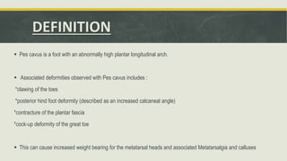
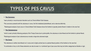
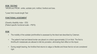
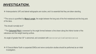
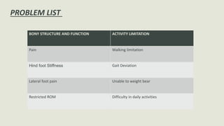
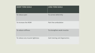
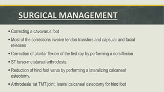
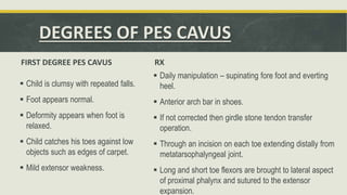
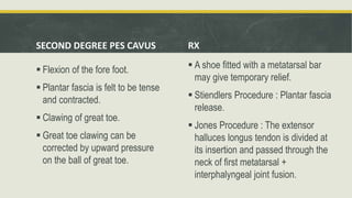
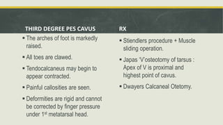
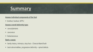
Deformities observed with Pes cavus includes :
*clawing of the toes
*posterior hind foot deformity (described as an increased calcaneal angle)
*contracture of the plantar fascia
*cock-up deformity of the great toe
Following References were used to prepare this powerpoint presentation which makes the slides accurate and relaible for studying purpose; Therapeutic Exrercise – Carolyn Kisner
Orthopaedic Physical Assessment – Magee
Orthopaedic Medicine – L. Ombregt
Campbell’s Operative Orthopaedics
Slides includes following headings;
DEFINITION
TYPES
ORTHOPAEDIC ASSESSMENT
MEDICAL MANAGGEMENT
PHYSIOTHERAPY MANAGEMENT
SURGICAL MANAGEMENT
Bicipital tendonitis is inflammation of long head of the biceps tendon under the bicipital groove.
In early stage, tendon becomes red and swollen, as tendonitis develops the tendon sheath can thicken.
In late stage, often become dark red in color due to inflammation.
As part of a class presentation, we attempted to make this to briefly explain what Torticollis meas, the Types of presentation of Torticollis, and Management strategies for a Physiotherapist for Congenital Torticollis especially.
I hope this helps. :)
The pictures and information had been taken from internet, complied to make a brief presentation for the purpose of class presentation.
I do not own any content.
Introduction:-
Hoffa's pad syndrome also known as Infrapatellar fat syndrome is an intrinsic disease of Hoffa's fat pad and a problem of knee joint which causes the pain at the front of knee joint so this pain is known as anterior knee pain.
Hoffa's fat pad contains pluripotent cells that can differentiate into osteoblasts and chondrocytes.
Hoffa's pad becomes inflamed or Damaged by The crushing of the pad between the femur and tibia during extension, causes inflammation of Hoffa's fat pad.
Nerve supply of fat pad is excellent( It receives branches of the femoral , common peroneal and saphenous nerves) so if it occurs any injury ,causes a sharp anterior knee pain.
The infrapatellar fat pad is a pad of adipose tissue underneath or deep to the patella tendon and the top of the fat pad attaches to the lower part of patella (knee cap).
hoffa's pad is a shock absorber ,when there is a direct force on the patella can result in pinching of the fat pad between femur and tibial plateau. The tibial plateau is the proximal tibial surface on which the femur rests.
HFP is surrounded anteriorly by the patellar tendon and the joint capsule, superiorly by the inferior pole of the patella, inferiorly by the proximal tibia and the deep infrapatellar bursa, and posteriorly by the joint synovium .
The main function of the HFP is to reduce friction between the patella, the patellar tendon, and the deep skeletal structures. In addition, it prevents pinching of the synovial membrane and it facilitates the vascularization of adjacent structures.
Causes:-
cause is usually due to single or repetitive traumatic episodes.
when you extend your knee the fat pad act as a cushion and reduces friction between outer patella facets and quadriceps tendons .
when you flex your knee ,upper part of fat pad becomes tensioned, it moves backwards in the knee.
it develops gradually over time if you repeatedly move your knee.
This is when your knee is forced to move forward from its completely straight normal position.
You may have always been able to over straighten your knee, which is called knee hyperextension or genu recurvatum .
hyperextension sports such as basketball, volleyball or high jumping may also cause inflammation of Hoffa's pad.
Hoffa's disease is more frequent in young women and the symptoms are anterior knee pain when upstairs and downstairs.
Sign and Symptoms:-
Complaints of anterior knee pain occurs when playing hyperextension sports such as basketball ,volleyball or high jumping .effusion and inflammation may be occurs and decreases the ROM of joint , stair negotiation .
Symptoms may worsen if the knee is overly straightened or bent for too long a period. Complications may include an inability to fully straighten the knee.
Diagnosis:-
Hoffa's syndrome completely diagnosed by MRI .we have requirement of an experienced orthopaedics to diagnose it.
primary Assessment have to check the Active and Passive Range Of Motion(AROM/PROM) of Hip joint and Knee joint.
Collapse of medial longitudinal arch, with the entire sole of the foot coming into complete or near-complete contact with the ground.
Books Refered :
Text Book Of ANATOMY - Vishram Singh
Joint Structure And Function – Cynthia Norkin
Therapeutic Exrercise – Carolyn Kisner
Orthopaedic Physical Assessment – Magee
Orthopaedic Medicine – L. Ombregt
Campbell’s Operative Orthopaedics
Brief discussion regarding management of physiotherapy, pharmacotherapy, orthosis, principles of orthopedic surgical managements, addressing problems at hip, knee and ankle, soft tissue release procedures, osteotomies, timing of surgery, complications, prognosis, hip at risk signs, birthday syndrome, role of botulinum toxin, upper extremity involvement, contracture release.
Global launch of the Healthy Ageing and Prevention Index 2nd wave – alongside...ILC- UK
The Healthy Ageing and Prevention Index is an online tool created by ILC that ranks countries on six metrics including, life span, health span, work span, income, environmental performance, and happiness. The Index helps us understand how well countries have adapted to longevity and inform decision makers on what must be done to maximise the economic benefits that comes with living well for longer.
Alongside the 77th World Health Assembly in Geneva on 28 May 2024, we launched the second version of our Index, allowing us to track progress and give new insights into what needs to be done to keep populations healthier for longer.
The speakers included:
Professor Orazio Schillaci, Minister of Health, Italy
Dr Hans Groth, Chairman of the Board, World Demographic & Ageing Forum
Professor Ilona Kickbusch, Founder and Chair, Global Health Centre, Geneva Graduate Institute and co-chair, World Health Summit Council
Dr Natasha Azzopardi Muscat, Director, Country Health Policies and Systems Division, World Health Organisation EURO
Dr Marta Lomazzi, Executive Manager, World Federation of Public Health Associations
Dr Shyam Bishen, Head, Centre for Health and Healthcare and Member of the Executive Committee, World Economic Forum
Dr Karin Tegmark Wisell, Director General, Public Health Agency of Sweden
CRISPR-Cas9, a revolutionary gene-editing tool, holds immense potential to reshape medicine, agriculture, and our understanding of life. But like any powerful tool, it comes with ethical considerations.
Unveiling CRISPR: This naturally occurring bacterial defense system (crRNA & Cas9 protein) fights viruses. Scientists repurposed it for precise gene editing (correction, deletion, insertion) by targeting specific DNA sequences.
The Promise: CRISPR offers exciting possibilities:
Gene Therapy: Correcting genetic diseases like cystic fibrosis.
Agriculture: Engineering crops resistant to pests and harsh environments.
Research: Studying gene function to unlock new knowledge.
The Peril: Ethical concerns demand attention:
Off-target Effects: Unintended DNA edits can have unforeseen consequences.
Eugenics: Misusing CRISPR for designer babies raises social and ethical questions.
Equity: High costs could limit access to this potentially life-saving technology.
The Path Forward: Responsible development is crucial:
International Collaboration: Clear guidelines are needed for research and human trials.
Public Education: Open discussions ensure informed decisions about CRISPR.
Prioritize Safety and Ethics: Safety and ethical principles must be paramount.
CRISPR offers a powerful tool for a better future, but responsible development and addressing ethical concerns are essential. By prioritizing safety, fostering open dialogue, and ensuring equitable access, we can harness CRISPR's power for the benefit of all. (2998 characters)
Struggling with intense fears that disrupt your life? At Renew Life Hypnosis, we offer specialized hypnosis to overcome fear. Phobias are exaggerated fears, often stemming from past traumas or learned behaviors. Hypnotherapy addresses these deep-seated fears by accessing the subconscious mind, helping you change your reactions to phobic triggers. Our expert therapists guide you into a state of deep relaxation, allowing you to transform your responses and reduce anxiety. Experience increased confidence and freedom from phobias with our personalized approach. Ready to live a fear-free life? Visit us at Renew Life Hypnosis..
QA Paediatric dentistry department, Hospital Melaka 2020Azreen Aj
QA study - To improve the 6th monthly recall rate post-comprehensive dental treatment under general anaesthesia in paediatric dentistry department, Hospital Melaka
We understand the unique challenges pickleball players face and are committed to helping you stay healthy and active. In this presentation, we’ll explore the three most common pickleball injuries and provide strategies for prevention and treatment.
How many patients does case series should have In comparison to case reports.pdfpubrica101
Pubrica’s team of researchers and writers create scientific and medical research articles, which may be important resources for authors and practitioners. Pubrica medical writers assist you in creating and revising the introduction by alerting the reader to gaps in the chosen study subject. Our professionals understand the order in which the hypothesis topic is followed by the broad subject, the issue, and the backdrop.
https://pubrica.com/academy/case-study-or-series/how-many-patients-does-case-series-should-have-in-comparison-to-case-reports/
4. DEFINITION
Pes cavus is a foot with an abnormally high plantar longitudinal arch.
Associated deformities observed with Pes cavus includes :
*clawing of the toes
*posterior hind foot deformity (described as an increased calcaneal angle)
*contracture of the plantar fascia
*cock-up deformity of the great toe
This can cause increased weight bearing for the metatarsal heads and associated Metatarsalgia and calluses
5. TYPES OF PES CAVUS
Pes Cavovarus :
*seen primarily in neuromuscular disorders such as Charcot-Marie-Tooth disease.
*Pes cavovarus presents with the calcaneus in varus, the first metatarsal plantarflexed, and a claw-toe deformity.
*Radiological analysis of pes cavus in Charcot-Marie-Tooth disease shows the forefoot is typically plantar flexed in relation to the rear foot.
Pes Calcaneocavus :
*which is seen primarily following paralysis of the Triceps Surae due to poliomyelitis, the calcaneus is dorsi-flexed and the forefoot is plantar-flexed.
*Radiological analysis of pes calcaneocavus reveals a large talo-calcaneal angle.
Pes Cavus :
*the calcaneus is neither dorsi-flexed nor in varus and is highly arched due to a plantar-flexed position of the forefoot on the rear-foot.
*A combination of any or all of these elements can also be seen in a ‘combined’ type of pes cavus that may be further categorized as flexible or rigid.
7. ORTHOPAEDIC ASSESSMENT
A] DEMOGRAPHIC DATA
B] CHIEF COMPLAINT
Patients complains of pain , instability , difficulty walking and problems with footwear.
The symptoms vary with the degree of deformity.
Also presents with lateral foot pain from increased weight bearing on the lateral foot
8. C] HISTORY
• The presentation for patients with pes cavus is highly variable, depending largely on the extent of the deformity.
• Patients can present with lateral foot pain from increased weightbearing on the lateral foot.
• Metatarsalgia is a frequent symptom. Ankle instability can be a presenting symptom, especially in patients with hindfoot
varus and weak peroneus brevis.
• Weakness and fatigue can be observed in patients with neuromuscular disease.
• Evaluation of a patient who presents with pes cavus begins with a thorough history and complete examination to
determine the etiology.
• Patients with a unilateral deformity frequently have a history of major trauma.
• Patients should be questioned about weakness/clumsiness, indicating intrinsic muscle involvement.
9. D] BODY CHART
Area – Foot and Ankle
Onset – Gradual / Insidious
Type – Dull Aching
Depth – Deep
Constancy – Intermittent
10. E] AGGRAVATING FACTOR – prolonged standing and during activities of daily living.
F] RELIEVING FACTOR – Rest
G] SEVERITY – Vas Scale
H] IRRITABILITY – Moderate
I] 24 HOURS PATTERN
J] PAST HISTORY – Ask for any h/o trauma
K] MEDICAL HISTORY – Charcot-Marie-Tooth disease
L] FAMILY HISTORY – Clinically significant
11. M] SOCIAL HISTORY – Work / Sports / Hobbies affected
N] ECONOMIC HISTORY – Modified KuppuSwamy Scale.
OBJECTIVE ASSESMENT
OBSERVATION
POSTURE –
• Observe foot posture in standing and arch posture
• Subtle evidence of foot drop may be evident, if there is calf wasting (stork leg deformity)
MOTOR EXAMINATION
• Rigidity maybe present [ Modified Ashworth Scale ]
12. GAIT
The feet should be examined –
with the patient walking and standing –
both from the front and behind.
• Recruitment of secondary ankle
dorsiflexors (cock-up toes in swing
phase).
During the gait cycle,
• The foot remains locked in hindfoot
inversion and forefoot varus throughout
the stance phase, causing less stress
dissipation.
• This can result in metatarsalgia, stress
fracture of the fifth metatarsal, plantar
fasciitis, medial longitudinal arch pain,
ilio-tibial band syndrome and instability
13. ROM TESTING
*AROM and PROM - ankle, subtalar joint, midfoot, forefoot and toes.
*Lower limb muscle length Test
FUNCTIONAL ASSESMENT
(Oswetry disability index - ODI)
(Patient specific functional scale – PSFS)
ROM
• The mobility of the subtalar joint/hindfoot is assessed by the block test described by Coleman.
• The patient’s heel and lateral border are placed on a block approximately 2.5 cm thick. The first to
third/fourth metatarsals are allowed to fall into pronation, eliminating their effect on the tripod.
• During weight bearing, the hindfoot that returns to valgus is flexible and those that do not are considered
rigid.
14. Anteroposterior (AP) and lateral radiographs are routine, and it is essential that they are taken standing.
**The cavus is quantified by Meary’s angle, the angle between the long axis of the first metatarsal and the long axis
of the talus.
This should normally be 0°
**The Calcaneal Pitch is assessed by the angle formed between a line drawn along the inferior border of the
calcaneus and the weight bearing surface.
An angle of greater than 30° is abnormal, and consistent with c a v o v a r u s / c a l c a n e o c a v u s
If Charcot-Marie-Tooth is suspected EMGs and nerve conduction studies should be performed as an initial
investigation.
15. PROBLEM LIST
BONY STRUCTURE AND FUNCTION ACTIVITY LIMITATION
Pain Walking limitation
Hind foot Stiffness Gait Deviation
Lateral foot pain Unable to weight bear
Restricted ROM Difficulty in daily activities
16. SHORT TERM GOALS LONG TERM GOALS
To reduce pain To correct deformity
To increase the ROM Pain free ambulation
To reduce stiffness To strengthen weak muscles
To reduce any muscle tightness Gait training and Argonomics
17. MEDICAL MANAGEMENT
The goal of treatment is to allow the patient to ambulate without symptoms.
The underlying cause must be identified in order to determine if the disorder is
progressive
18. Physiotherapy management
Conservative management is appropriate in the minimally symptomatic mild case or in cases where surgical treatment is
contraindicated.
Well-fitting, pliable shoes, pliable metatarsal bar orthoses, and ankle-foot orthoses may all be appropriate.
Conservative management of patients with painful pes cavus typically involves strategies to reduce and redistribute
plantar pressure loading, with use of foot orthoses and specialized cushioned footwear.
The orthoses for pes cavus needs to accomplish to several specific goals:
Increasing plantar surface contact area.
The overload on the metatarsal heads is a result of limited plantar surface contact due to high arch and limited ankle-joint
dorsiflexion.
Increasing the plantar surface contact ensures the foot to bear more weight in the arch while the metatarsal heads bear less
weight during activity.
19. Resisting against excessive supination
Lateral ankle stability and laterally deviated subtalar joint axis (STJ) are frequently associated with high-arched feet.
This position results in an excessive supinator torque around the subtalar joint axis.
Resisting against recessive pronation and supination forces
Rearfoot instability is caused by an extension of the laterally deviated subtalar axis.
In flexible pes cavus, midtarsal flexibility complicates the later portion of the stance pgase of gait.
The forefoot pathology produces midtarsal joint supination, that leads to excessive pronation of the rearfoot.
Some pes cavus suffer from both lateral ankle instability at midstance and rearfoot pronation at late midstance. stretching and
strengthening of tight and weak muscles, debridement of plantar callosities, osseous mobilization, massage, manipulation of the foot
and ankle, and strategies to improve balance
Orthotics with extra-depth shoes to offload bony prominences and prevent rubbing of the toes may improve symptoms.
For varus deformities, a lateral wedge sole modification can improve function.
Bracing for supple deformities or foot drop may allow patients to ambulate
In patients with sensation deficits, Plastazote linings in the brace are required and frequent inspection of the skin for ulceration is
warranted.
20. INDICATIONS FOR SURGICAL RX
progressive deformity
painful callosities or ulceration
symptomatic clawing
ankle instability.
Feet with supple deformities require only soft tissue release with or without tendon transfer.
In the younger patient in whom deformities may change, several procedures may be necessary to
accommodate growth or compensate for altering neurology.
Where a specific fixed bony deformity exists in an otherwise supple foot, which prevents
planti-grade loading, then an osteotomy is done to correct this deformity along with the appropriate soft
tissue corrections is indicated.
21. SURGICAL MANAGEMENT
Correcting a cavovarus foot
Most of the corrections involve tendon transfers and capsular and facial
releases
Correction of plantar flexion of the first ray by performing a dorsiflexion
ST tarso-metatarsal arthrodesis.
Reduction of hind foot varus by performing a lateralizing calcaneal
osteotomy.
Arthrodesis 1st TMT joint, lateral calcaneal osteotomy for hind foot
22. DEGREES OF PES CAVUS
FIRST DEGREE PES CAVUS
Child is clumsy with repeated falls.
Foot appears normal.
Deformity appears when foot is
relaxed.
Child catches his toes against low
objects such as edges of carpet.
Mild extensor weakness.
RX
Daily manipulation – supinating fore foot and everting
heel.
Anterior arch bar in shoes.
If not corrected then girdle stone tendon transfer
operation.
Through an incision on each toe extending distally from
metatarsophalyngeal joint.
Long and short toe flexors are brought to lateral aspect
of proximal phalynx and sutured to the extensor
expansion.
23. SECOND DEGREE PES CAVUS
Flexion of the fore foot.
Plantar fascia is felt to be tense
and contracted.
Clawing of great toe.
Great toe clawing can be
corrected by upward pressure
on the ball of great toe.
RX
A shoe fitted with a metatarsal bar
may give temporary relief.
Stiendlers Procedure : Plantar fascia
release.
Jones Procedure : The extensor
halluces longus tendon is divided at
its insertion and passed through the
neck of first metatarsal +
interphalyngeal joint fusion.
24. THIRD DEGREE PES CAVUS
The arches of foot is markedly
raised.
All toes are clawed.
Tendocalcaneus may begin to
appear contracted.
Painful callosities are seen.
Deformities are rigid and cannot
be corrected by finger pressure
under 1st metatarsal head.
RX
Stiendlers procedure + Muscle
sliding operation.
Japas ‘V’osteotomy of tarsus :
Apex of V is proximal and
highest point of cavus.
Dwayers Calcaneal Otetomy.
25. FOURTH DEGREE PES CAVUS
In addition to cavus and claw
toes.
Adduction at tarsometatarsal
joints resulting in varus
deformity.
Rigid and painful foot.
Walking becomes painful and
difficult.
FIFTH DEGREE PES CAVUS
Seen on paralytic conditions
(poliomyelitis)
Whole foot is contracted into
rigid equino varus with high
arch.
Tender callosities.
The patient is very disabled.