Download to read offline
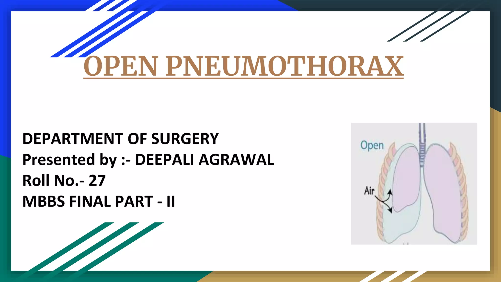


Open pneumothorax is characterized by a large defect in the thorax causing direct communication between the pleura and atmosphere, resulting in impaired lung function and hypoxia. This condition occurs in about 80% of penetrating chest wounds, with diagnosis made through clinical assessment and imaging techniques. Management includes high-flow oxygen, sterile occlusive dressings, and possibly surgical intervention to address underlying injuries.


