Downloaded 377 times

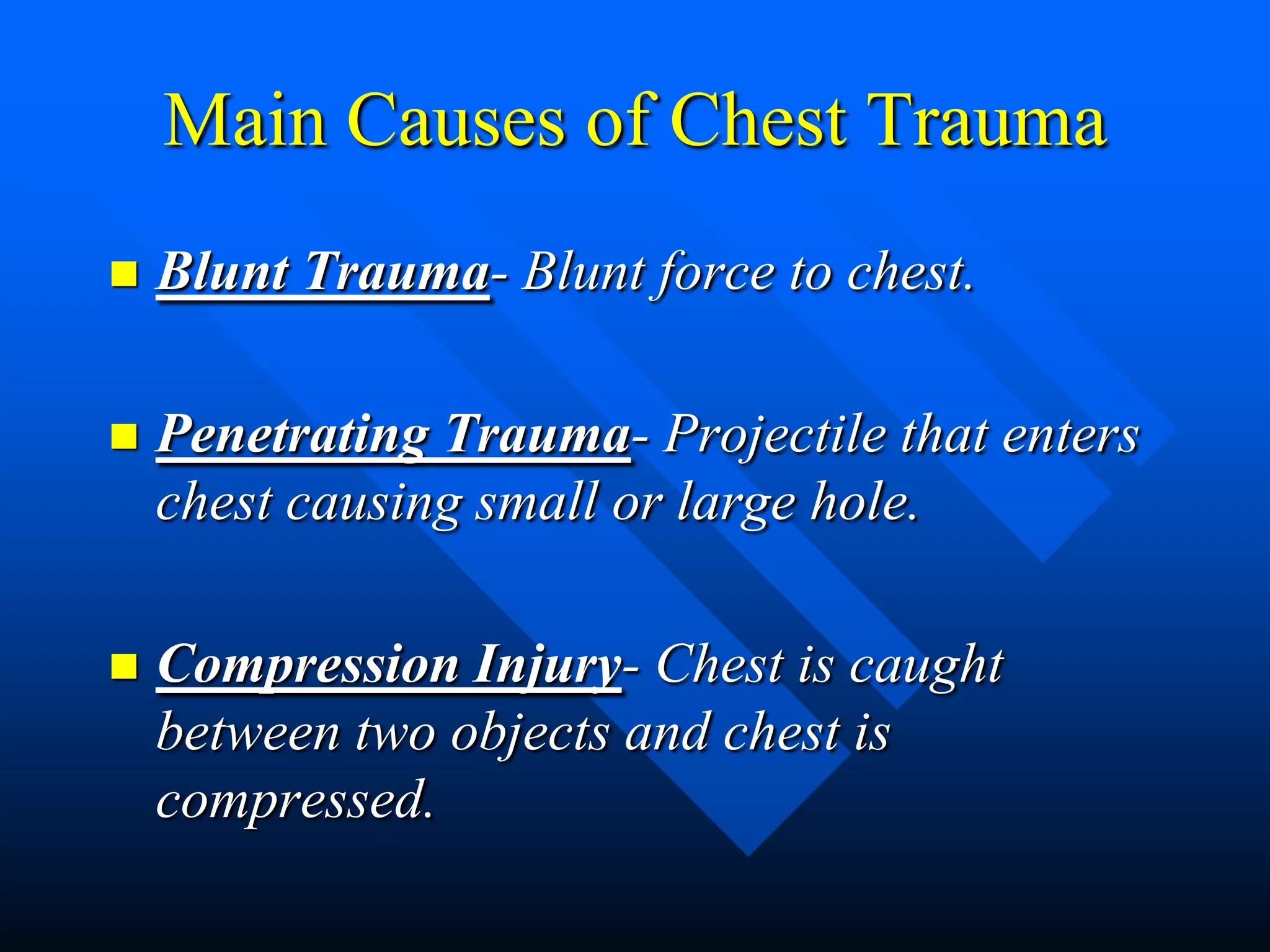


Chest injuries are the second leading cause of trauma deaths each year. The majority of chest trauma can be managed without surgery. Common causes include blunt trauma from force to the chest, penetrating trauma from projectiles entering the chest, and compression injuries. Injuries include rib fractures, flail chest, pneumothoraces, pulmonary contusions, and others. Tension pneumothorax is a life-threatening condition where air builds up in the pleural space with no way to escape, resulting in collapsed lungs and compressed heart and blood vessels. Needle decompression is immediately needed to relieve pressure in the chest and prevent further deterioration.


