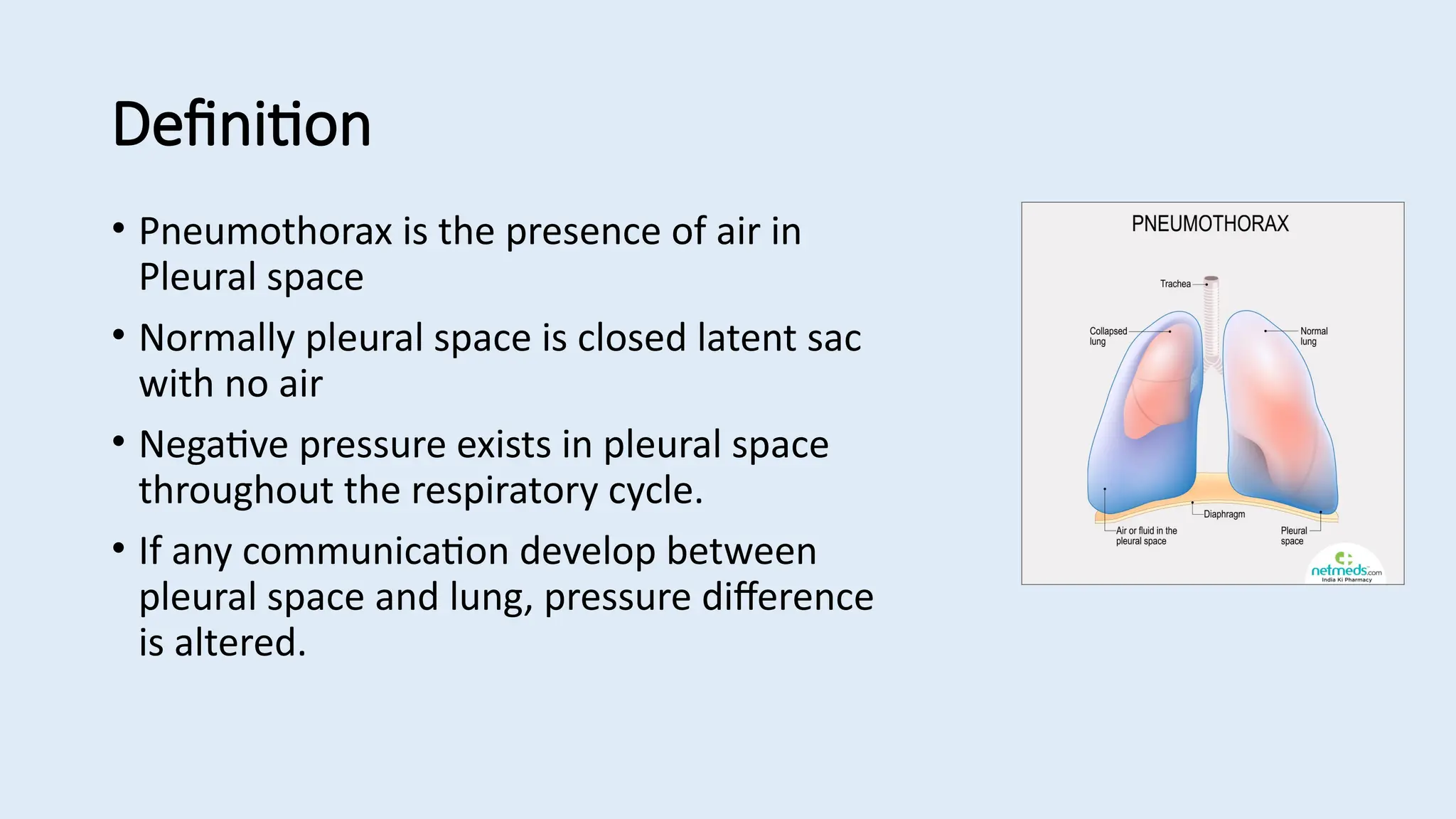
Pneumothorax
Dr. Irfat Mahjabeen
Clinical fellow (Respiratory Medicine)
Worcestershire Acute Hospitals NHS Trust
Definition
• Pneumothorax is the presence of air in
Pleural space
• Normally pleural space is closed latent sac
with no air
• Negative pressure exists in pleural space
throughout the respiratory cycle.
• If any communication develop between
pleural space and lung, pressure difference
is altered.
Classification
1. Spontaneous/ simple 2. Traumatic
Primary :
• no evidence of lung disease
• smoking, tall stature are
additional risk factors.
• usually seen in young age <30
years
• usually rupture of apical or sub
pleural blebs
Secondary :
• Occurs due to Underlying
diseases : COPD and Pulmonary
TB mostly
• Others : asthma, (ILD), necrotizing
pneumonia, cystic fibrosis (CF),
Marfan’s syndrome, lung cancer,
pulmonary infarction
• Usually seen in males above 55
years
• Mortality is 10%
Iatrogenic or traumatic chest injury :
• percutaneous biopsy
• endoscopic perforation of the
esophagus
• nasogastric tube placement
• esophageal rupture
• post CPR
• Can be associated with
haemothorax or
haemopneumothoraz
Tension Pneumothorax :
• Life threatening condition
• One way valve , air enters the pleural space on inspiration, but unable to escape on expiration
• The progressive increase in pleural pressure > atmospheric pressure
• Shifting of mediastinum compresses opposite lung and vessels result in impairment of venous
return & CVS compromise
Clinical features:
Symptoms :
• Pleuritic chest pain and/or
• Breathlessness , more severe in
2° pneumothorax
• In tension pneumothorax,
features of shock or cardiac
arrest or acute deterioration,
increasing inflation pressures in
mechanical ventilation
Signs:
• tachycardia, tachypnoea, hyoxia,
cyanosis, accessory muscles of
respiration, raised JVP,
• Lung : tracheal deviation,
reduced expansion, hyper-
resonant percussion note,
reduced air entry, diminished
breath sounds .
Investigation:
1. CXR :
• visceral pleural edge is seen as a very thin, sharp white line.
• Absent lung markings peripheral to the line
• Mediastinal shift +/-,
• lung collapse +/-
• Peripheral space is radiolucent compared to the adjacent lung
2. Computed tomography (CT):
• More accurate in detrmining size of
pneumothorax
• Differentiate pneumothorax from bullous
disease/ associated underlying lung disease
3.Ultrasound (US) chest :
4. Arterial blood gases (ABGs) - hypoxia and
or hypercapnia in 2° pneumothorax
Management:
1. degree of breathlessness and hypoxia, haemodynamic compromise.
2. background lung disease/respiratory reserve :
3. size of the pneumothorax :
• Width of the rim of air, measured from chest wall to lung edge at the level of the
hilum , <2 cm: small & ≥2 cm: large (BTS guideline 2010)
• A 2 cm rim of air = to a 50% pneumothorax in volume (approx)
Management of Tension Pneumothorax
• Medical emergency
• Do not wait for a CXR if the diagnosis is clinically certain / patient
seriously compromised or cardiac arrest
• Immediate decompression - insert large-bore cannula (14- 16 G) into
second intercostal space in mid-clavicular line
• Cannula shoula be left in place untill ICD can be inserted.
Intercostal drain
• inserted in 4th, 5th. 6th intercostal space in midaxillary line
• should be removed 24 hrs after lung has fully reinflated and bubbling
stopped
• usually 10-14 F in most cases,
• never clamp a bubbling chest drain
Surgical management
Aim is to repair the apical hole or bleb and close the pleural space.
Indications for cardiothoracic surgical referral
• Bilateral spontaneous pneumothorax
• Persistent air leak or failure of lung to re-expand (3–5 days of drainage)
• Spontaneous haemothorax
• 1st contralateral and 2nd ipsilateral pneumothorax
• Professions at risk (e.g. pilots, divers) after first pneumothorax.
• pneumothorax in pregnancy
Plurodesis
• Talc commonly used, failure rate 10-20%
• via intercostal drain , placed between the pleural layers to decrease
pleural space and prevent fluid build up
• only as a last resort in older patients with recurrent pneumothorax in
whom surgery would be high risk ( severe COPD)
• in an incompletely re-expanded lung with a persistent air leak,
plurodesis may be attempted if surgery is not an option.
Advice during discharge
• Diving- never unless surgically corrected , normal lung function
testand a chest CT postoperatively
• Flying - 1 week from complete resolution
• smoking cessation
• F/up in 4 to 6 weeks time
• Risk of recurrence is around 30% after a 1st pneumothorax, 40% after
a 2nd, and > 50% after a 3rd incidence
Thank You

Pneumothorax PPT -----topic presentation

  • 1.
    Pneumothorax Dr. Irfat Mahjabeen Clinicalfellow (Respiratory Medicine) Worcestershire Acute Hospitals NHS Trust
  • 2.
    Definition • Pneumothorax isthe presence of air in Pleural space • Normally pleural space is closed latent sac with no air • Negative pressure exists in pleural space throughout the respiratory cycle. • If any communication develop between pleural space and lung, pressure difference is altered.
  • 3.
    Classification 1. Spontaneous/ simple2. Traumatic Primary : • no evidence of lung disease • smoking, tall stature are additional risk factors. • usually seen in young age <30 years • usually rupture of apical or sub pleural blebs Secondary : • Occurs due to Underlying diseases : COPD and Pulmonary TB mostly • Others : asthma, (ILD), necrotizing pneumonia, cystic fibrosis (CF), Marfan’s syndrome, lung cancer, pulmonary infarction • Usually seen in males above 55 years • Mortality is 10% Iatrogenic or traumatic chest injury : • percutaneous biopsy • endoscopic perforation of the esophagus • nasogastric tube placement • esophageal rupture • post CPR • Can be associated with haemothorax or haemopneumothoraz
  • 4.
    Tension Pneumothorax : •Life threatening condition • One way valve , air enters the pleural space on inspiration, but unable to escape on expiration • The progressive increase in pleural pressure > atmospheric pressure • Shifting of mediastinum compresses opposite lung and vessels result in impairment of venous return & CVS compromise
  • 5.
    Clinical features: Symptoms : •Pleuritic chest pain and/or • Breathlessness , more severe in 2° pneumothorax • In tension pneumothorax, features of shock or cardiac arrest or acute deterioration, increasing inflation pressures in mechanical ventilation Signs: • tachycardia, tachypnoea, hyoxia, cyanosis, accessory muscles of respiration, raised JVP, • Lung : tracheal deviation, reduced expansion, hyper- resonant percussion note, reduced air entry, diminished breath sounds .
  • 6.
    Investigation: 1. CXR : •visceral pleural edge is seen as a very thin, sharp white line. • Absent lung markings peripheral to the line • Mediastinal shift +/-, • lung collapse +/- • Peripheral space is radiolucent compared to the adjacent lung
  • 9.
    2. Computed tomography(CT): • More accurate in detrmining size of pneumothorax • Differentiate pneumothorax from bullous disease/ associated underlying lung disease 3.Ultrasound (US) chest : 4. Arterial blood gases (ABGs) - hypoxia and or hypercapnia in 2° pneumothorax
  • 10.
    Management: 1. degree ofbreathlessness and hypoxia, haemodynamic compromise. 2. background lung disease/respiratory reserve : 3. size of the pneumothorax : • Width of the rim of air, measured from chest wall to lung edge at the level of the hilum , <2 cm: small & ≥2 cm: large (BTS guideline 2010) • A 2 cm rim of air = to a 50% pneumothorax in volume (approx)
  • 12.
    Management of TensionPneumothorax • Medical emergency • Do not wait for a CXR if the diagnosis is clinically certain / patient seriously compromised or cardiac arrest • Immediate decompression - insert large-bore cannula (14- 16 G) into second intercostal space in mid-clavicular line • Cannula shoula be left in place untill ICD can be inserted.
  • 13.
    Intercostal drain • insertedin 4th, 5th. 6th intercostal space in midaxillary line • should be removed 24 hrs after lung has fully reinflated and bubbling stopped • usually 10-14 F in most cases, • never clamp a bubbling chest drain
  • 14.
    Surgical management Aim isto repair the apical hole or bleb and close the pleural space. Indications for cardiothoracic surgical referral • Bilateral spontaneous pneumothorax • Persistent air leak or failure of lung to re-expand (3–5 days of drainage) • Spontaneous haemothorax • 1st contralateral and 2nd ipsilateral pneumothorax • Professions at risk (e.g. pilots, divers) after first pneumothorax. • pneumothorax in pregnancy
  • 15.
    Plurodesis • Talc commonlyused, failure rate 10-20% • via intercostal drain , placed between the pleural layers to decrease pleural space and prevent fluid build up • only as a last resort in older patients with recurrent pneumothorax in whom surgery would be high risk ( severe COPD) • in an incompletely re-expanded lung with a persistent air leak, plurodesis may be attempted if surgery is not an option.
  • 16.
    Advice during discharge •Diving- never unless surgically corrected , normal lung function testand a chest CT postoperatively • Flying - 1 week from complete resolution • smoking cessation • F/up in 4 to 6 weeks time • Risk of recurrence is around 30% after a 1st pneumothorax, 40% after a 2nd, and > 50% after a 3rd incidence
  • 17.