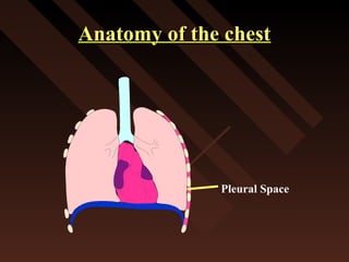
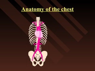
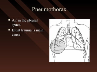
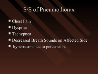
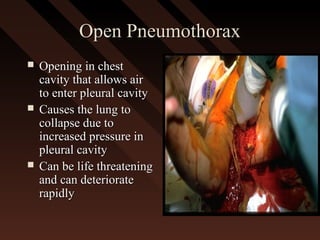
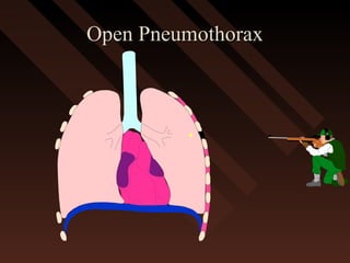
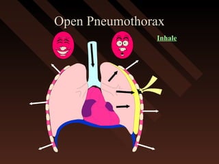
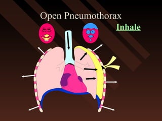
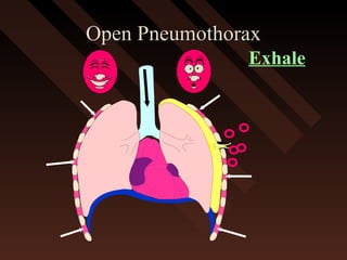
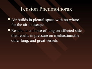
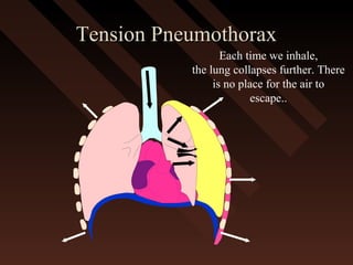
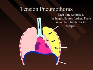
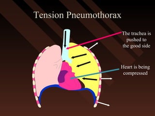
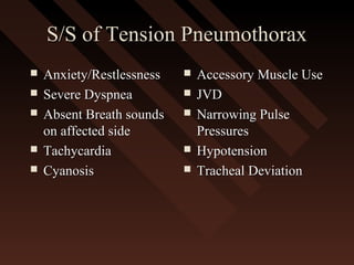
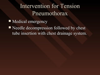
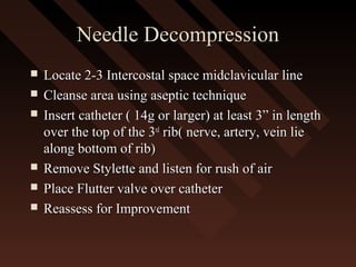
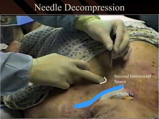
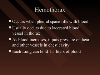
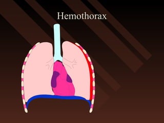
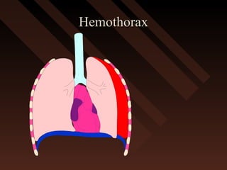
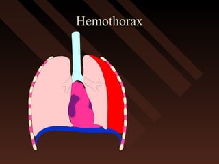
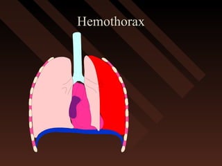
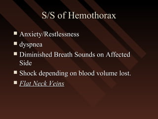
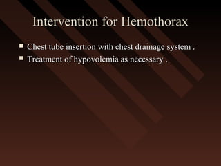
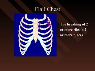
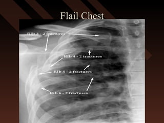
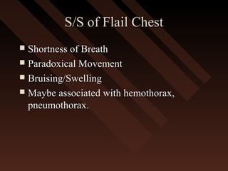
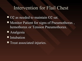
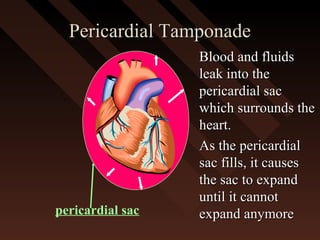
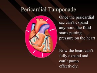
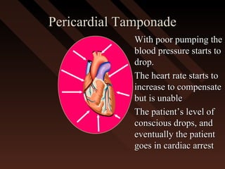
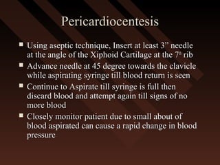
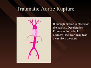
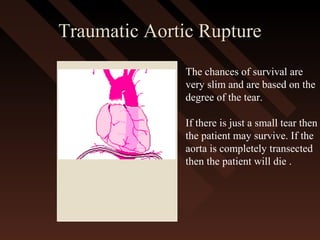
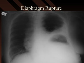
This document provides an overview of chest trauma, including the anatomy of the chest, common causes of injury, signs and symptoms of different types of chest injuries, and treatment approaches. It describes various chest injuries such as pneumothorax, hemothorax, open pneumothorax, tension pneumothorax, flail chest, cardiac tamponade, and diaphragmatic rupture. For each injury, it outlines the mechanism of injury, associated signs and symptoms, and recommended interventions. The goal is to educate on evaluating and managing different chest trauma presentations.
1. Chest trauma accounts for 25% of all trauma deaths, with two-thirds occurring after reaching the hospital. Thoracic injuries are common in major trauma patients.
2. Life-threatening injuries requiring immediate treatment include tension pneumothorax, massive hemothorax, flail chest, and cardiac tamponade. Other injuries like rib fractures, lung contusions, and aortic injuries also commonly occur from blunt chest trauma.
3. Penetrating chest injuries can damage the heart, blood vessels, lungs and other internal organs. Gunshot wounds and stab wounds are common causes of penetrating chest injuries.
This document discusses various types of chest trauma, including pneumothorax, hemothorax, flail chest, diaphragmatic injury, aortic tear, esophageal rupture, pulmonary contusion, and cardiac tamponade. It provides details on the procedures used to treat chest trauma, such as intercostal drains and tracheostomy. Chest trauma can be caused by blunt or penetrating mechanisms and accounts for 20-25% of trauma deaths. Clinical features, investigations, and management are described for each type of chest injury.
Chest trauma can cause significant morbidity and mortality if not managed promptly and effectively. The document discusses the pathophysiology of chest trauma from both blunt and penetrating mechanisms. It emphasizes the importance of the primary survey approach, including assessing the airway, breathing, and circulation (ABCs). For patients who are unstable, interventions like intubation, chest tube insertion, and fluid resuscitation may be required during the initial assessment to stabilize their condition.
This document provides an overview of chest trauma, including the leading causes, types of injuries, clinical presentation, investigations, and management strategies. It discusses specific injuries such as pneumothorax, hemothorax, flail chest, cardiac tamponade, and aortic rupture. The summary emphasizes that chest trauma is a leading cause of trauma deaths, often involves multiple injuries, and requires rapid diagnosis and treatment of life-threatening conditions like tension pneumothorax to prevent death.
This document provides an overview of chest trauma, including the anatomy of the chest, causes of chest injuries, types of chest injuries, and treatments. It begins with the objectives and anatomy of the thorax. The main causes of chest trauma are then discussed as blunt trauma, penetrating trauma, and compression injuries. Various chest injuries are described in detail such as rib fractures, flail chest, pneumothorax, hemothorax, pulmonary contusion, and pericardial tamponade. The treatments for each type of injury are also outlined. The document provides a comprehensive review of chest trauma for medical professionals.
Chest trauma can cause injuries to the chest wall, heart, lungs and blood vessels in the chest. Common injuries include rib fractures, flail chest, pneumothorax, hemothorax and cardiac tamponade. Initial management involves stabilizing the patient, assessing for life-threatening injuries like tension pneumothorax, and treating hypoxia and hypotension. Further management may include chest tube insertion, thoracotomy or other surgeries depending on the specific injuries present. Complications can include respiratory failure, pneumonia and acute respiratory distress syndrome. Proper evaluation, timely intervention and supportive care are needed to optimize outcomes from chest trauma.
This document provides an overview of chest trauma and its management. It discusses that chest trauma accounts for 25% of all trauma deaths and is the second leading cause of trauma deaths after head injuries. It then covers the epidemiology, types, causes, pathophysiology and initial assessment of chest injuries. It describes important life-threatening injuries to assess for such as tension pneumothorax, massive hemothorax, flail chest and cardiac tamponade. It outlines the questions to ask and signs to look for when initially evaluating a patient with chest trauma. It also discusses monitoring the patient, management approaches, and complications to watch out for with chest trauma patients.
chest trauma is one of the leading cause of death in poly trauma patients. ER doctor should be aware of how to suspect and how to deal with life threatening conditions resulting from chest trauma
This document outlines the anatomy of the chest and describes various types of chest trauma, including blunt and penetrating injuries. It discusses the initial assessment and management of life-threatening injuries like tension pneumothorax, massive hemothorax, and cardiac tamponade. The document also covers procedures like needle decompression, tube thoracostomy, and emergency department thoracotomy that may be used to treat severe chest trauma. Overall, the document emphasizes the importance of promptly identifying and treating life-threatening injuries to the chest.
This document provides an overview of chest trauma. It begins by defining chest trauma as any injury to the chest, including the ribs, heart and lungs. Chest injuries are categorized as open or closed. Common causes are discussed, including blunt trauma from accidents or penetrating trauma from objects. Signs and symptoms, diagnostic tests, and specific injuries like pneumothorax are described. Treatment focuses on ABCs - airway, breathing and circulation while performing tests to evaluate cardiac and pulmonary function.
Trauma is a leading cause of death and disability. Chest injuries from blunt or penetrating trauma can cause life-threatening conditions like tension pneumothorax, massive hemothorax, and pericardial tamponade if not recognized and treated promptly. The primary survey focuses on identifying and correcting issues like airway obstruction, hypoxemia, and hypovolemia. Chest x-rays and further tests in secondary survey can identify additional injuries like simple pneumothorax, pulmonary contusion, and cardiac injuries that require specific management. Proper evaluation and treatment of chest trauma aims to prevent complications and reduce mortality from uncontrolled hemorrhage, hypoxia, or delayed interventions.
Chest injuries are a leading cause of trauma deaths. Immediate life-threatening chest injuries include airway obstruction, tension pneumothorax, open pneumothorax, massive hemothorax, flail chest, and cardiac tamponade. These injuries can cause death within seconds if not recognized and treated promptly. Potentially life-threatening injuries include cardiac contusion, aortic disruption, diaphragmatic rupture, esophageal injury, pulmonary contusion, and tracheobronchial injuries. Prompt assessment and management following ATLS guidelines is essential to stabilize vital functions and prevent complications such as respiratory failure, shock, and death from chest injuries.
This document provides an overview of chest trauma. It begins with the anatomy of the thorax and then discusses various types of chest injuries including pneumothorax, hemothorax, flail chest, cardiac tamponade, and traumatic aortic rupture. For each type of injury, the document describes the mechanism of injury, signs and symptoms, and treatment approaches. It emphasizes the life-threatening nature of many chest injuries and stresses the importance of rapid diagnosis and management.
This document summarizes various types of thoracic trauma. It covers epidemiology, injuries to the chest wall including rib fractures and flail chest. It also discusses pulmonary injuries such as pulmonary contusion, pneumothorax, hemothorax, and tracheobronchial injuries. Cardiovascular injuries addressed include myocardial contusion, myocardial rupture, penetrating cardiac injury, and acute pericardial tamponade. Management strategies are provided for each type of injury.
1) Penetrating chest injuries can damage the chest wall, lungs, heart, blood vessels, diaphragm or mediastinum. Signs and symptoms depend on the specific organ injured but may include pain, difficulty breathing, shock or abdominal symptoms.
2) For penetrating injuries to the heart (cardiac tamponade or wound), management depends on the patient's hemodynamic stability. Unstable patients require immediate thoracotomy while stable patients can be evaluated first with ultrasound or surgery.
3) Surgical techniques for penetrating cardiac injury include median sternotomy or left lateral thoracotomy. The pericardium is opened to access the heart. Bleeding from ventricles is controlled manually or with s
Thoracic injuries account for a significant proportion of trauma deaths. Chest injuries can be immediately life-threatening or potentially life-threatening. Early recognition and management are essential to patient survival. A flail chest occurs when two or more consecutive ribs are fractured in two or more places, leading to paradoxical chest wall motion. Ventilation may be needed for patients with flail chest who have shock, multiple injuries, or pulmonary disease. Surgical fixation can help in cases of progressive chest wall collapse during ventilator weaning.
Chest trauma, especially blunt chest trauma, can cause many serious injuries from rib fractures to life-threatening conditions like tension pneumothorax. It is the second leading cause of trauma deaths. Immediate life-threatening injuries include tension pneumothorax, massive hemothorax, flail chest, and cardiac tamponade which must be quickly diagnosed and treated to prevent death. Other potential injuries include pulmonary contusions, pneumothorax, and aortic disruption which require close monitoring and treatment.
This document discusses the management of chest trauma. It describes various types of chest injuries including rib fractures, flail chest, pneumothorax, hemothorax, and tension pneumothorax. For each injury, it outlines the signs and symptoms and recommended treatment approaches. It emphasizes the importance of assessing airway, breathing, and circulation when treating chest trauma patients. It also provides details on procedures for needle chest decompression to treat tension pneumothorax.
This document provides information on key topics in the management of chest trauma:
- Tension pneumothorax is a medical emergency diagnosed clinically and treated with needle or tube thoracostomy without delay.
- Hemothorax over 1.5L or drainage over 200mL/hr requires tube thoracostomy or thoracotomy. Cardiac tamponade is diagnosed clinically and with ultrasound.
- Pulmonary contusions are managed with oxygenation, fluids restriction, and analgesia. Blunt cardiac injuries may cause hypotension or arrhythmias.
Chest injuries are the second leading cause of trauma deaths each year. The majority of chest trauma can be managed without surgery. Common causes include blunt trauma from force to the chest, penetrating trauma from projectiles entering the chest, and compression injuries. Injuries include rib fractures, flail chest, pneumothoraces, pulmonary contusions, and others. Tension pneumothorax is a life-threatening condition where air builds up in the pleural space with no way to escape, resulting in collapsed lungs and compressed heart and blood vessels. Needle decompression is immediately needed to relieve pressure in the chest and prevent further deterioration.
This document summarizes various types of thoracic trauma, including mechanisms of injury, anatomical injuries, pathophysiology, assessment findings, and management strategies. Some key injuries discussed include rib fractures, flail chest, pneumothorax, hemothorax, pulmonary contusion, myocardial contusion, pericardial tamponade, aortic dissection/rupture, and traumatic asphyxia.
1. The document provides guidelines for evaluating and treating chest trauma, with a focus on the ABCs (airway, breathing, circulation). It discusses assessing and securing the airway, treating tension pneumothorax, hemothorax, and cardiac tamponade.
2. The secondary survey involves using imaging like CXR, CT, and ultrasound to identify injuries like aortic tears, tracheal lacerations, and pulmonary contusions. Procedures like bronchoscopy and thoracotomy may be needed. Outcomes of various injuries are discussed.
3. Complications of chest trauma like empyema are outlined. Treatment depends on imaging and test findings. Pneumatoceles may become infected and
This document provides tips for using a PowerPoint presentation (ppt). It recommends:
1. Freely downloading, editing, modifying and adding your name to the ppt.
2. Not worrying about the number of slides, as half will be blank except for the title to facilitate active learning sessions.
3. Showing the blank slides, asking students what they know, and then showing the content slide to fill gaps in knowledge.
4. Repeating this process of blank slide + student response + content slide three times for reinforcement.
It also notes the ppt can be used for self-study and refers the user to notes for bibliographic references.
The document discusses various life-threatening chest injuries that may occur from thoracic trauma. It identifies injuries like tension pneumothorax, open pneumothorax, flail chest, massive hemothorax, and cardiac tamponade as immediately life-threatening injuries that must be addressed during the primary survey. It also discusses potentially life-threatening injuries like tracheobronchial injuries, pulmonary contusions, hemothorax, and blunt cardiac injuries that require identification and treatment. Physical exams, chest x-rays, and other tests can help identify these injuries, which then often require simple procedures or surgeries to treat. The overall goals are to restore normal breathing and blood flow.
This document discusses the management of chest trauma. Some key points:
- Chest injuries are a leading cause of death from trauma, accounting for 25% of trauma-related deaths.
- Types of chest injuries include rib fractures, pneumothorax, hemothorax, lung contusions, and injuries to the heart or great vessels.
- Life-threatening injuries requiring immediate treatment are termed the "lethal six" and include tension pneumothorax, massive hemothorax, open pneumothorax, flail chest, cardiac tamponade, and airway obstruction.
- Assessment involves identifying these injuries through physical exam, chest x-ray, and CT scan. Tube
Thoracic trauma can cause life-threatening injuries like tension pneumothorax, cardiac tamponade, and uncontrolled hemorrhage. Pre-hospital management focuses on identifying and treating these immediately life-threatening conditions through assessment of airway, breathing, and circulation. Additional assessments identify injuries like rib fractures, lung contusions, and injuries to major blood vessels. Proper positioning, splinting of injuries, analgesia, and rapid transport to the hospital can help manage thoracic trauma in the pre-hospital setting.
This document provides an overview of chest trauma, including:
- Common injuries like pneumothorax, hemothorax, flail chest and their signs and symptoms.
- Assessment involves clinical exam, chest x-ray and ultrasound to detect injuries. Chest tube insertion can be both therapeutic and diagnostic.
- "Deadly dozen" life-threatening injuries from chest trauma include tension pneumothorax, cardiac tamponade, aortic disruption and others that often require emergency procedures or surgery.
- Management involves airway control, ventilation, chest tube drainage, analgesia and monitoring for complications like respiratory failure. Operative treatment is indicated for severe injuries or those not responding to initial management.
This document discusses the management of chest injuries. It begins with an introduction stating that chest trauma is a significant cause of morbidity and mortality worldwide. It then covers the epidemiology, relevant anatomy, causes, pathophysiology, investigations, management, and complications of various chest injuries. The pathophysiology section describes the mechanisms and types of injuries that can occur to the chest wall, pleura, lungs, heart, and major vessels. Management involves following ATLS protocols, administering analgesics and antibiotics, and performing procedures such as tube thoracostomy or thoracotomy when needed to treat injuries such as hemothorax, flail chest, or cardiac tamponade. Complications include wound infections, dehiscence,
Chest trauma can result from blunt or penetrating injuries and cause serious internal injuries like rib fractures, flail chest, pulmonary contusions, pneumothorax, and hemothorax. Flail chest involves two or more broken ribs in multiple places, creating a detached rib segment that moves independently. Pneumothorax is air in the pleural space that can cause lung collapse, and tension pneumothorax is life-threatening as pressure builds within the chest. Hemothorax occurs when blood fills the pleural space, potentially compressing the heart and lungs. Treatment depends on the specific injuries but may include oxygen, analgesia, chest tube insertion, and surgery.
This document summarizes various types of thoracic trauma. It discusses chest injuries such as rib fractures, flail chest, pneumothorax, hemothorax, and cardiac injuries including myocardial contusion and cardiac tamponade. It provides details on signs, symptoms, and management of each condition. Thoracic trauma can range from minor rib fractures to life-threatening injuries like aortic rupture or cardiac tamponade and requires prompt evaluation and treatment.
This document contains terms related to thoracic trauma and injuries to the chest. It lists various pathologies such as pneumothorax, hemothorax, flail chest, and pulmonary contusion. It also outlines topics that will be covered such as rib fractures, tension pneumothorax, ventilation/perfusion mismatch, and traumatic aortic injuries.
Chest trauma is common, with the most life-threatening injuries including tension pneumothorax, massive haemothorax, and cardiac tamponade. Simple injuries are typically diagnosed and treated with clinical exams, imaging, chest decompression, analgesia and physiotherapy. Severe chest trauma may require advanced life support like emergency thoracotomy or transfer to the operating room.
- Blunt and penetrating chest trauma are leading causes of trauma death, accounting for approximately 25% of trauma fatalities in the US each year.
- Common injuries from chest trauma include rib fractures, flail chest, pulmonary contusion, pneumothorax, hemothorax, and cardiac/vascular injuries. Aggressive management is needed to support ventilation and treat underlying injuries.
- Tension pneumothorax is a life-threatening emergency requiring immediate needle decompression to relieve pressure on the heart and great vessels. Other critical treatments include securing the airway, administering oxygen, performing needle decompression if indicated, giving IV fluids, and rapid transport to a trauma center.
Chest tubes are plastic tubes inserted through the chest wall to drain fluid or air from the pleural space. Chest tubes connect to a drainage canister which collects the contents and maintains a water seal. Chest tubes are used after surgery, trauma, or to treat pneumothorax, hemothorax, chylothorax, or empyema. Drainage canisters come in one, two, or three bottle systems to collect and regulate drainage and suction. Nurses provide dressing changes using sterile technique and monitor patients and drainage systems for any signs of complications.
This document discusses the evaluation and treatment of thoracic emergencies. It outlines the importance of identifying the mechanism of injury and treating early to prevent deaths. The history, physical exam, resuscitation, lab/radiological investigations, and classification of various conditions are described. Specific conditions like tension pneumothorax, massive hemothorax, cardiac tamponade and aortic disruption are evaluated and treatments like chest tube insertion, thoracotomy, and pericardiocentesis are presented. Myocardial contusion is also discussed.
The document provides an overview of thoracic trauma, including anatomy and physiology of the thorax, pathophysiology of various thoracic injuries from blunt and penetrating trauma, and management of conditions like pneumothorax, hemothorax, and cardiac injuries. It describes the vital structures in the thorax, mechanisms of injury, signs and symptoms of conditions like flail chest and tension pneumothorax, and general management principles like ensuring airway, breathing, and circulation while anticipating potential myocardial compromise.
A chest tube is a catheter inserted into the pleural cavity to drain air and fluid and allow the lung to re-expand. It is used to treat various conditions that cause pneumothorax or pleural effusion. The tube is placed through an incision and connected to a drainage system. Nurses monitor the patient's respiratory status, tube placement and function, and drainage output. They also provide comfort measures and teach the patient self-care. The chest tube is usually removed once the lung is fully re-expanded and drainage decreases.
This ppt was done for a case presentation competetion in SRM, Trichy for managing trauma based on anatomical perspective. It belongs to cardiothoracic trauma. It gives a complete idea of how to present a cardiothoracic trauma case. It is comprehensive as well as compact.
1) Chest injuries can involve external trauma, damage to internal organs like the heart and lungs, and possible spinal injury. They account for 25% of trauma-related deaths.
2) The assessment and treatment of chest injuries follows the ABCDE approach - Airway, Breathing, Circulation, Disability, Exposure. This includes treating life-threatening conditions like tension pneumothorax immediately.
3) Specific injuries like rib fractures, pneumothorax, and open chest wounds require stabilization, positioning the patient for comfort, applying dressings, and providing supplemental oxygen while seeking urgent medical care.
A rib fracture is a break in one or more of the 12 ribs on either side of the chest. It often involves injury to the soft tissues between the ribs as well. Ribs 4-9 are most commonly broken due to being less protected. Symptoms include severe pain at the fracture site, tenderness, difficulty breathing deeply, and pain when coughing or sneezing. Rib fractures are usually caused by direct blows to the chest or compression of the chest and can increase risks for lung puncture or injury to internal organs. Treatment focuses on pain management, breathing exercises, and seeking medical care if symptoms worsen.
Rib fractures are commonly caused by blunt chest trauma and are often seen following motor vehicle crashes and falls. While usually not life-threatening on their own, they can indicate more severe underlying injuries to the chest or abdomen. Treatment focuses on pain management to prevent respiratory complications and complications are more common in elderly patients and those with multiple rib fractures.
Chest injuries ranks 3rd after head injuries and extremity injuries in a case of multisystem trauma.It is of two types blunt chest trauma and peneterating chest trauma.The main cause of blunt chest trauma is road side accidents due to vehicles. Peneterating chest trauma is more dangerous and is common in war injuries and civilian terroism.In this ppp I have discussed some useful uncommon and important aspects of chest injuries
1) Chest injuries account for 20-25% of all trauma deaths and are a leading cause of death worldwide. Life-threatening conditions include tension pneumothorax, open pneumothorax, massive hemothorax, flail chest, and cardiac tamponade.
2) Tension pneumothorax requires immediate needle decompression without waiting for imaging if suspected clinically. Open pneumothorax is managed with an occlusive dressing.
3) Flail chest involves fractures of 3 or more ribs in two places, leading to paradoxical chest wall movement and impaired ventilation. Massive hemothorax involves over 1.5L of blood drained by chest tube or more than 200cc/hour
Chest trauma can involve injuries to multiple structures in the chest cavity. Common injuries include pneumothorax, hemothorax, rib fractures, lung contusions and lacerations. On chest x-ray, a pneumothorax appears as a thin white line along the edge of the lung with no lung markings extending past it. A tension pneumothorax causes mediastinal shift away from the affected side. CT is useful for evaluating lung injuries like contusions, which appear as non-segmental areas of opacity, and lacerations, which may form pneumatoceles. Proper diagnosis requires understanding the mechanisms and radiographic appearances of various chest trauma injuries.
This document provides information on managing respiratory emergencies. It defines respiratory emergencies as medical situations involving difficulty or inability to breathe. The physiology of respiration is described. Common causes of respiratory emergencies include chronic lung diseases, infections, and failure of ventilation, diffusion, or perfusion. Assessment involves evaluating breathing rate, effort, and oxygen saturation. Specific emergencies discussed include status asthmaticus, acute exacerbation of COPD, acute respiratory distress syndrome, and acute pulmonary edema. Treatment priorities are oxygen therapy, ventilation support, fluids, corticosteroids, bronchodilators, and antibiotics as needed.
This document discusses pneumothorax and hemothorax. It defines pneumothorax as a collection of air in the pleural space, which can be spontaneous or traumatic. Tension pneumothorax is a life-threatening condition where air builds up pressure in the pleural space. Hemothorax is defined as a collection of blood in the pleural space. The document covers causes, pathogenesis, clinical presentations, and treatment approaches for pneumothorax and hemothorax.
El documento describe el hemotórax, la presencia de sangre en el espacio pleural. Las principales causas son trauma torácico y enfermedad maligna metastásica de la pleura. El diagnóstico se establece mediante toracocentesis y análisis del líquido pleural. Los criterios quirúrgicos incluyen drenaje inicial mayor a 1,500 ml o pérdida de 200 ml/hora. Las complicaciones más comunes son hemotórax retenido, empiema, derrame pleural residual y fibrotórax. La tasa de
This document provides details on performing a cardiopulmonary assessment through palpation and examination. Key areas assessed by palpation include the sinuses, lymph nodes in the neck, thyroid gland, trachea, chest, pulses, swelling, and apex beat. Examination involves percussion of the chest to evaluate underlying lung tissue and auscultation of the lungs and heart. Auscultation of the lungs assesses breath sounds and adventitious sounds like wheezes and crackles.
The document discusses the anatomy and divisions of the mediastinum. It is divided into superior, anterior, middle and posterior compartments by imaginary lines. The superior mediastinum contains structures like the thymus, great vessels and nerves. The anterior mediastinum contains the thymus and lymph nodes. The middle mediastinum contains the heart and great vessels. The posterior mediastinum contains the esophagus and descending aorta. Common mediastinal tumors are discussed along with their locations.
This document summarizes several types of chest injuries, including flail chest, penetrating chest wounds, fractured ribs, collapsed lungs, and sucking chest wounds. It describes the nature of each injury, signs and symptoms, and current primary first aid management techniques. For injuries like penetrating chest wounds, fractured ribs, flail chest, and collapsed lungs, first aid focuses on positioning the casualty comfortably and calling emergency services. Sucking chest wounds are treated by covering the wound with an airtight dressing taped on three sides to allow trapped air to escape without being sucked back in.
1) The document discusses various types of thoracic trauma including rib fractures, pneumothorax, flail chest, and pulmonary embolism.
2) For rib fractures, treatment involves rest, ice, and pain medication while x-rays are required for diagnosis. Tension pneumothorax requires immediate needle decompression if breathing is compromised.
3) Open pneumothorax is treated by applying an occlusive dressing to create a one-way valve allowing air to escape on exhalation. Prompt oxygen supplementation and transport to the hospital are emphasized for all serious thoracic injuries.
This document provides information on performing a local examination of the chest. It describes the key components of inspection, palpation, percussion, and auscultation. Inspection involves examining the shape of the chest and spine for any deformities. Palpation is used to confirm respiratory movements and feel for pulsations, adventitious sounds, and tracheal position. Percussion determines the lung borders and areas of dullness or resonance. Auscultation identifies breath sounds and adventitious sounds such as rhonchi or pleural rubs. Performing a thorough local chest exam provides important clinical information.
Chest physiotherapy involves mobilizing pulmonary secretions through techniques like percussion, vibration, and postural drainage. Percussion involves clapping on the chest wall to force secretions into larger airways for expectoration. Vibration applies gentle shaking pressure to the chest wall to move secretions. Postural drainage uses gravity by positioning the patient to drain secretions from different lung segments into larger airways. The goal is to improve ventilation and clearance of secretions from conditions like cystic fibrosis and bronchiectasis. Precautions are needed for those with unstable injuries, increased intracranial pressure, or uncontrolled hypertension.
This document provides instructions for performing various clinical skills assessments including: resuscitation, assessing peripheral pulses, measuring blood pressure, examining the cardiovascular and respiratory systems, performing an electrocardiogram, assessing peak flow, and using a vitalograph machine. Key steps are outlined for each skill, such as procedures for opening an airway, performing chest compressions, locating and feeling different pulse points, correctly applying a blood pressure cuff, examining heart sounds and murmurs, and interpreting vital sign readings.
1) Flail chest occurs when three or more ribs are broken in two or more places, causing a segment of the rib cage to become detached from the rest.
2) The detached or "flail" segment loses its attachment to the chest wall and becomes free floating.
3) A characteristic finding is paradoxical movement, where the flail segment moves in the opposite direction of the rest of the chest wall during breathing.
4) This paradoxical movement can significantly impair breathing and cause injury to the underlying lung. Aggressive pain management and respiratory support are usually required for treatment.
1) Chest injuries can range from fractured ribs to collapsed lungs (pneumothorax) to sections of detached rib cage (flail segment) to open chest wounds.
2) Fractured ribs are the most common chest injury and are managed by positioning the victim in comfort, stabilizing the fracture, seeking medical aid, and monitoring for breathing issues.
3) A collapsed lung (pneumothorax) occurs when air enters the space between the lungs and chest wall, causing pain and breathing difficulty. Immediate medical help is needed along with oxygen support.
Clinical examination of Respiratory System by Pandian M, tutor, Dept of Physi...Pandian M
This document provides information on examining the respiratory system through inspection, palpation, percussion, and auscultation. It details the specific procedures and findings for each examination technique, including normal breath sounds, positions of structures like the trachea and apex beat, tactile vocal fremitus, and abnormal sounds. Key areas of the chest are identified for percussion. The lower borders of the lungs and types of breath sounds are also described.
This document discusses various types of chest injuries including fractured ribs, flail segments, collapsed lungs, and sucking chest wounds. It provides information on recognizing each type of injury and the appropriate first aid management. Recognition signs include pain, bruising, deformity, difficulty breathing, and sounds of air entering open chest wounds. Management generally involves conducting an initial assessment, calling for emergency help if needed, stabilizing injuries, and providing supplemental oxygen.
PHYSICAL ASSESSMENT OF THORAX AND LUNGS..pptxMilkaM1
This document provides an overview of assessing the thorax and lungs through physical examination. It discusses the anatomy and physiology of the lungs and thorax, then describes the process of inspection, palpation, percussion, and auscultation. Inspection involves observing the patient's breathing patterns and chest shape. Palpation feels for masses, tenderness, and tracheal position. Percussion distinguishes lung fullness, and auscultation listens for normal and abnormal breath sounds to identify potential respiratory issues. The document aims to demonstrate a thorough physical assessment of the chest and lungs.
PHYSICAL ASSESSMENT OF THORAX AND LUNGS..pptxMilkaM1
This document provides an overview of assessing the lungs and thorax. It discusses the anatomy and physiology of the respiratory system briefly. It demonstrates the process of inspecting, palpating, percussing, and auscultating the lungs and thorax. Normal and abnormal findings are identified for each assessment technique. The goal is to assess the lungs and thorax in a caring manner that respects the patient.
This document provides guidelines for conducting a thoracic and lung assessment, including subjective and objective components. The objective assessment involves inspection, palpation, percussion, and auscultation of the chest. Inspection assesses shape, expansion, respiration, and pulsations. Palpation evaluates tactile vocal fremitus and expansion. Percussion compares resonance between sides. Auscultation listens to breath sounds and altered voice sounds over the lungs. The document also notes variations for pediatric and geriatric populations.
Introduction,Goals,Muscles of ventilation,Inspiration,Expiration ,Mechanics of ventilation,Lungs and pleurae,Lobes of lungs,Lung volumes and capacities,Total lung capacity,Analysis of chest shape,Barrel chest ,Pectus excavatum (funnel chest),Chest mobility,Palpation,Mediastinal shift,Auscultation of breath sounds,Normal Breath sound,Adventitious Breath sound.
The document provides details on inspecting and examining the chest through various techniques including inspection of shape and movements, palpation, percussion, and auscultation. It describes assessing respiratory rate and patterns, intercostal retractions, areas of dullness or resonance on percussion over different regions of the chest, and listening for breath sounds and adventitious sounds by auscultation. The examination aims to evaluate the lungs, pleura, mediastinum, and underlying cardiac and skeletal structures.
This document provides information on respiratory emergencies including causes, types of chest injuries, signs and symptoms, assessments, and treatments. It discusses specific chest injuries like rib fractures, flail chest, pneumothorax, hemothorax, and cardiac tamponade. For each injury, it describes definitions, risk factors, manifestations, diagnostic procedures, nursing care, and complications. The overall document aims to define respiratory emergencies, list causes, describe patient presentations, discuss assessments, and examine various chest injuries in detail.
This document summarizes the mechanics of breathing. It describes normal breathing rates and types of abnormal breathing. It discusses the boundaries of the thoracic cage and the two pleura layers. Breathing involves both positive pressure from inspiration and negative pressure from expiration. Inspiration is an active process using the diaphragm and intercostal muscles while expiration is usually passive. Gas exchange occurs through pressure gradients in the lungs. The document outlines the muscles, pressures, and mechanics involved in inspiration and expiration.
A workshop hosted by the South African Journal of Science aimed at postgraduate students and early career researchers with little or no experience in writing and publishing journal articles.
The simplified electron and muon model, Oscillating Spacetime: The Foundation...RitikBhardwaj56
Discover the Simplified Electron and Muon Model: A New Wave-Based Approach to Understanding Particles delves into a groundbreaking theory that presents electrons and muons as rotating soliton waves within oscillating spacetime. Geared towards students, researchers, and science buffs, this book breaks down complex ideas into simple explanations. It covers topics such as electron waves, temporal dynamics, and the implications of this model on particle physics. With clear illustrations and easy-to-follow explanations, readers will gain a new outlook on the universe's fundamental nature.
Main Java[All of the Base Concepts}.docxadhitya5119
This is part 1 of my Java Learning Journey. This Contains Custom methods, classes, constructors, packages, multithreading , try- catch block, finally block and more.
How to Build a Module in Odoo 17 Using the Scaffold MethodCeline George
Odoo provides an option for creating a module by using a single line command. By using this command the user can make a whole structure of a module. It is very easy for a beginner to make a module. There is no need to make each file manually. This slide will show how to create a module using the scaffold method.
it describes the bony anatomy including the femoral head , acetabulum, labrum . also discusses the capsule , ligaments . muscle that act on the hip joint and the range of motion are outlined. factors affecting hip joint stability and weight transmission through the joint are summarized.
Exploiting Artificial Intelligence for Empowering Researchers and Faculty, In...Dr. Vinod Kumar Kanvaria
Exploiting Artificial Intelligence for Empowering Researchers and Faculty,
International FDP on Fundamentals of Research in Social Sciences
at Integral University, Lucknow, 06.06.2024
By Dr. Vinod Kumar Kanvaria
This presentation includes basic of PCOS their pathology and treatment and also Ayurveda correlation of PCOS and Ayurvedic line of treatment mentioned in classics.
How to Make a Field Mandatory in Odoo 17Celine George
In Odoo, making a field required can be done through both Python code and XML views. When you set the required attribute to True in Python code, it makes the field required across all views where it's used. Conversely, when you set the required attribute in XML views, it makes the field required only in the context of that particular view.
3. ObjectivesObjectives
Anatomy of ThoraxAnatomy of Thorax
Main Causes of Chest InjuriesMain Causes of Chest Injuries
S/S of Chest InjuriesS/S of Chest Injuries
Different Types of Chest InjuriesDifferent Types of Chest Injuries
Treatments of Chest InjuriesTreatments of Chest Injuries
4. Anatomy of the chestAnatomy of the chest
Two Lungs (right and left)
Heart
Diaphragm
7. Main Causes of Chest TraumaMain Causes of Chest Trauma
Blunt TraumaBlunt Trauma- the chest strikes by an- the chest strikes by an
object.object.
Penetrating TraumaPenetrating Trauma- is an open injury in- is an open injury in
which a foreign body passes through thewhich a foreign body passes through the
body tissue.body tissue.
8. Blunt traumaBlunt trauma
Motor vehicle accident, fall, assault withMotor vehicle accident, fall, assault with
blunt object, compression.blunt object, compression.
Assessment:Assessment:
-dyspnea, respiratory distress.-dyspnea, respiratory distress.
-cough with or without hemoptysis.-cough with or without hemoptysis.
-trachea deviation.-trachea deviation.
-decrease O2 sat.-decrease O2 sat.
-decrease breath sounds on the side of injury.-decrease breath sounds on the side of injury.
9. Intervention :Intervention :
-ensure patent airway.-ensure patent airway.
-administer O2 to keep O2 sat more than-administer O2 to keep O2 sat more than
90%.90%.
-begin fluid resuscitation.-begin fluid resuscitation.
-remove clothes to assess injury.-remove clothes to assess injury.
-apply bulky dressing if there is an open-apply bulky dressing if there is an open
wound.wound.
-place pt on semi-fowlers position or position-place pt on semi-fowlers position or position
pt on the injured side if breathing is easierpt on the injured side if breathing is easier
after c-spine has been ruled out.after c-spine has been ruled out.
14. S/S of PneumothoraxS/S of Pneumothorax
Chest PainChest Pain
DyspneaDyspnea
TachypneaTachypnea
Decreased Breath Sounds on Affected SideDecreased Breath Sounds on Affected Side
hyperresonance to percussion.hyperresonance to percussion.
15. InterventionIntervention
chest tube insertion with flutter valve orchest tube insertion with flutter valve or
chest drainage system.chest drainage system.
16. Open PneumothoraxOpen Pneumothorax
Opening in chestOpening in chest
cavity that allows aircavity that allows air
to enter pleural cavityto enter pleural cavity
Causes the lung toCauses the lung to
collapse due tocollapse due to
increased pressure inincreased pressure in
pleural cavitypleural cavity
Can be life threateningCan be life threatening
and can deteriorateand can deteriorate
rapidlyrapidly
24. S/S of Open PneumothoraxS/S of Open Pneumothorax
DyspneaDyspnea
Sudden sharp painSudden sharp pain
Subcutaneous EmphysemaSubcutaneous Emphysema
Decreased lung sounds on affected sideDecreased lung sounds on affected side
Red Bubbles on Exhalation from woundRed Bubbles on Exhalation from wound
( a.k.a. Sucking chest wound)( a.k.a. Sucking chest wound)
25. Subcutaneous EmphysemaSubcutaneous Emphysema
Air collects in subcutaneous fat fromAir collects in subcutaneous fat from
pressure of air in pleural cavitypressure of air in pleural cavity
Feels like rice crispies or bubble wrapFeels like rice crispies or bubble wrap
Can be seen from neck to groin areaCan be seen from neck to groin area
27. Treatment for OpenTreatment for Open
PneumothoraxPneumothorax
High Flow oxygen.High Flow oxygen.
Apply occlusive dressing to wound.Apply occlusive dressing to wound.
29. Tension PneumothoraxTension Pneumothorax
Air builds in pleural space with no whereAir builds in pleural space with no where
for the air to escapefor the air to escape
Results in collapse of lung on affected sideResults in collapse of lung on affected side
that results in pressure on mediastium,thethat results in pressure on mediastium,the
other lung, and great vesselsother lung, and great vessels
33. S/S of Tension PneumothoraxS/S of Tension Pneumothorax
Anxiety/RestlessnessAnxiety/Restlessness
Severe DyspneaSevere Dyspnea
Absent Breath soundsAbsent Breath sounds
on affected sideon affected side
TachycardiaTachycardia
CyanosisCyanosis
Accessory Muscle UseAccessory Muscle Use
JVDJVD
Narrowing PulseNarrowing Pulse
PressuresPressures
HypotensionHypotension
Tracheal DeviationTracheal Deviation
34. Intervention for TensionIntervention for Tension
PneumothoraxPneumothorax
Medical emergencyMedical emergency
Needle decompression followed by chestNeedle decompression followed by chest
tube insertion with chest drainage system.tube insertion with chest drainage system.
35. Needle DecompressionNeedle Decompression
Locate 2-3 Intercostal space midclavicular lineLocate 2-3 Intercostal space midclavicular line
Cleanse area using aseptic techniqueCleanse area using aseptic technique
Insert catheter ( 14g or larger) at least 3” in lengthInsert catheter ( 14g or larger) at least 3” in length
over the top of the 3over the top of the 3rdrd
rib( nerve, artery, vein lierib( nerve, artery, vein lie
along bottom of rib)along bottom of rib)
Remove Stylette and listen for rush of airRemove Stylette and listen for rush of air
Place Flutter valve over catheterPlace Flutter valve over catheter
Reassess for ImprovementReassess for Improvement
37. HemothoraxHemothorax
Occurs when pleural space fills with bloodOccurs when pleural space fills with blood
Usually occurs due to lacerated bloodUsually occurs due to lacerated blood
vessel in thoraxvessel in thorax
As blood increases, it puts pressure on heartAs blood increases, it puts pressure on heart
and other vessels in chest cavityand other vessels in chest cavity
Each Lung can hold 1.5 liters of bloodEach Lung can hold 1.5 liters of blood
45. S/S of HemothoraxS/S of Hemothorax
Anxiety/RestlessnessAnxiety/Restlessness
dyspneadyspnea
Diminished Breath Sounds on AffectedDiminished Breath Sounds on Affected
SideSide
Shock depending on blood volume lost.Shock depending on blood volume lost.
Flat Neck VeinsFlat Neck Veins
46. Intervention for HemothoraxIntervention for Hemothorax
Chest tube insertion with chest drainage system .Chest tube insertion with chest drainage system .
Treatment of hypovolemia as necessary .Treatment of hypovolemia as necessary .
47. Flail ChestFlail Chest
The breaking of 2The breaking of 2
or more ribs in 2or more ribs in 2
or more placesor more places
49. S/S of Flail ChestS/S of Flail Chest
Shortness of BreathShortness of Breath
Paradoxical MovementParadoxical Movement
Bruising/SwellingBruising/Swelling
Maybe associated with hemothorax,Maybe associated with hemothorax,
pneumothorax.pneumothorax.
50. Intervention for Flail ChestIntervention for Flail Chest
O2 as needed to maintain O2 sat.O2 as needed to maintain O2 sat.
Monitor Patient for signs of Pneumothorax ,Monitor Patient for signs of Pneumothorax ,
hemothorax or Tension Pneumothorax.hemothorax or Tension Pneumothorax.
AnalgesiaAnalgesia
IntubationIntubation
Treat associated injuries.Treat associated injuries.
51. Pericardial TamponadePericardial Tamponade
Blood and fluidsBlood and fluids
leak into theleak into the
pericardial sacpericardial sac
which surrounds thewhich surrounds the
heart.heart.
As the pericardialAs the pericardial
sac fills, it causessac fills, it causes
the sac to expandthe sac to expand
until it cannotuntil it cannot
expand anymoreexpand anymorepericardial sac
52. Pericardial TamponadePericardial Tamponade
Once the pericardialOnce the pericardial
sac can’t expandsac can’t expand
anymore, the fluidanymore, the fluid
starts puttingstarts putting
pressure on the heartpressure on the heart
Now the heart can’tNow the heart can’t
fully expand andfully expand and
can’t pumpcan’t pump
effectively.effectively.
53. Pericardial TamponadePericardial Tamponade
With poor pumping theWith poor pumping the
blood pressure starts toblood pressure starts to
drop.drop.
The heart rate starts toThe heart rate starts to
increase to compensateincrease to compensate
but is unablebut is unable
The patient’s level ofThe patient’s level of
conscious drops, andconscious drops, and
eventually the patienteventually the patient
goes in cardiac arrestgoes in cardiac arrest
54. S/S of Pericardial TamponadeS/S of Pericardial Tamponade
Distended Neck VeinsDistended Neck Veins
Respiratory Rate increasesRespiratory Rate increases
HypotensionHypotension
Increase central venous pressure .Increase central venous pressure .
55. Intervention of cardiacIntervention of cardiac
tamponadetamponade
Medical emergency.Medical emergency.
Cardiac MonitorCardiac Monitor
pericardiocentesis, with surgical repair aspericardiocentesis, with surgical repair as
appropriate.appropriate.
56. PericardiocentesisPericardiocentesis
Using aseptic technique, Insert at least 3” needleUsing aseptic technique, Insert at least 3” needle
at the angle of the Xiphoid Cartilage at the 7at the angle of the Xiphoid Cartilage at the 7thth
ribrib
Advance needle at 45 degree towards the clavicleAdvance needle at 45 degree towards the clavicle
while aspirating syringe till blood return is seenwhile aspirating syringe till blood return is seen
Continue to Aspirate till syringe is full thenContinue to Aspirate till syringe is full then
discard blood and attempt again till signs of nodiscard blood and attempt again till signs of no
more bloodmore blood
Closely monitor patient due to small about ofClosely monitor patient due to small about of
blood aspirated can cause a rapid change in bloodblood aspirated can cause a rapid change in blood
pressurepressure
57. Traumatic Aortic RuptureTraumatic Aortic Rupture
If enough motion is placed on
the heart (.. Deceleration
From a motor vehicle
accident) the heart may tear
away from the aorta.
58. Traumatic Aortic RuptureTraumatic Aortic Rupture
The chances of survival are
very slim and are based on the
degree of the tear.
If there is just a small tear then
the patient may survive. If the
aorta is completely transected
then the patient will die .
59. S/S Of Traumatic Aortic RuptureS/S Of Traumatic Aortic Rupture
Burning or Tearing Sensation in chest orBurning or Tearing Sensation in chest or
shoulder bladesshoulder blades
Rapidly dropping Blood PressureRapidly dropping Blood Pressure
Pulse Rapidly IncreasingPulse Rapidly Increasing
Rapid Loss of ConsciousnessRapid Loss of Consciousness
60. Treatment of Traumatic AorticTreatment of Traumatic Aortic
RuptureRupture
Pain relief.Pain relief.
Blood transfusion if necessary .Blood transfusion if necessary .
Surgical aortic resection and repairSurgical aortic resection and repair
endovascular aortic dissection repair.endovascular aortic dissection repair.
61. Diaphragmatic RuptureDiaphragmatic Rupture
A tear in the Diaphragm that allows theA tear in the Diaphragm that allows the
abdominal organs enter the chest cavityabdominal organs enter the chest cavity
More common on Left side due to liverMore common on Left side due to liver
helps protect the right side of diaphragmhelps protect the right side of diaphragm
Associated with multipile injury patientsAssociated with multipile injury patients
63. S/S of Diaphragmatic RuptureS/S of Diaphragmatic Rupture
Abdominal PainAbdominal Pain
Shortness of breathShortness of breath
Decreased Breath Sounds on side of ruptureDecreased Breath Sounds on side of rupture
Bowel Sounds heard in chest cavityBowel Sounds heard in chest cavity
64. Treatment of DiaphragmaticTreatment of Diaphragmatic
RuptureRupture
Airway management including IntubationAirway management including Intubation
Cardiac MonitorCardiac Monitor
Observe for Pneumothorax due toObserve for Pneumothorax due to
compression on lung by abdominal contentscompression on lung by abdominal contents
Treat Associated InjuriesTreat Associated Injuries
Possible insertion of NG tube to helpPossible insertion of NG tube to help
decompress the stomach to relieve pressuredecompress the stomach to relieve pressure
65. SummarySummary
Chest Injuries are common and often life threateningChest Injuries are common and often life threatening
in trauma patients. So, Rapid identification andin trauma patients. So, Rapid identification and
treatment of these patients is paramount to patienttreatment of these patients is paramount to patient
survival. Airway management is very important andsurvival. Airway management is very important and
aggressive management is sometimes needed foraggressive management is sometimes needed for
proper management of most chest injuries.proper management of most chest injuries.