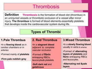
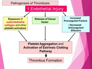
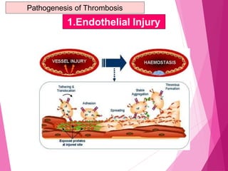
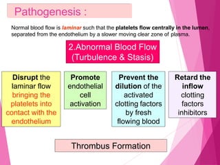
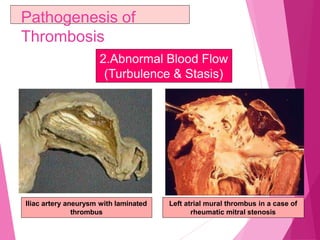
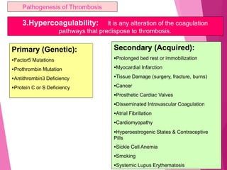
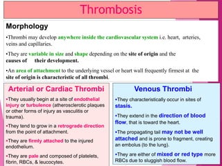
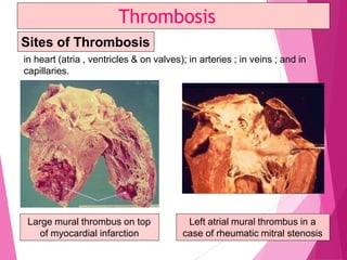
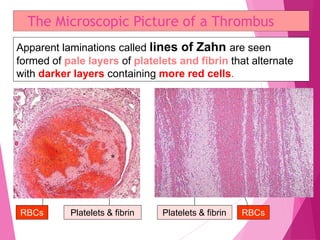
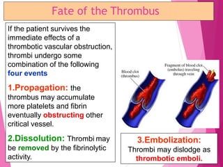
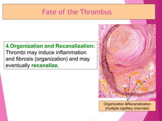
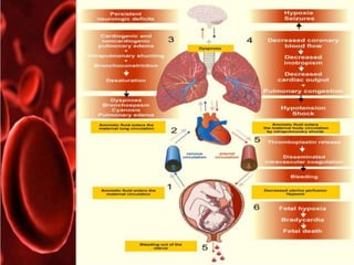
OEDEMA,THROMBOSIS
AND EMBOLISM
CONTENTS:
 INTRODUCTION
 OEDEMA
DEFINITION
TYPES
PATHOGENESIS
 THROMBOSIS
DEFINITION
TYPES
PATHOGENESIS
MORPHOLOGY
MICROSCOPIC PICTURE
FATE
 EMBOLISM
TYPES ,
CAUSES
 CONCLUSION
 REFERENCES
 PREVIOUS YEAR QUESTIONS
CCiircurcullaattooryryDDiissttuurbrbaanncecess
((DisDisttuurrbbaanncecessooffBBloloododaandndBBoodydy
FFlluuidids)s)The health of cells and tissues depends not only on an intact circulation to
deliver oxygen and remove wastes but also on normal fluid balance.
Normal fluid homeostasis includes maintenance of vessel wall integrity
(intact circulation) as well as intravascular pressure, blood volume, and protein
content (osmolarity) within certain physiologic ranges. Therefore any change in
one of these factors will affect the tissue homeostasis and may result in oedema or
congestion.
Normal fluid homeostasis also means maintaining blood as a liquid until
such time as injury necessitates clot formation. Clotting at inappropriate sites
(thrombosis) or migration of clots (embolism) obstructs blood flow to tissues &
leads to cell death (infarction).
Conversely inability to clot after injury results in haemorrhage. Extensive
haemorrhage can result in shock.
INTRODUCTION
HOMEOSTASIS
• The mechanism by which the constancy of
the internal environment is maintained and
ensured is called the homeostasis.
• Claude Bernarde (1949) – internal environment
or milieu interieur
NORMAL FLUID
EXCHANGE;
OEDEMA- DEFINITION
•The greek word ‘oidema’ means swelling.
•Oedema is defined as abnormal and excessive accumulation of
“free fluid ‘’ in the interstititial tissue spaces and serous cavities
-
•Occurs when there is free fluid i.e in body cavities or in
interstitial spaces
-
-
 BASED ON DISTRIBUTION
LOCALIZED
GENERALIZED
 BASED ON FLUID COMPOSITION
TRANSUDATE
EXUDATE
TYPES
PATHOGENESIS
 Decreased plasma oncotic pressure
 Increased capillary hydrostatic pressure
 Lymphatic obstruction
 Tissue factors
 Increased capillary permeability
 Sodium and water retention.
PATHOPHYSIOLOGICAL CATEGORIES OF
EDEMA
A) ↑ed hydrostatic pressure :
• OF CARDIAC DISEASES - CHF,constructive pericarditis
•ASCITIS OF LIVER
•PASSIVE CONGESTION
•POSTURAL
B) ↓ed plasma oncotic pressure:
•Oedema of renal diseases
•Ascitis
•hypoproteinemia
c.Lymphatic obstruction:
•Post surgical eg:following radical mastectomy
•Pressure due to tumors
•Inflammatory –elephantiasis
•Occlusion of lymphatic channels via malignant cells
•MILROY’S DISEASE OR HERIDITARY LYMPHOEDEMA
D.TISSUE FACTORS:
•quite small and insignificant
•May occur via elevation of oncotic pressure of IF or lowering tissue
Tension eg: in eye lids and external genitalia
E.Increased capillary permeability:
•An intact endothelium acts as a semi permable membrane .
•When it is injured via ‘capillary poisons ‘ such as toxins ,
histamines etc leads to development of gaps and leakage of
plasma fluids leading to edema
Eg:
Generalized edema
Localized –inflammatory,angioneurotic
F.Mechanisms involved in oedema by sodium and water retention
PATHOGENESIS
Depending on the composition of fluid:
transudate & exudate oedema
TRANSUDATE
Definition Filtrate of blood plasma
without changes in
endothelial permeability
Character Non-inflammatory
oedema
Protein Low (less than 1 gm/dl);
content mainly albumin, low
fibrinogen; hence no
tendency to coagulate
Glucose Same as in plasma
content
EXUDATE
Oedema of inflamed
tissue associated with
increased vascular
permeability
Inflammatory oedema
High ( 2.5-3.5 gm/dl),
readily coagulates due
to high content of
fibrinogen and other
Coagulation factors
Low (less than 60
mg/dl)
TRANSUDATE EXUDATE
Specific gravity Low (less than 1.015) High (more than 1.018)
pH > 7.3 < 7.3
LDH Low High
Effusion LDH/ Serum < 0.6 > 0.6
LDH ratio
Cells Few cells, mainly Many cells, inflammatory
mesothelial cells as well as parenchymal
and cellular debris
Examples Oedema in congestive Purulent exudate such
cardiac failure as pus
SPECIAL FORMS
RENAL OEDEMA
• Generalised oedema occurs in certain
diseases of renal origin- such as in
nephrotic syndrome, some types of
glomerulonephritis, and in renal failure
due to acute tubular injury.
• Initially manifests in tissues with loose
connective tissue matrix - eyelids
• Periorbital edema - characteristic
TYPES
 In nephrotic syndrome
 In nephritic syndrome
 Acute tubular injury
Differences between Nephrotic and Nephritic
Oedema
Feature Nephrotic Nephritic
Cause
Proteinuria
Mechanism
Degree of oedema
Distribution
Nephrotic syndrome
Heavy
↓Plasma oncotic
Pressure and
Na+ and water
retention
Severe, generalised
Subcutaneous tissues
as well as visceral
organs
Glomerulonephritis
(acute, rapidly
progressive)
Mild to Moderate
Na+ and water
retention
Mild
Loose tissues mainly
(face, eyes, ankles,
genitalia)
• OEDEMA IN ACUTE TUBULAR INJURY-
damaged tubules lose their capacity for selective
reabsorption and concentration of glomerular filtrate
resulting in increased reabsorption.
PULMONARY OEDEMA
• CAUSES - left ventricular failure, renal
failure, acute respiratory distress
syndrome and pulmonary inflammation or
infection
• CONSEQUENCES - impede oxygen
diffusion- hypoxia - hypercapnia
-favorable environment – bacterial infection
Mechanisms involved in the pathogenesis of pulmonary oedema. A, Normal fluid
exchange at the alveolocapillary membrane (capillary endothelium and alveolar
epithelium). B, Pulmonary oedema via elevated pulmonary hydrostatic pressure. C,
Pulmonary oedema via increased vascular permeability.
CHF EDEMA
• INCREASED VENOUS PRESSURE
DUE TO FAILURE
• DECREASED RENAL PERFUSION,
triggering of RENIN-
ANGIOTENSION-ALDOSTERONE
complex, resulting ultimately in
SODIUM RETENTION
HEPATIC OEDEMA
• i) Hypoproteinaemia - impaired synthesis of proteins
• ii) Portal hypertension - increased venous pressure in
the abdomen - raised hydrostatic pressure.
• iii) Failure of inactivation of aldosterone
-hyperaldosteronism.
• iv) Secondary stimulation of RAAS- sodium and water
retention.
CEREBRAL OEDEMA
• Brain edema -localized or generalized - nature extent
-pathologic process or injury. 3 types-
• VASOGENIC OEDEMA : increased filtration pressure or
increased capillary permeability
• CYTOTOXIC OEDEMA : disturbance in the cellular
osmoregulation - response to cell injury
• INTERSTITIAL OEDEMA : hydrocephalus
MISCELLANEOUS
• Nutritional Oedema-
• Due to nutritional deficiency of Proteins (Kwashiorkor, prolonged
starvation, famine, fasting), Vitamins (beri-beri due to vitamin B1
deficiency) and Chronic alcoholism
• Main contributing factors- Hypoproteinaemia & Sodium-water
retention
• Myxoedema-
• Hypothyroidism -non pitting oedema occuring on skin of face and
internal organs due to excessive deposition of glycosaminoglycans
in the interstitium
• Microscopically -basophilic mucopolysaccharides.
Thrombosis
Definition Thrombosis is the formation of blood clot (thrombus) in
an uninjuried vessels or thrombotic occlusion of a vessel after minor
injury. The thrombus is formed of blood elements essentially platelets
that develops inside the cardiovascular system during life.
Types of Thrombi
1.Pale Thrombus
•In a flowing blood as in
cardiac chambers or in
arteries
•Formed mainly of platelets
•Firm pale reddish grey
2. Red Thrombus
•In a stagnant blood
adjacent to complete
vascular occlusion
•Formed of fibrin
entrapping RBCs,
leucocytes& platelets
•Soft dark red and
gelatinous
3.Mixed Thrombus
•In a slowly flowing blood
usually in veins & arteries
•Formed of alternating
layers of platelets and
fibrin entrapping RBCs
and leucocytes.
•Alternating red &pale
layers
Thrombosis
Pathogenesis (Causes= Predisposing Factors)
Three primary influences predispose to thrombus formation called Virchow’s triad
Thrombosis
Endothelial Injury
Abnormal Blood Flow Hypercoagulability
Myocardial infarction or
valvulitis (cardiac chambers),
atherosclerosis, vasculitis,
hypertension, smoking,
radiation, chemical irritation
(arteries), prolonged recumbency &
inflammation (veins)
chambers)
Turbulence (arteries) Stasis (veins)
&(cardiac
Atherosclerosis, aneurysm,
dilated atria, atrial fibrillation,
venous stasis, varicose veins
Primary causes (Genetic)
Secondary Causes (Acquired):
High Risk Causes & Low Risk
Causes
1.Endothelial Injury
Exposure of
subendothelial
collagen and other
platelet activators
Release of tissue
factor
• Increased
Procoagulant Factors
• Decreased
Anticoagulant
Effectors
Platelet Aggregation and
Activation of Extrinsic Clotting
Pathway
Thrombus Formation
Pathogenesis of Thrombosis
1.Endothelial Injury
2.Abnormal Blood Flow
(Turbulence & Stasis)
Pathogenesis :
Normal blood flow is laminar such that the platelets flow centrally in the lumen,
separated from the endothelium by a slower moving clear zone of plasma.
Disrupt the
laminar flow
bringing the
platelets into
contact with the
endothelium
Prevent the
dilution of the
activated
clotting factors
by fresh
flowing blood
Retard the
inflow
clotting
factors
inhibitors
Promote
endothelial
cell
activation
Thrombus Formation
Pathogenesis of
Thrombosis
2.Abnormal Blood Flow
(Turbulence & Stasis)
Left atrial mural thrombus in a case of
rheumatic mitral stenosis
Iliac artery aneurysm with laminated
thrombus
Pathogenesis of Thrombosis
3.Hypercoagulability: It is any alteration of the coagulation
pathways that predispose to thrombosis.
Primary (Genetic):
Factor5 Mutations
Prothrombin Mutation
Antithrombin3 Deficiency
Protein C or S Deficiency
Secondary (Acquired):
Prolonged bed rest or immobilization
Myocardial Infarction
Tissue Damage (surgery, fracture, burns)
Cancer
Prosthetic Cardiac Valves
Disseminated Intravascular Coagulation
Atrial Fibrillation
Cardiomyopathy
Hyperoestrogenic States & Contraceptive
Pills
Sickle Cell Anemia
Smoking
Systemic Lupus Erythematosis
Thrombosis
Morphology
•Thrombi may develop anywhere inside the cardiovascular system i.e. heart, arteries,
veins and capillaries.
•They are variable in size and shape depending on the site of origin and the
causes of their development.
•An area of attachment to the underlying vessel or heart wall frequently firmest at the
site of origin is characteristic of all thrombi.
Arterial or Cardiac Thrombi
They usually begin at a site of endothelial
injury or turbulence (atherosclerotic plaques
or other forms of injury as vasculitis or
trauma).
They tend to grow in a retrograde direction
from the point of attachment.
They are firmly attached to the injured
endothelium.
They are pale and composed of platelets,
fibrin, RBCs, & leucocytes.
Venous Thrombi
They characteristically occur in sites of
stasis.
They extend in the direction of blood
flow, that is toward the heart.
The propagating tail may not be well
attached and is prone to fragment, creating
an embolus (to the lung).
They are either of mixed or red type more
RBCs due to sluggish blood flow.
Venous Thrombosis
It commonly occurs in veins of
lower limbs following operations,
congestive heart failure, delivery,
and severe injury due to:
Slowing of circulation as a result
of lack of muscular activity
(stasis).
Damage to the intima by
pressure on calf muscles in
recumbency (endothelial injury&
change in blood flow).
Increased number and
adhesiveness of the platelets
(hypercoagulability).
Phlebothrombosis Thrombophlebitis
It results from damage of endothelium
due to inflammation of the vein, either
bacterial or nonbacterial:
Septic or bacterial thrombophlebitis
The thrombus is invaded by
microorganisms from the vessel wall,
then become fragmented and circulate
in blood steam as septic emboli to form
pyaemic abscesses wherever they settle.
Nonseptic (nonbacterial)
thrombophlebitis which is induced by
ionizing radiation or chemicals.
Venous Thrombosis
3.Hypercoagulability: It is any alteration of the coagulation
pathways that predispose to thrombosis.
A case of deep venous thrombosis
(DVT) in a patient suffering from
systemic lupus erythematosus
Swollen, painful, dusky
red left lower limb
Venous Thrombosis
Due to stasis, hypercoagulability
&endothelial injury
Migratory Thrombophlebitis
(Trousseau’s Syndrome)
Disseminated cancers or certain types of
malignancy as pancreatic carcinoma are
sometimes associated with repeated attacks
of multiple venous thrombosis at different
and changing sites due to the procoagulant
factors formed by cancer cells. This is referred
to as migratory thrombophlebitis or
Trousseau’s syndrome.
Capillary Thrombosis
It is formed mainly of fused RBCs and are seen in some types
of vasculitis and in disseminated intravascular coagulopathy
(DIC).
Disseminated Intravascular Coagulopathy
A variety of disorders ranging from obstetric complications to
advanced malignancy may be complicated by DIC, the sudden
onset of widespread fibrin thrombi in the microcirculation.
With the development of the multiple thrombi, there is rapid
concurrent consumption of platelets & coagulation proteins
(consumption coagulopathy); at the same time the fibrinolytic
mechanisms are activated, and as a result an initially
thrombotic disorder can evolve into a serious bleeding
disorder.
in heart (atria , ventricles & on valves); in arteries ; in veins ; and in
capillaries.
Thrombosis
Large mural thrombus on top
of myocardial infarction
Left atrial mural thrombus in a
case of rheumatic mitral stenosis
Sites of Thrombosis
The Microscopic Picture of a Thrombus
Apparent laminations called lines of Zahn are seen
formed of pale layers of platelets and fibrin that alternate
with darker layers containing more red cells.
RBCs Platelets & fibrin Platelets & fibrin RBCs
Fate of the Thrombus
If the patient survives the
immediate effects of a
thrombotic vascular obstruction,
thrombi undergo some
combination of the following
four events
1.Propagation: the
thrombus may accumulate
more platelets and fibrin
eventually obstructing other
critical vessel.
2.Dissolution: Thrombi may
be removed by the fibrinolytic
activity.
3.Embolization:
Thrombi may dislodge as
thrombotic emboli.
Fate of the Thrombus
4.Organization and Recanalization:
Thrombi may induce inflammation
and fibrosis (organization) and may
eventually recanalize.
Organization &Recanalization
(multiple capillary channels)
To conclude
REFERENCES:
•Essential pathology for dental students –Harsh mohan
•Robbin’s basic pathology
•Web references and images
PREVIOUS YEAR QUESTIONS:
•SHORT ESSAY
HEMOTHROMBOSIS(RGUHS 2010)
4.oedema,thrombosis,embolism

4.oedema,thrombosis,embolism

  • 2.
  • 3.
    CONTENTS:  INTRODUCTION  OEDEMA DEFINITION TYPES PATHOGENESIS THROMBOSIS DEFINITION TYPES PATHOGENESIS MORPHOLOGY
  • 4.
    MICROSCOPIC PICTURE FATE  EMBOLISM TYPES, CAUSES  CONCLUSION  REFERENCES  PREVIOUS YEAR QUESTIONS
  • 5.
    CCiircurcullaattooryryDDiissttuurbrbaanncecess ((DisDisttuurrbbaanncecessooffBBloloododaandndBBoodydy FFlluuidids)s)The health ofcells and tissues depends not only on an intact circulation to deliver oxygen and remove wastes but also on normal fluid balance. Normal fluid homeostasis includes maintenance of vessel wall integrity (intact circulation) as well as intravascular pressure, blood volume, and protein content (osmolarity) within certain physiologic ranges. Therefore any change in one of these factors will affect the tissue homeostasis and may result in oedema or congestion. Normal fluid homeostasis also means maintaining blood as a liquid until such time as injury necessitates clot formation. Clotting at inappropriate sites (thrombosis) or migration of clots (embolism) obstructs blood flow to tissues & leads to cell death (infarction). Conversely inability to clot after injury results in haemorrhage. Extensive haemorrhage can result in shock. INTRODUCTION
  • 6.
    HOMEOSTASIS • The mechanismby which the constancy of the internal environment is maintained and ensured is called the homeostasis. • Claude Bernarde (1949) – internal environment or milieu interieur
  • 8.
  • 9.
    OEDEMA- DEFINITION •The greekword ‘oidema’ means swelling. •Oedema is defined as abnormal and excessive accumulation of “free fluid ‘’ in the interstititial tissue spaces and serous cavities - •Occurs when there is free fluid i.e in body cavities or in interstitial spaces - -
  • 10.
     BASED ONDISTRIBUTION LOCALIZED GENERALIZED  BASED ON FLUID COMPOSITION TRANSUDATE EXUDATE TYPES
  • 11.
    PATHOGENESIS  Decreased plasmaoncotic pressure  Increased capillary hydrostatic pressure  Lymphatic obstruction  Tissue factors  Increased capillary permeability  Sodium and water retention.
  • 12.
    PATHOPHYSIOLOGICAL CATEGORIES OF EDEMA A)↑ed hydrostatic pressure : • OF CARDIAC DISEASES - CHF,constructive pericarditis •ASCITIS OF LIVER •PASSIVE CONGESTION •POSTURAL B) ↓ed plasma oncotic pressure: •Oedema of renal diseases •Ascitis •hypoproteinemia
  • 13.
    c.Lymphatic obstruction: •Post surgicaleg:following radical mastectomy •Pressure due to tumors •Inflammatory –elephantiasis •Occlusion of lymphatic channels via malignant cells •MILROY’S DISEASE OR HERIDITARY LYMPHOEDEMA D.TISSUE FACTORS: •quite small and insignificant •May occur via elevation of oncotic pressure of IF or lowering tissue Tension eg: in eye lids and external genitalia
  • 14.
    E.Increased capillary permeability: •Anintact endothelium acts as a semi permable membrane . •When it is injured via ‘capillary poisons ‘ such as toxins , histamines etc leads to development of gaps and leakage of plasma fluids leading to edema Eg: Generalized edema Localized –inflammatory,angioneurotic
  • 15.
    F.Mechanisms involved inoedema by sodium and water retention
  • 16.
  • 17.
    Depending on thecomposition of fluid: transudate & exudate oedema TRANSUDATE Definition Filtrate of blood plasma without changes in endothelial permeability Character Non-inflammatory oedema Protein Low (less than 1 gm/dl); content mainly albumin, low fibrinogen; hence no tendency to coagulate Glucose Same as in plasma content EXUDATE Oedema of inflamed tissue associated with increased vascular permeability Inflammatory oedema High ( 2.5-3.5 gm/dl), readily coagulates due to high content of fibrinogen and other Coagulation factors Low (less than 60 mg/dl)
  • 18.
    TRANSUDATE EXUDATE Specific gravityLow (less than 1.015) High (more than 1.018) pH > 7.3 < 7.3 LDH Low High Effusion LDH/ Serum < 0.6 > 0.6 LDH ratio Cells Few cells, mainly Many cells, inflammatory mesothelial cells as well as parenchymal and cellular debris Examples Oedema in congestive Purulent exudate such cardiac failure as pus
  • 19.
  • 20.
    RENAL OEDEMA • Generalisedoedema occurs in certain diseases of renal origin- such as in nephrotic syndrome, some types of glomerulonephritis, and in renal failure due to acute tubular injury. • Initially manifests in tissues with loose connective tissue matrix - eyelids • Periorbital edema - characteristic
  • 21.
    TYPES  In nephroticsyndrome  In nephritic syndrome  Acute tubular injury
  • 22.
    Differences between Nephroticand Nephritic Oedema Feature Nephrotic Nephritic Cause Proteinuria Mechanism Degree of oedema Distribution Nephrotic syndrome Heavy ↓Plasma oncotic Pressure and Na+ and water retention Severe, generalised Subcutaneous tissues as well as visceral organs Glomerulonephritis (acute, rapidly progressive) Mild to Moderate Na+ and water retention Mild Loose tissues mainly (face, eyes, ankles, genitalia)
  • 23.
    • OEDEMA INACUTE TUBULAR INJURY- damaged tubules lose their capacity for selective reabsorption and concentration of glomerular filtrate resulting in increased reabsorption.
  • 24.
    PULMONARY OEDEMA • CAUSES- left ventricular failure, renal failure, acute respiratory distress syndrome and pulmonary inflammation or infection • CONSEQUENCES - impede oxygen diffusion- hypoxia - hypercapnia -favorable environment – bacterial infection
  • 25.
    Mechanisms involved inthe pathogenesis of pulmonary oedema. A, Normal fluid exchange at the alveolocapillary membrane (capillary endothelium and alveolar epithelium). B, Pulmonary oedema via elevated pulmonary hydrostatic pressure. C, Pulmonary oedema via increased vascular permeability.
  • 26.
    CHF EDEMA • INCREASEDVENOUS PRESSURE DUE TO FAILURE • DECREASED RENAL PERFUSION, triggering of RENIN- ANGIOTENSION-ALDOSTERONE complex, resulting ultimately in SODIUM RETENTION
  • 28.
    HEPATIC OEDEMA • i)Hypoproteinaemia - impaired synthesis of proteins • ii) Portal hypertension - increased venous pressure in the abdomen - raised hydrostatic pressure. • iii) Failure of inactivation of aldosterone -hyperaldosteronism. • iv) Secondary stimulation of RAAS- sodium and water retention.
  • 29.
    CEREBRAL OEDEMA • Brainedema -localized or generalized - nature extent -pathologic process or injury. 3 types- • VASOGENIC OEDEMA : increased filtration pressure or increased capillary permeability • CYTOTOXIC OEDEMA : disturbance in the cellular osmoregulation - response to cell injury • INTERSTITIAL OEDEMA : hydrocephalus
  • 30.
    MISCELLANEOUS • Nutritional Oedema- •Due to nutritional deficiency of Proteins (Kwashiorkor, prolonged starvation, famine, fasting), Vitamins (beri-beri due to vitamin B1 deficiency) and Chronic alcoholism • Main contributing factors- Hypoproteinaemia & Sodium-water retention • Myxoedema- • Hypothyroidism -non pitting oedema occuring on skin of face and internal organs due to excessive deposition of glycosaminoglycans in the interstitium • Microscopically -basophilic mucopolysaccharides.
  • 31.
    Thrombosis Definition Thrombosis isthe formation of blood clot (thrombus) in an uninjuried vessels or thrombotic occlusion of a vessel after minor injury. The thrombus is formed of blood elements essentially platelets that develops inside the cardiovascular system during life. Types of Thrombi 1.Pale Thrombus •In a flowing blood as in cardiac chambers or in arteries •Formed mainly of platelets •Firm pale reddish grey 2. Red Thrombus •In a stagnant blood adjacent to complete vascular occlusion •Formed of fibrin entrapping RBCs, leucocytes& platelets •Soft dark red and gelatinous 3.Mixed Thrombus •In a slowly flowing blood usually in veins & arteries •Formed of alternating layers of platelets and fibrin entrapping RBCs and leucocytes. •Alternating red &pale layers
  • 32.
    Thrombosis Pathogenesis (Causes= PredisposingFactors) Three primary influences predispose to thrombus formation called Virchow’s triad Thrombosis Endothelial Injury Abnormal Blood Flow Hypercoagulability Myocardial infarction or valvulitis (cardiac chambers), atherosclerosis, vasculitis, hypertension, smoking, radiation, chemical irritation (arteries), prolonged recumbency & inflammation (veins) chambers) Turbulence (arteries) Stasis (veins) &(cardiac Atherosclerosis, aneurysm, dilated atria, atrial fibrillation, venous stasis, varicose veins Primary causes (Genetic) Secondary Causes (Acquired): High Risk Causes & Low Risk Causes
  • 33.
    1.Endothelial Injury Exposure of subendothelial collagenand other platelet activators Release of tissue factor • Increased Procoagulant Factors • Decreased Anticoagulant Effectors Platelet Aggregation and Activation of Extrinsic Clotting Pathway Thrombus Formation Pathogenesis of Thrombosis
  • 34.
  • 35.
    2.Abnormal Blood Flow (Turbulence& Stasis) Pathogenesis : Normal blood flow is laminar such that the platelets flow centrally in the lumen, separated from the endothelium by a slower moving clear zone of plasma. Disrupt the laminar flow bringing the platelets into contact with the endothelium Prevent the dilution of the activated clotting factors by fresh flowing blood Retard the inflow clotting factors inhibitors Promote endothelial cell activation Thrombus Formation
  • 36.
    Pathogenesis of Thrombosis 2.Abnormal BloodFlow (Turbulence & Stasis) Left atrial mural thrombus in a case of rheumatic mitral stenosis Iliac artery aneurysm with laminated thrombus
  • 37.
    Pathogenesis of Thrombosis 3.Hypercoagulability:It is any alteration of the coagulation pathways that predispose to thrombosis. Primary (Genetic): Factor5 Mutations Prothrombin Mutation Antithrombin3 Deficiency Protein C or S Deficiency Secondary (Acquired): Prolonged bed rest or immobilization Myocardial Infarction Tissue Damage (surgery, fracture, burns) Cancer Prosthetic Cardiac Valves Disseminated Intravascular Coagulation Atrial Fibrillation Cardiomyopathy Hyperoestrogenic States & Contraceptive Pills Sickle Cell Anemia Smoking Systemic Lupus Erythematosis
  • 38.
    Thrombosis Morphology •Thrombi may developanywhere inside the cardiovascular system i.e. heart, arteries, veins and capillaries. •They are variable in size and shape depending on the site of origin and the causes of their development. •An area of attachment to the underlying vessel or heart wall frequently firmest at the site of origin is characteristic of all thrombi. Arterial or Cardiac Thrombi They usually begin at a site of endothelial injury or turbulence (atherosclerotic plaques or other forms of injury as vasculitis or trauma). They tend to grow in a retrograde direction from the point of attachment. They are firmly attached to the injured endothelium. They are pale and composed of platelets, fibrin, RBCs, & leucocytes. Venous Thrombi They characteristically occur in sites of stasis. They extend in the direction of blood flow, that is toward the heart. The propagating tail may not be well attached and is prone to fragment, creating an embolus (to the lung). They are either of mixed or red type more RBCs due to sluggish blood flow.
  • 39.
    Venous Thrombosis It commonlyoccurs in veins of lower limbs following operations, congestive heart failure, delivery, and severe injury due to: Slowing of circulation as a result of lack of muscular activity (stasis). Damage to the intima by pressure on calf muscles in recumbency (endothelial injury& change in blood flow). Increased number and adhesiveness of the platelets (hypercoagulability). Phlebothrombosis Thrombophlebitis It results from damage of endothelium due to inflammation of the vein, either bacterial or nonbacterial: Septic or bacterial thrombophlebitis The thrombus is invaded by microorganisms from the vessel wall, then become fragmented and circulate in blood steam as septic emboli to form pyaemic abscesses wherever they settle. Nonseptic (nonbacterial) thrombophlebitis which is induced by ionizing radiation or chemicals.
  • 40.
    Venous Thrombosis 3.Hypercoagulability: Itis any alteration of the coagulation pathways that predispose to thrombosis. A case of deep venous thrombosis (DVT) in a patient suffering from systemic lupus erythematosus Swollen, painful, dusky red left lower limb
  • 41.
    Venous Thrombosis Due tostasis, hypercoagulability &endothelial injury
  • 42.
    Migratory Thrombophlebitis (Trousseau’s Syndrome) Disseminatedcancers or certain types of malignancy as pancreatic carcinoma are sometimes associated with repeated attacks of multiple venous thrombosis at different and changing sites due to the procoagulant factors formed by cancer cells. This is referred to as migratory thrombophlebitis or Trousseau’s syndrome.
  • 43.
    Capillary Thrombosis It isformed mainly of fused RBCs and are seen in some types of vasculitis and in disseminated intravascular coagulopathy (DIC). Disseminated Intravascular Coagulopathy A variety of disorders ranging from obstetric complications to advanced malignancy may be complicated by DIC, the sudden onset of widespread fibrin thrombi in the microcirculation. With the development of the multiple thrombi, there is rapid concurrent consumption of platelets & coagulation proteins (consumption coagulopathy); at the same time the fibrinolytic mechanisms are activated, and as a result an initially thrombotic disorder can evolve into a serious bleeding disorder.
  • 44.
    in heart (atria, ventricles & on valves); in arteries ; in veins ; and in capillaries. Thrombosis Large mural thrombus on top of myocardial infarction Left atrial mural thrombus in a case of rheumatic mitral stenosis Sites of Thrombosis
  • 45.
    The Microscopic Pictureof a Thrombus Apparent laminations called lines of Zahn are seen formed of pale layers of platelets and fibrin that alternate with darker layers containing more red cells. RBCs Platelets & fibrin Platelets & fibrin RBCs
  • 46.
    Fate of theThrombus If the patient survives the immediate effects of a thrombotic vascular obstruction, thrombi undergo some combination of the following four events 1.Propagation: the thrombus may accumulate more platelets and fibrin eventually obstructing other critical vessel. 2.Dissolution: Thrombi may be removed by the fibrinolytic activity. 3.Embolization: Thrombi may dislodge as thrombotic emboli.
  • 47.
    Fate of theThrombus 4.Organization and Recanalization: Thrombi may induce inflammation and fibrosis (organization) and may eventually recanalize. Organization &Recanalization (multiple capillary channels)
  • 62.
  • 63.
    REFERENCES: •Essential pathology fordental students –Harsh mohan •Robbin’s basic pathology •Web references and images
  • 64.
    PREVIOUS YEAR QUESTIONS: •SHORTESSAY HEMOTHROMBOSIS(RGUHS 2010)