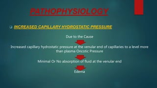
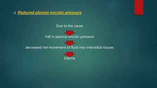
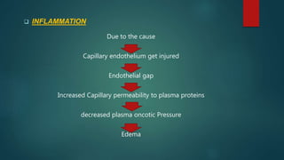
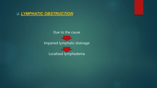
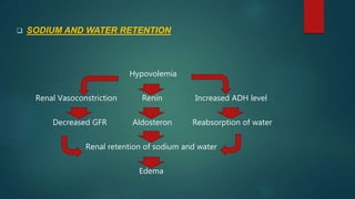
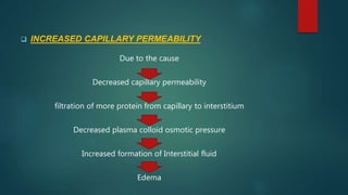
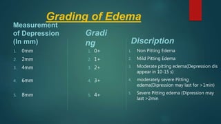
EDEMA
AHAMMED NASEEM
BSc Nursing Student
Introduction
 EDEMA is a normal response of body to inflammation or injury
(For example; a twisted angle, a bee sting or a skin infection this
may result in edema in the involved area)
 In some cases such as infection this may be bacterial. Increased
fluid from the blood vessels allows more infection-fighting with
blood cells to enter the affected area
DEFINITION
 Edema is defined as "the accumulation of excessive
body fluids in the interstitial space or serous body
cavity, which is a pathogenic process caused by
disease rather than a disease entry "
ETIOLOGY
 INCREASED CAPILLARY HYDROSTATIC PRESSURE
• Elevated plasma volume
• increased venous pressure (Eg:- congestive heart failure, Venous thrombosis)
• Arteriolar dilation (Eg:- acute inflammatio)
 REDUCED PLASMA ONCOTIC PRESSURE
Plasma albumin content decreased due to;
• Decreased Protein production (Eg:-hepatic cirrhosis, malnutrition)
• Excess loss of protein (Eg :- Nephrosis)
• Elevated Catabolism of protein (Eg:-Chronic debilitating disease such as malignant
tumors)
 Inflammation
Capillary endothelium injured from;
• Toxins
• Histamine
• Anoxia
• Drugs
 Lymphatic Obstruction
• Blockage by cancer
• Blockage by infection (especially with filariasis)
• Pressure on main lymphatic duct
• Inflammation of lymphatics
 Sodium & Water Retention
 Increased Capillary permeability
• infection
• Burns
• Allergic response
• Trauma
• Anoxia
• Acidosis
• Inflammation
CLASSIFICATION
 Depending upon site of edema;
 Generalized Edema:-
• It is a condition in which notice that swelling, puffiness and water retention
in various body parts including abdomen arms legs face & feel
• Symptoms:- Bloating, tightening of skin & skin that hold an imprint or
looks pitted
• Conditions:- congestive heart failure, Nephrotic syndrome, Cirrhosis of
liver, Systematic infection
 Localised Edema:-
• It is a type of edema confined to a particular part or organ of the body
• Conditions:- Trauma, Infection, Lymphatic and venous obstruction
 Edema can also classified as:
 Putting Edema/Cutaneous edema
• It is a condition in which after a pressure applied on a small area of edema the
indentation persist after release of pressure
• Causes:- water retention, systematic disease, pregnancy, heart failure, vericose
vein, Thrombophlebitis, insect bite & Dermatitis can also cause the pitting
edema
 Non Pitting Edema
• In this condition the indentation doesn't persist
• It is associated with conditions such as lymphedema, lipidemia & myxedema
 Other Major Types of Edema
 Peripheral Edema
• It mainly occurs in the legs, feets, ankles
• This is the most common type of edema it cause swelling in lower extremities
• It may caused by increasing age, hypertension, pregnancy, Congestive Heart
Failure, Kidney problems and other health conditions
• It may also experiance when standing/sitting for extended hours
• Some medications like NSAID's (Ibuprofen, Naproxen), Corticosteroids
(Prednisone, Methyl Prednisolone)can also cure
 Pulmonary Edema
• It is the fluid accumulation in the lungs due to the blockage of pulmonary veins.
As blood pressure rises inthe blood vessels of the lungs fluid rush into fill the
lungs
• The pleural cavity can also be filled with fluid in such cases pulmonary edema is
said to also present with pleural Effusion
• It can be caused by malfunction of left ventricle, acute respiratory distress
syndrome & also hypersensitivity reaction
 Cerebral Edema
• It is the accumulation of fluid in the intracellular and extracellular spaces of brain
• It can be cause by metabolic abnormalities due to an underlying diseases Or as a
response to O2 deprivation at high altitude
• It is very serious condition & can also leads to unconsciousness and brain
damage
• It is devided into 3 sub types; Vasogenic, Cytogenic & Interstitial ceribral edema
PATHOPHYSIOLOGY
 INCREASED CAPILLARY HYDROSTATIC PRESSURE
Due to the Cause
Increased capillary hydrostatic pressure at the venular end of capillaries to a level more
than plasma Oncotic Pressure
Minimal Or No absorption of fluid at the venular end
Edema
 Reduced plasma oncotic pressure
Due to the cause
Fall in plasma oncotic pressure
decreased net movement of fluid into interstitial tissues
Edema
 INFLAMMATION
Due to the cause
Capillary endothelium get injured
Endothelial gap
Increased Capillary permeability to plasma proteins
decreased plasma oncotic Pressure
Edema
 LYMPHATIC OBSTRUCTION
Due to the cause
Impaired lymphatic drainage
Localised lymphedema
 SODIUM AND WATER RETENTION
Hypovolemia
Renal Vasoconstriction Renin Increased ADH level
Decreased GFR Aldosteron Reabsorption of water
Renal retention of sodium and water
Edema
 INCREASED CAPILLARY PERMEABILITY
Due to the cause
Decreased capillary permeability
filtration of more protein from capillary to interstitium
Decreased plasma colloid osmotic pressure
Increased formation of Interstitial fluid
Edema
Signs & Symptoms
 Increase in the total body water cause weight gain in short period of time
 Peripheral Edema
 Excess Fluid in the Interstitial space
 Distended Neck and peripheral Veins
 Slow emptying peripheral Veins
 CVP over 11cm
 Crackles and wheezes in lungs
 Poly urea (if renal function is normal)
 Ascites
 Pleural Effusion
 Decreased Blood Urea Nitrogen (BUN) Due to plasma dilution
 Bounding Pulse
 Pulmonary Edema (if severe)
DIAGNOSIS
 Health History
 Diagnostic tests like ;
• monitor serum osmolarity
• Hemoglobin & Hematocrit
• Urine specific gravity and Osmolality
• CVP Reading
 In Physical Examination
• Daily weight
• Lung sounds
• Extremity measurement
• Abdominal Measurement
• grading of edema
Grading of Edema
Measurement
of Depression
(In mm)
1. 0mm
2. 2mm
3. 4mm
4. 6mm
5. 8mm
Gradi
ng
1. 0+
2. 1+
3. 2+
4. 3+
5. 4+
Discription
1. Non Pitting Edema
2. Mild Pitting Edema
3. Moderate pitting edema(Depression dis
appear in 10-15 s)
4. moderately severe Pitting
edema(Dipression may last for >1min)
5. Severe Pitting edema (Dipression may
last >2min
COMPLICATIONS
Untreated edema can leads to ;
 Painful Swelling, Pain that gets worse
 Stiffness and difficulties in walking
 Stretched & itchy skin
 Infection in the area of swelling
 Scarring Between layers of tissue
 Poor blood circulation
 Loss of elasticity in Artery, Veins & Joints
 Ulceration on skin
MEDICAL MANAGEMENT
 Treat the underlying Cause
 Treat the sign and symptoms
 Restrict salt intake
 Provide Oxygen in pulmonary edema
PHARMACOLOGICAL
MANAGEMENT
 Analgesics
 Diuretics
 Antibiotics
NURSING MANAGEMENT
 Assess the presence of worsening edema
 Encourage adherence to Sodium Restriction to avoid over counter drugs
 When indicated encourage rest periods
 monitor patients Response to Diuretics, Check the daily weight
 Monitor rate of parental fluids & Client response
 Teach the self monitoring of weight & I/O measurement (in cases such as
CCF, Renal failure, Cirrhosis of liver)
 Turn the position of patient frequently
 Help the patient to Relax & promote oxygenation
 Place the patient in high Fowler's position to enhance lungs
expansion
 Administer O2 As prescribed
 Assess patients condition frequently
 Auscultate Lung field for breath sounds & be alert for crackles
 Watch for complications of treatment such as electrolyte
depletion
 Monitor ABG results to identify Hypoxemia & hypercapnia

edemabyahammednaseem-210628024050.pdf

  • 1.
  • 2.
    Introduction  EDEMA isa normal response of body to inflammation or injury (For example; a twisted angle, a bee sting or a skin infection this may result in edema in the involved area)  In some cases such as infection this may be bacterial. Increased fluid from the blood vessels allows more infection-fighting with blood cells to enter the affected area
  • 3.
    DEFINITION  Edema isdefined as "the accumulation of excessive body fluids in the interstitial space or serous body cavity, which is a pathogenic process caused by disease rather than a disease entry "
  • 4.
    ETIOLOGY  INCREASED CAPILLARYHYDROSTATIC PRESSURE • Elevated plasma volume • increased venous pressure (Eg:- congestive heart failure, Venous thrombosis) • Arteriolar dilation (Eg:- acute inflammatio)  REDUCED PLASMA ONCOTIC PRESSURE Plasma albumin content decreased due to; • Decreased Protein production (Eg:-hepatic cirrhosis, malnutrition) • Excess loss of protein (Eg :- Nephrosis) • Elevated Catabolism of protein (Eg:-Chronic debilitating disease such as malignant tumors)
  • 5.
     Inflammation Capillary endotheliuminjured from; • Toxins • Histamine • Anoxia • Drugs  Lymphatic Obstruction • Blockage by cancer • Blockage by infection (especially with filariasis) • Pressure on main lymphatic duct • Inflammation of lymphatics  Sodium & Water Retention
  • 6.
     Increased Capillarypermeability • infection • Burns • Allergic response • Trauma • Anoxia • Acidosis • Inflammation
  • 7.
    CLASSIFICATION  Depending uponsite of edema;  Generalized Edema:- • It is a condition in which notice that swelling, puffiness and water retention in various body parts including abdomen arms legs face & feel • Symptoms:- Bloating, tightening of skin & skin that hold an imprint or looks pitted • Conditions:- congestive heart failure, Nephrotic syndrome, Cirrhosis of liver, Systematic infection  Localised Edema:- • It is a type of edema confined to a particular part or organ of the body • Conditions:- Trauma, Infection, Lymphatic and venous obstruction
  • 8.
     Edema canalso classified as:  Putting Edema/Cutaneous edema • It is a condition in which after a pressure applied on a small area of edema the indentation persist after release of pressure • Causes:- water retention, systematic disease, pregnancy, heart failure, vericose vein, Thrombophlebitis, insect bite & Dermatitis can also cause the pitting edema  Non Pitting Edema • In this condition the indentation doesn't persist • It is associated with conditions such as lymphedema, lipidemia & myxedema
  • 9.
     Other MajorTypes of Edema  Peripheral Edema • It mainly occurs in the legs, feets, ankles • This is the most common type of edema it cause swelling in lower extremities • It may caused by increasing age, hypertension, pregnancy, Congestive Heart Failure, Kidney problems and other health conditions • It may also experiance when standing/sitting for extended hours • Some medications like NSAID's (Ibuprofen, Naproxen), Corticosteroids (Prednisone, Methyl Prednisolone)can also cure
  • 10.
     Pulmonary Edema •It is the fluid accumulation in the lungs due to the blockage of pulmonary veins. As blood pressure rises inthe blood vessels of the lungs fluid rush into fill the lungs • The pleural cavity can also be filled with fluid in such cases pulmonary edema is said to also present with pleural Effusion • It can be caused by malfunction of left ventricle, acute respiratory distress syndrome & also hypersensitivity reaction  Cerebral Edema • It is the accumulation of fluid in the intracellular and extracellular spaces of brain • It can be cause by metabolic abnormalities due to an underlying diseases Or as a response to O2 deprivation at high altitude • It is very serious condition & can also leads to unconsciousness and brain damage • It is devided into 3 sub types; Vasogenic, Cytogenic & Interstitial ceribral edema
  • 11.
    PATHOPHYSIOLOGY  INCREASED CAPILLARYHYDROSTATIC PRESSURE Due to the Cause Increased capillary hydrostatic pressure at the venular end of capillaries to a level more than plasma Oncotic Pressure Minimal Or No absorption of fluid at the venular end Edema
  • 12.
     Reduced plasmaoncotic pressure Due to the cause Fall in plasma oncotic pressure decreased net movement of fluid into interstitial tissues Edema
  • 13.
     INFLAMMATION Due tothe cause Capillary endothelium get injured Endothelial gap Increased Capillary permeability to plasma proteins decreased plasma oncotic Pressure Edema
  • 14.
     LYMPHATIC OBSTRUCTION Dueto the cause Impaired lymphatic drainage Localised lymphedema
  • 15.
     SODIUM ANDWATER RETENTION Hypovolemia Renal Vasoconstriction Renin Increased ADH level Decreased GFR Aldosteron Reabsorption of water Renal retention of sodium and water Edema
  • 16.
     INCREASED CAPILLARYPERMEABILITY Due to the cause Decreased capillary permeability filtration of more protein from capillary to interstitium Decreased plasma colloid osmotic pressure Increased formation of Interstitial fluid Edema
  • 17.
    Signs & Symptoms Increase in the total body water cause weight gain in short period of time  Peripheral Edema  Excess Fluid in the Interstitial space  Distended Neck and peripheral Veins  Slow emptying peripheral Veins  CVP over 11cm  Crackles and wheezes in lungs  Poly urea (if renal function is normal)  Ascites  Pleural Effusion  Decreased Blood Urea Nitrogen (BUN) Due to plasma dilution  Bounding Pulse  Pulmonary Edema (if severe)
  • 18.
    DIAGNOSIS  Health History Diagnostic tests like ; • monitor serum osmolarity • Hemoglobin & Hematocrit • Urine specific gravity and Osmolality • CVP Reading  In Physical Examination • Daily weight • Lung sounds • Extremity measurement • Abdominal Measurement • grading of edema
  • 19.
    Grading of Edema Measurement ofDepression (In mm) 1. 0mm 2. 2mm 3. 4mm 4. 6mm 5. 8mm Gradi ng 1. 0+ 2. 1+ 3. 2+ 4. 3+ 5. 4+ Discription 1. Non Pitting Edema 2. Mild Pitting Edema 3. Moderate pitting edema(Depression dis appear in 10-15 s) 4. moderately severe Pitting edema(Dipression may last for >1min) 5. Severe Pitting edema (Dipression may last >2min
  • 20.
    COMPLICATIONS Untreated edema canleads to ;  Painful Swelling, Pain that gets worse  Stiffness and difficulties in walking  Stretched & itchy skin  Infection in the area of swelling  Scarring Between layers of tissue  Poor blood circulation  Loss of elasticity in Artery, Veins & Joints  Ulceration on skin
  • 21.
    MEDICAL MANAGEMENT  Treatthe underlying Cause  Treat the sign and symptoms  Restrict salt intake  Provide Oxygen in pulmonary edema
  • 22.
  • 23.
    NURSING MANAGEMENT  Assessthe presence of worsening edema  Encourage adherence to Sodium Restriction to avoid over counter drugs  When indicated encourage rest periods  monitor patients Response to Diuretics, Check the daily weight  Monitor rate of parental fluids & Client response  Teach the self monitoring of weight & I/O measurement (in cases such as CCF, Renal failure, Cirrhosis of liver)  Turn the position of patient frequently  Help the patient to Relax & promote oxygenation
  • 24.
     Place thepatient in high Fowler's position to enhance lungs expansion  Administer O2 As prescribed  Assess patients condition frequently  Auscultate Lung field for breath sounds & be alert for crackles  Watch for complications of treatment such as electrolyte depletion  Monitor ABG results to identify Hypoxemia & hypercapnia