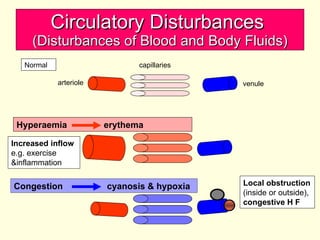
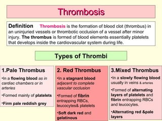
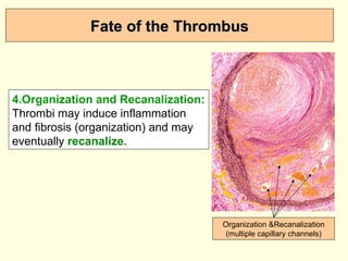
The document discusses circulatory disturbances, specifically hyperaemia, congestion, and thrombosis. It defines hyperaemia as an increase in blood flow to a tissue, which can be active via artery/arteriole dilation or passive via vein/venule engorgement. Congestion is passive hyperaemia. Thrombosis is the formation of a blood clot within a vessel and can be pale, red, or mixed depending on location and flow. Key causes of thrombosis are endothelial injury, abnormal blood flow, and hypercoagulability. Chronic venous congestion can cause organ damage over time due to increased pressure.
The study of the blood flow is called hemodynamics.
Thus hemodynamics deals with the dynamics of blood flow. The circulatory system is controlled by homeostatic mechanisms, much as hydraulic circuits and are controlled by control systems.
Hemodynamic response continuously monitors and adjusts to conditions in the body and its environment. Thus hemodynamics explains the physical laws that govern the flow of blood in the blood vessels.
2. Circulatory Disturbances (Disturbances of Blood and Body Fluids) The health of cells and tissues depends not only on an intact circulation to deliver oxygen and remove wastes but also on normal fluid balance. Normal fluid homeostasis includes maintenance of vessel wall integrity (intact circulation) as well as intravascular pressure , blood volume , and protein content (osmolarity) within certain physiologic ranges . Therefore any change in one of these factors will affect the tissue homeostasis and may result in oedema or congestion . Normal fluid homeostasis also means maintaining blood as a liquid until such time as injury necessitates clot formation. Clotting at inappropriate sites (thrombosis) or migration of clots (embolism) obstructs blood flow to tissues & leads to cell death (infarction). Conversely inability to clot after injury results in haemorrhage . Extensive haemorrhage can result in shock. Circulatory Disturbances (Disturbances of Blood and Body Fluids)
3.
4. Hyperaemia and Congestion Definition : Each can be defined as an increase in blood volume in a particular tissue. Hyperaemia is an increase in blood flow in an organ or tissue due to dilatation of arteries or arterioles i.e. active hyperaemia , while congestion is passive hyperaemia due to engorgement of veins and venules by blood. Active Hyperaemia Physiological Condition e.g. in skeletal &cardiac muscles in muscular exercise, in splanchnic area after eating. Pathological Condition e.g. in acute inflammation (dilatation of arteries &arterioles) The affected tissue is redder because of engorgement with oxygenated blood.
5. Passive Hyperaemia (Venous Congestion) Systemic Venous Congestion In which blood accumulates in veins allover the body as in case of congestive heart failure or sparing the lung in case of right side heart failure Pulmonary Venous Congestion In which blood accumulates in pulmonary veins and their tributaries in case of left side heart failure or mitral stenosis Localized V C It is due to obstruction to blood flow in a limited area or an organ: 1.Acute as in mechanical obstruction or venous thrombosis 2.Chronic by pressure on a vein by a tumour or by constriction of a vein by fibrosis .
6. Passive Hyperaemia (Venous Congestion) In rt. side heart failure: there is systemic venous congestion with sparing of the lung while in congestive heart failure (both sides) there is congestion of all the organs including the lung. In lt. side heart failure: there is pulmonary venous congestion but later it may cause pulmonary hypertension leading to rt. side heart failure i.e. congestive ht. failure.
7. Circulatory Disturbances (Disturbances of Blood and Body Fluids) Normal arteriole venule Increased inflow e.g. exercise &inflammation Hyperaemia erythema Congestion cyanosis & hypoxia Local obstruction (inside or outside), congestive H F capillaries
8.
9. Pathological Features of Chronic Venous Congestion in Various Organs (Liver) 1.Liver Gross Picture: The liver is enlarged , firm and the cut surface shows alternating dark areas of congestion with pale areas of fatty change , giving the liver the nutmeg appearance. Nutmeg cut surface of the liver (dark areas of congestion alternating with pale areas of fatty change)
10. Pathological Features of Chronic Venous Congestion in Various Organs (Liver) Microscopic Picture of Liver in Chronic Venous Congestion 1. The central vein in hepatic lobule is congested as well as the hepatic sinusoids in the central area. 2. The central hepatocytes will show atrophy and necrosis . 3. The mid zonal hepatocytes may show fatty change due to relative hypoxia. 4. The peripheral hepatocytes are normal. Early CVC of liver (congestion of central vein ¢ral hepatic sinusoids)
11. Pathological Features of Chronic Venous Congestion in Various Organs (Liver) Congested central vein & hepatic sinusoids Atrophied central hepatocytes (due to pressure necrosis, hypoxia & anoxia) Fatty change in midzonal hepatocytes (due to relative hypoxia) Normal peripheral hepatocytes (better nourished as they are near the portal v &hepatic a) Hepatic Lobule
12. Pathological Features of Chronic Venous Congestion in Various Organs (Spleen) Chronic venous congestion can cause a form of splenic enlargement referred to as congestive splenomegaly. Causes of Splenic Venous Congestion: 1.Systemic or central venous congestion is encountered in right side heart failure. It produces moderate enlargement of the spleen that rarely exceeds 500gms. 2.Intrahepatic causes that retard portal venous drainage as in various forms of cirrhosis or in bilharzial periportal fibrosis . These cause striking congestive splenomegaly. The weight of the spleen can reach to 5000gms. 3.Extrahepatic disorders that obstruct the portal or splenic veins. A cirrhotic liver
13. Pathological Features of Chronic Venous Congestion in Various Organs (Spleen) 2.Spleen Gross Picture: It is moderately to markedly enlarged, firm with deep red to grey red cut surface (depending on the degree of fibrosis) . The capsule is thickened . Microscopic Picture: The sinusoids of red pulp are dilated and engorged with blood &their walls are thickened by fibrous tissue. Haemorrhage may occur that will result in liberation of haemosiderin granules from haemolysed RBCs that will undergo fibrosis with formation of fibrosiderotic nodules called Gamma-Gandy nodules . The white pulp (lymphoid follicles) is first hyperplastic then later becomes atrophied due to pressure by areas of hge & fibrosis.
14. Pathological Features of Chronic Venous Congestion in Various Organs (Lung) 3.Lung Gross Picture: The lungs are enlarged, heavy, firm and deep red in colour (brown induration). Frothy blood oozes from the cut surface on squeezing. Microscopic Exam.: The alveolar septa are thickened by congested dilated capillaries and oedema fluid followed later by fibrosis. As a result of microhaemorrhages, the alveoli contain oedema fluid, RBCs either intact or haemolysed and haemosiderin laden macrophage (heart failure cells). Congested alveolar wall heart failure cells Thickened alveolar septa (fibrosis)
15. Pathological Features of Chronic Venous Congestion in Various Organs (Kidney) 4.Kidney Gross Picture: The kidneys are slightly enlarged, firm with congested cut surface. Microscopic Picture: the convoluted tubule cells show cloudy change or fatty change due to hypoxia. 5. Other organs as brain, adrenals stomach and intestines also show congestion.
20. 2.Abnormal Blood Flow (Turbulence & Stasis) Pathogenesis of Thrombosis Normal blood flow is laminar such that the platelets flow centrally in the lumen , separated from the endothelium by a slower moving clear zone of plasma. Disrupt the laminar flow bringing the platelets into contact with the endothelium Prevent the dilution of the activated clotting factors by fresh flowing blood Retard the inflow clotting factors inhibitors Promote endothelial cell activation Thrombus Formation
21. Pathogenesis of Thrombosis 2.Abnormal Blood Flow (Turbulence & Stasis) Left atrial mural thrombus in a case of rheumatic mitral stenosis Iliac artery aneurysm with laminated thrombus
26. Venous Thrombosis 3.Hypercoagulability: It is any alteration of the coagulation pathways that predispose to thrombosis. A case of deep venous thrombosis (DVT) in a patient suffering from systemic lupus erythematosus Swollen, painful, dusky red left lower limb
27. Migratory Thrombophlebitis (Trousseau’s Syndrome) Disseminated cancers or certain types of malignancy as pancreatic carcinoma are sometimes associated with repeated attacks of multiple venous thrombosis at different and changing sites due to the procoagulant factors formed by cancer cells. This is referred to as migratory thrombophlebitis or Trousseau’s syndrome.
28. Capillary Thrombosis It is formed mainly of fused RBCs and are seen in some types of vasculitis and in disseminated intravascular coagulopathy (DIC). Disseminated Intravascular Coagulopathy A variety of disorders ranging from obstetric complications to advanced malignancy may be complicated by DIC, the sudden onset of widespread fibrin thrombi in the microcirculation. With the development of the multiple thrombi, there is rapid concurrent consumption of platelets & coagulation proteins (consumption coagulopathy); at the same time the fibrinolytic mechanisms are activated , and as a result an initially thrombotic disorder can evolve into a serious bleeding disorder.
29. in heart (atria , ventricles & on valves); in arteries ; in veins ; and in capillaries. Thrombosis Large mural thrombus on top of myocardial infarction Left atrial mural thrombus in a case of rheumatic mitral stenosis Sites of Thrombosis
32. The Microscopic Picture of a Thrombus Apparent laminations called lines of Zahn are seen formed of pale layers of platelets and fibrin that alternate with darker layers containing more red cells . RBCs Platelets & fibrin Platelets & fibrin RBCs
33. Fate of the Thrombus If the patient survives the immediate effects of a thrombotic vascular obstruction, thrombi undergo some combination of the following four events 1.Propagation: the thrombus may accumulate more platelets and fibrin eventually obstructing other critical vessel. 2.Dissolution: Thrombi may be removed by the fibrinolytic activity. 3.Embolization: Thrombi may dislodge as thrombotic emboli.
34. Fate of the Thrombus 4.Organization and Recanalization: Thrombi may induce inflammation and fibrosis (organization) and may eventually recanalize. Organization &Recanalization (multiple capillary channels)