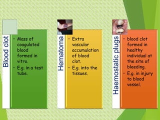
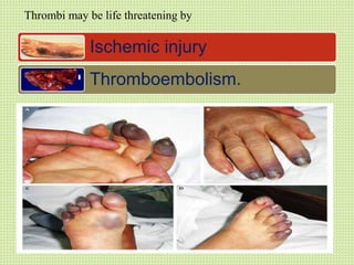
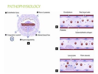
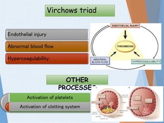
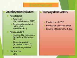
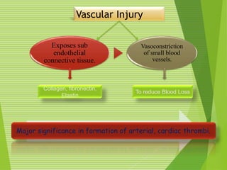
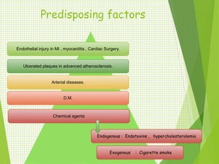
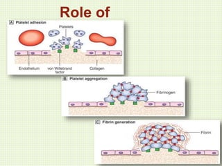
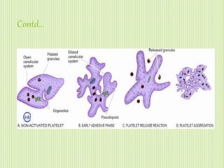
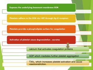
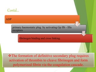
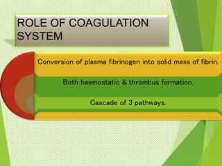
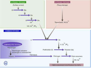
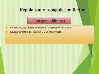
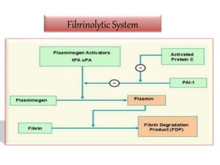
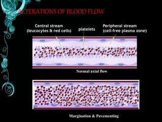
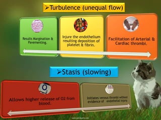
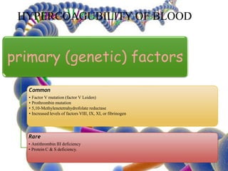
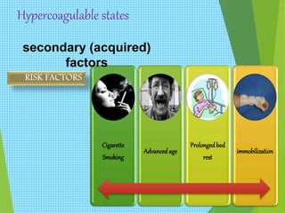
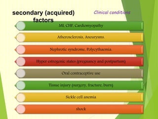
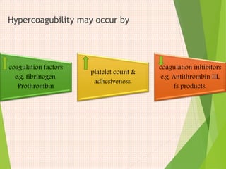
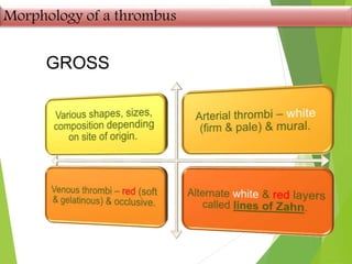
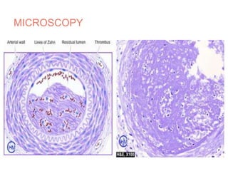
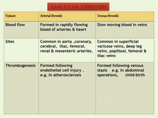
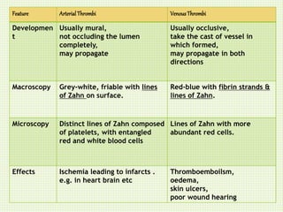
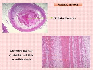
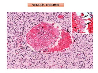
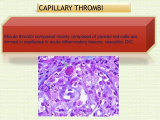
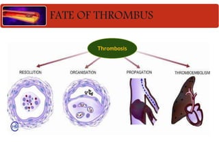
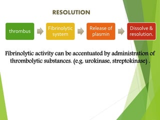
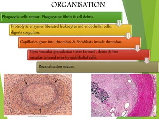
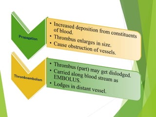
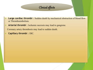
GOOD
AFTERNOON
THROMBOSIS
By ,
Pawan Kumar B
Process of formation of solid mass
in circulation from the constituents
of flowing blood.
The mass is called as THROMBUS.
Bloodclot
• Mass of
coagulated
blood
formed in
vitro.
• E.g. in a test
tube.
Hematoma
• Extra
vascular
accumulation
of blood
clot.
• E.g. into the
tissues.
Haemostaticplugs
• blood clot
formed in
healthy
individual at
the site of
bleeding.
• E.g. in injury
to blood
vessel.
Ischemic injury
Thromboembolism.
Thrombi may be life threatening by
PATHOPHYSIOLOGY
Virchows triad
Endothelial injury
Abnormal blood flow
Hypercoagulability.
OTHER
PROCESSES
Activation of platelets
Activation of clotting system
Endothelium
 Antithrombotic factors
 Antiplatelet
 Adenosine
diphosphatase ( ADP)
 Prostacyclin and nitric
oxide (also
vasodilatation)
 Anticoagulant
 Heparin-like molecules
(activate antithrombin
III)
 Thrombomodulin
(activates protein C)
 Protein S synthesis
 Fibrinolytic
 t-PA
 Procoagulant factors
 Production of vWF
 Production of tissue factor
 Binding of factors IXa & Xa.
Vascular Injury
Major significance in formation of arterial, cardiac thrombi.
Exposes sub
endothelial
connective tissue.
Vasoconstriction
of small blood
vessels.
Collagen, fibronectin,
Elastin
To reduce Blood Loss
Predisposing factors
Endothelial injury in MI , myocarditis , Cardiac Surgery.
Ulcerated plaques in advanced atherosclerosis.
Arterial diseases.
D.M.
Chemical agents
Endogenous : Endotoxins , hypercholesterolemia
Exogenous : Cigarette smoke
Role of
platelets
Contd…
Exposes the underlying basement membrane ECM
Platelets adhere to the ECM via vWF through Gp1B receptors
Platelets provide a phospholipids surface for coagulation
Activation of platelet cause degranulation , secretes
calcium that activates coagulation proteins
ADP which mediates further platelet aggregation
TXA2 which increases platelet activation and cause
vasoconstriction.
Contd…
ADP
primary haemostatic plug by activating Gp IIb - IIIa
receptors
fibrinogen binding and cross linking.
The formation of definitive secondary plug requires
activation of thrombin to cleave fibrinogen and form
polymerized fibrin via the coagulation cascade.
ROLE OF COAGULATION
SYSTEM
Conversion of plasma fibrinogen into solid mass of fibrin.
Both haemostatic & thrombus formation.
Cascade of 3 pathways.
 Act on clotting factors to oppose formation of thrombin.
 e.g.antithrombin III, Protein C , C1 inactivator.
Regulation of coagulation factor
Protease inhibitors
Fibrinolytic System
ALTERATIONS OF BLOOD FLOW
Central stream
(leucocytes & red cells) platelets
Peripheral stream
(cell-free plasma zone)
Normal axial flow
Margination & Pavementing
Turbulence (unequal flow)
Stasis (slowing)
HYPERCOAGUBILITY OF BLOOD
primary (genetic) factors
Common
• Factor V mutation (factor V Leiden)
• Prothrombin mutation
• 5,10-Methylenetetrahydrofolate reductase
• Increased levels of factors VIII, IX, XI, or fibrinogen
Rare
• Antithrombin III deficiency
• Protein C & S deficiency.
Hypercoagulable states
Cigarette
Smoking
Advancedage
Prolongedbed
rest
immobilization
secondary (acquired)
factors
RISK FACTORS
MI, CHF, Cardiomyopathy
Atherosclerosis, Aneurysms.
Nephrotic syndrome, Polycythaemia.
Hyper estrogenic states (pregnancy and postpartum)
Oral contraceptive use
Tissue injury (surgery, fracture, burn)
Sickle cell anemia
shock
secondary (acquired)
factors
Clinical conditions
Hypercoagubility may occur by
coagulation factors
e.g. fibrinogen,
Prothrombin
platelet count &
adhesiveness.
coagulation inhibitors
e.g. Antithrombin III,
fs products.
Morphology of a thrombus
GROSS
MICROSCOPY
ORIGIN OF THROMBI
Common in atrial appendages (right atrium, mitral, aortic valves) –
Infective Endocarditis
Mural & Non-Occlusive.
Ball-valve thrombus : large round thrombus obstructing mitral valve
Agonal thrombi – shortly before death – occur either or both ventricles.
CARDIAC THROMBI
Feature Arterialthrombi Venousthrombi
Blood flow Formed in rapidly flowing
blood of arteries & heart
Slow moving blood in veins
Sites Common in aorta ,coronary,
cerebral, iliac, femoral,
renal & mesenteric arteries.
Common in superficial
varicose veins, deep leg
veins, popliteal, femoral &
iliac veins
Thrombogenesis Formed following
endothelial cell injury .
e.g. in atherosclerosis
Formed following venous
stasis e.g. in abdominal
operations, child-birth
VASCULAR THROMBI
Feature ArterialThrombi VenousThrombi
Developmen
t
Usually mural,
not occluding the lumen
completely,
may propagate
Usually occlusive,
take the cast of vessel in
which formed,
may propagate in both
directions
Macroscopy Grey-white, friable with lines
of Zahn on surface.
Red-blue with fibrin strands &
lines of Zahn.
Microscopy Distinct lines of Zahn composed
of platelets, with entangled
red and white blood cells
Lines of Zahn with more
abundant red cells.
Effects Ischemia leading to infarcts .
e.g. in heart brain etc
Thromboemboilsm,
oedema,
skin ulcers,
poor wound hearing
Occlusive thrombus
Alternating layers of
a) platelets and fibrin
b) red blood cells
ARTERIAL THROMBI
VENOUS THROMBI
CAPILLARY THROMBI
Minute thrombi composed mainly composed of packed red cells are
formed in capillaries in acute inflammatory lesions, vasculitis, DIC.
FATE OF THROMBUS
Thrombosis
thrombus
Fibrinolytic
system
Release of
plasmin
Dissolve &
resolution.
RESOLUTION
Fibrinolytic activity can be accentuated by administration of
thrombolytic substances. (e.g. urokinase, streptokinase) .
ORGANISATION
Phagocytic cells appear. Phagocytose fibrin & cell debris.
Proteolytic enzymes liberated leukocytes and endothelial cells,
digests coagulum.
Capillaries grow into thrombus & fibroblasts invade thrombus.
Fibro vascular granulation tissue formed , dense & less
vascular covered over by endothelial cells.
Recanalisation occurs.
 Large cardiac thrombi : Sudden death by mechanical obstruction of blood flow
or Thromboembolism.
 Arterial thrombi : Ischemic necrosis may lead to gangrene.
Coronary artery thrombosis may lead to sudden death.
 Capillary thrombi : DIC.
Clinical effects
 Venous thrombi (Phlebothrombosis)
Thromboembolism
Oedema
Poor wound healing
Skin ulcer
Painful thrombosed veins (Thrombophlebitis)
Painful white leg (phlegma alba dolens)
THANK YOU… 

Thrombosis - Pathology by Dr.Pawan

  • 1.
  • 2.
  • 3.
    Process of formationof solid mass in circulation from the constituents of flowing blood. The mass is called as THROMBUS.
  • 4.
    Bloodclot • Mass of coagulated blood formedin vitro. • E.g. in a test tube. Hematoma • Extra vascular accumulation of blood clot. • E.g. into the tissues. Haemostaticplugs • blood clot formed in healthy individual at the site of bleeding. • E.g. in injury to blood vessel.
  • 5.
  • 6.
  • 7.
    Virchows triad Endothelial injury Abnormalblood flow Hypercoagulability. OTHER PROCESSES Activation of platelets Activation of clotting system
  • 8.
    Endothelium  Antithrombotic factors Antiplatelet  Adenosine diphosphatase ( ADP)  Prostacyclin and nitric oxide (also vasodilatation)  Anticoagulant  Heparin-like molecules (activate antithrombin III)  Thrombomodulin (activates protein C)  Protein S synthesis  Fibrinolytic  t-PA  Procoagulant factors  Production of vWF  Production of tissue factor  Binding of factors IXa & Xa.
  • 9.
    Vascular Injury Major significancein formation of arterial, cardiac thrombi. Exposes sub endothelial connective tissue. Vasoconstriction of small blood vessels. Collagen, fibronectin, Elastin To reduce Blood Loss
  • 10.
    Predisposing factors Endothelial injuryin MI , myocarditis , Cardiac Surgery. Ulcerated plaques in advanced atherosclerosis. Arterial diseases. D.M. Chemical agents Endogenous : Endotoxins , hypercholesterolemia Exogenous : Cigarette smoke
  • 11.
  • 12.
  • 13.
    Exposes the underlyingbasement membrane ECM Platelets adhere to the ECM via vWF through Gp1B receptors Platelets provide a phospholipids surface for coagulation Activation of platelet cause degranulation , secretes calcium that activates coagulation proteins ADP which mediates further platelet aggregation TXA2 which increases platelet activation and cause vasoconstriction.
  • 14.
    Contd… ADP primary haemostatic plugby activating Gp IIb - IIIa receptors fibrinogen binding and cross linking. The formation of definitive secondary plug requires activation of thrombin to cleave fibrinogen and form polymerized fibrin via the coagulation cascade.
  • 15.
    ROLE OF COAGULATION SYSTEM Conversionof plasma fibrinogen into solid mass of fibrin. Both haemostatic & thrombus formation. Cascade of 3 pathways.
  • 17.
     Act onclotting factors to oppose formation of thrombin.  e.g.antithrombin III, Protein C , C1 inactivator. Regulation of coagulation factor Protease inhibitors
  • 18.
  • 19.
    ALTERATIONS OF BLOODFLOW Central stream (leucocytes & red cells) platelets Peripheral stream (cell-free plasma zone) Normal axial flow Margination & Pavementing
  • 20.
  • 21.
    HYPERCOAGUBILITY OF BLOOD primary(genetic) factors Common • Factor V mutation (factor V Leiden) • Prothrombin mutation • 5,10-Methylenetetrahydrofolate reductase • Increased levels of factors VIII, IX, XI, or fibrinogen Rare • Antithrombin III deficiency • Protein C & S deficiency.
  • 22.
  • 23.
    MI, CHF, Cardiomyopathy Atherosclerosis,Aneurysms. Nephrotic syndrome, Polycythaemia. Hyper estrogenic states (pregnancy and postpartum) Oral contraceptive use Tissue injury (surgery, fracture, burn) Sickle cell anemia shock secondary (acquired) factors Clinical conditions
  • 24.
    Hypercoagubility may occurby coagulation factors e.g. fibrinogen, Prothrombin platelet count & adhesiveness. coagulation inhibitors e.g. Antithrombin III, fs products.
  • 25.
    Morphology of athrombus GROSS
  • 26.
  • 27.
    ORIGIN OF THROMBI Commonin atrial appendages (right atrium, mitral, aortic valves) – Infective Endocarditis Mural & Non-Occlusive. Ball-valve thrombus : large round thrombus obstructing mitral valve Agonal thrombi – shortly before death – occur either or both ventricles. CARDIAC THROMBI
  • 28.
    Feature Arterialthrombi Venousthrombi Bloodflow Formed in rapidly flowing blood of arteries & heart Slow moving blood in veins Sites Common in aorta ,coronary, cerebral, iliac, femoral, renal & mesenteric arteries. Common in superficial varicose veins, deep leg veins, popliteal, femoral & iliac veins Thrombogenesis Formed following endothelial cell injury . e.g. in atherosclerosis Formed following venous stasis e.g. in abdominal operations, child-birth VASCULAR THROMBI
  • 29.
    Feature ArterialThrombi VenousThrombi Developmen t Usuallymural, not occluding the lumen completely, may propagate Usually occlusive, take the cast of vessel in which formed, may propagate in both directions Macroscopy Grey-white, friable with lines of Zahn on surface. Red-blue with fibrin strands & lines of Zahn. Microscopy Distinct lines of Zahn composed of platelets, with entangled red and white blood cells Lines of Zahn with more abundant red cells. Effects Ischemia leading to infarcts . e.g. in heart brain etc Thromboemboilsm, oedema, skin ulcers, poor wound hearing
  • 30.
    Occlusive thrombus Alternating layersof a) platelets and fibrin b) red blood cells ARTERIAL THROMBI
  • 31.
  • 32.
    CAPILLARY THROMBI Minute thrombicomposed mainly composed of packed red cells are formed in capillaries in acute inflammatory lesions, vasculitis, DIC.
  • 33.
  • 34.
    thrombus Fibrinolytic system Release of plasmin Dissolve & resolution. RESOLUTION Fibrinolyticactivity can be accentuated by administration of thrombolytic substances. (e.g. urokinase, streptokinase) .
  • 35.
    ORGANISATION Phagocytic cells appear.Phagocytose fibrin & cell debris. Proteolytic enzymes liberated leukocytes and endothelial cells, digests coagulum. Capillaries grow into thrombus & fibroblasts invade thrombus. Fibro vascular granulation tissue formed , dense & less vascular covered over by endothelial cells. Recanalisation occurs.
  • 38.
     Large cardiacthrombi : Sudden death by mechanical obstruction of blood flow or Thromboembolism.  Arterial thrombi : Ischemic necrosis may lead to gangrene. Coronary artery thrombosis may lead to sudden death.  Capillary thrombi : DIC. Clinical effects
  • 39.
     Venous thrombi(Phlebothrombosis) Thromboembolism Oedema Poor wound healing Skin ulcer Painful thrombosed veins (Thrombophlebitis) Painful white leg (phlegma alba dolens)
  • 40.