DISORDERS OF PERFUSION
Dr. Jyothi Reshma S
Tutor
Dept of Pathology
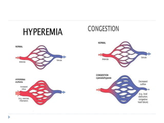
HYPEREMIA, CONGESTION,
HEMORRHAGE
HEMODYNAMICS - 2
ACTIVE HYPEREMIA
 Active hyperemia : increased supply of blood from
the arterial system
 Physiologic response to increased functional demand
 Eg: heart and skeletal muscle during exercise,
inflammation
 Neurogenic and hormonal influences
 Reactive hyperemia - temporary interruption of blood
PASSIVE HYPEREMIA
 Passive hyperemia or congestion : impaired
exit of blood through venous pathways
 Increased hydrostatic pressure - edema.
 Lack of blood flow - chronic hypoxia - ischemic
tissue injury and scarring.
 Capillary rupture - small hemorrhages -
catabolism of extravasated red cells -
hemosiderin-laden macrophages - Chronic
venous congestion (CVC)
Consequences of Impaired Venous
Outflow
Increased
intravascular
pressure
Stasis
HAEMORRHAGE
NECROSIS
CONGESTION
OEDEMA
HYPOXIA
FIBROSIS
Capillary rupture
Acute Chronic
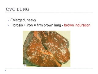
CVC LUNG
 Enlarged, heavy
 Fibrosis + iron = firm brown lung - brown induration
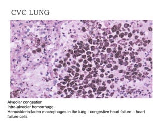
CVC LUNG
Alveolar congestion
Intra-alveolar hemorrhage
Hemosiderin-laden macrophages in the lung - congestive heart failure – heart
failure cells
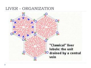
LIVER - ORGANIZATION
CVC LIVER Distension - central
vein and sinusoids
Centrilobular area is
at the distal end of
the hepatic blood
supply - ischemic
necrosis
Periportal
hepatocytes - better
oxygenated
because of
proximity to hepatic
arterioles — fatty
change.
CVC LIVER – NUTMEG LIVER
Dark foci of centrilobular congestion surrounded by paler zones of unaffected
peripheral portions of the lobules
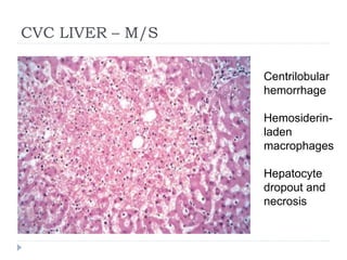
CVC LIVER – M/S
Centrilobular
hemorrhage
Hemosiderin-
laden
macrophages
Hepatocyte
dropout and
necrosis
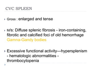
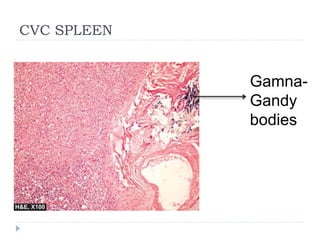
CVC SPLEEN
 Gross : enlarged and tense
 M/s: Diffuse splenic fibrosis - iron-containing,
fibrotic and calcified foci of old hemorrhage
Gamna-Gandy bodies
 Excessive functional activity—hypersplenism
- hematologic abnormalities -
thrombocytopenia
CVC SPLEEN
Gamna-
Gandy
bodies
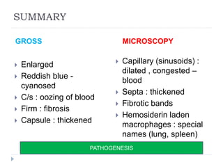
SUMMARY
GROSS MICROSCOPY
 Enlarged
 Reddish blue -
cyanosed
 C/s : oozing of blood
 Firm : fibrosis
 Capsule : thickened
 Capillary (sinusoids) :
dilated , congested –
blood
 Septa : thickened
 Fibrotic bands
 Hemosiderin laden
macrophages : special
names (lung, spleen)
PATHOGENESIS
HEMORRHAGE
Escape of blood
from the
vasculature into
surrounding
tissues, a hollow
organ or body
cavity, or to the
outside
ETIOLOGY, TYPES, CONSEQUENCES
HEMATOMA
 Localized hemorrhage -
within a tissue or organ
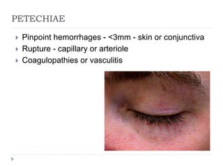
PETECHIAE
 Pinpoint hemorrhages - <3mm - skin or conjunctiva
 Rupture - capillary or arteriole
 Coagulopathies or vasculitis
PURPURA
 Diffuse superficial hemorrhages in the skin
 Up to 1 cm in diameter
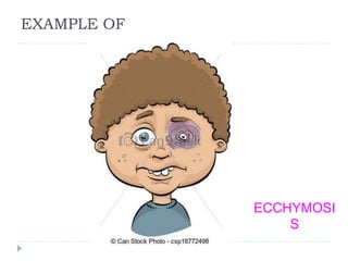
ECCHYMOSIS
 Large superficial hemorrhage in the skin
 > 1cm
 Purple – green – yellow (progressive oxidation of
bilirubin released from the hemoglobin of degraded
erythrocytes)
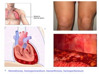
Hemothorax, hemopericardium, hemarthrosis, hemoperitoneum
COMPREHENSION QUESTIONS
 A 40-year-old woman dies after a long history of an illness characterized
by dyspnea, orthopnea, hepatomegaly, distended neck veins, and
peripheral edema. The cut surface of the liver as it appears at autopsy is
shown in the first panel. The second panel shows the microscopic
appearance of the liver. What is the most likely cause of these findings?
 A 36-year-old man dies during cardiac surgery. He
had a history of long-standing rheumatic heart
disease with mitral stenosis. At autopsy, the
pathologist reports findings consistent with mitral
stenosis and noted the presence of “heart failure
cells.” This finding results from
(A) activation of the coagulation cascade.
(B) chronic passive congestion of the lungs.
(C) hypoxic myocardial injury.
(D) myocardial hyperemia.
EXAMPLE OF
ECCHYMOSI
S
T
H
A
N
K
Y
O
U