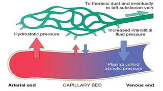
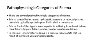
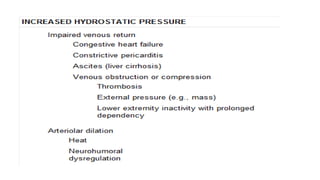
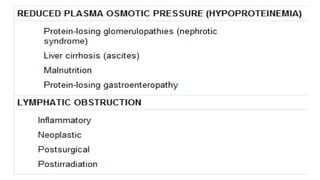
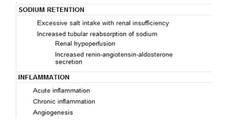
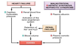
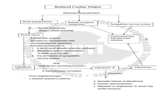
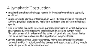
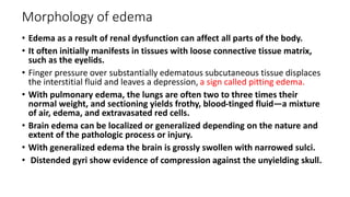
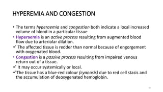
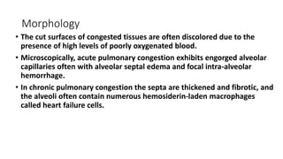
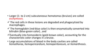
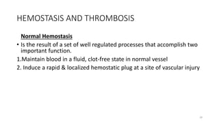
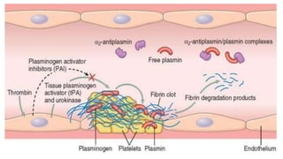
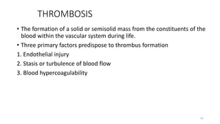
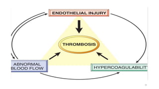
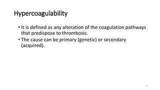
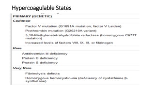
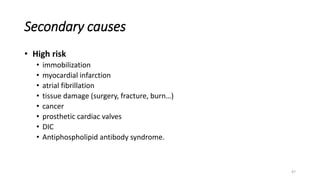
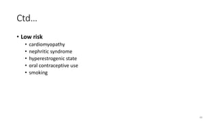
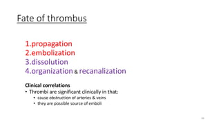
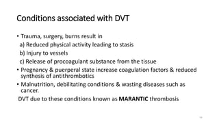
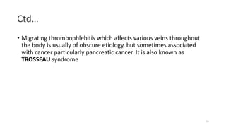
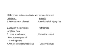
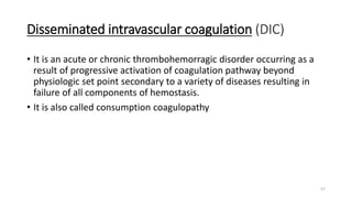
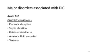
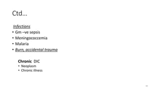
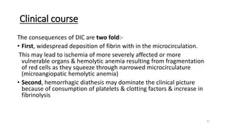
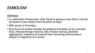
3. • Under normal conditions, as blood passes through capillary beds,
proteins in the plasma are retained within the vasculature and there is
little net movement of water and electrolytes into the tissues.
This balance is often disturbed by pathologic conditions that alter
endothelial function, increase vascular hydrostatic pressure, or
decrease plasma protein content, all of which promote edema—the
accumulation of fluid in tissues resulting from a net movement of
water into extravascular spaces.
4. Introduction
• Cardiovascular diseases are the most important cause of morbidity
and mortality in Western society.
• Which were responsible for 35% to 40% of deaths.
• Three major components of the cardiovascular system: the heart; the
blood vessels; and the blood itself are involved.
• Blood is composed of water, salts, a wide variety of proteins, elements
that regulate clotting (the coagulation factors and platelets), and other
formed elements (red cells and white cells).
• Here, we focus on disorders of hemodynamics (edema, congestion,
and shock) and hemostasis (hemorrhage and thrombosis), as well as
various forms of embolism.
5. • An abnormal increase in interstitial fluid within tissues is called
edema.
• While fluid collections in the different body cavities are variously
designated hydrothorax, hydropericardium, and hydroperitoneum.
• Anasarca is a severe and generalized edema with widespread
subcutaneous tissue swelling.
6. • There are four primary forces that determine fluid movement across
the capillary membrane.
• Each of them can be listed under the above two basic categories, the
hydrostatic pressure & the oncotic pressure.
• These four primary forces are known as Starling forces & they are:-
1. The capillary hydrostatic pressure
2. The interstial fluid hydrostatic pressure
3. The plasma colloid osmotic (oncotic) pressure
4. The interstial fluid colloid osmotic (oncotic) pressure
7.
8. Pathophysiologic Categories of Edema
• There are several pathophysiologic categories of edema.
• Edema caused by increased hydrostatic pressure or reduced plasma
protein is typically a protein-poor fluid called a transudate.
• Edema fluid of this type is seen in patients suffering from heart failure,
renal failure, hepatic failure, and certain forms of malnutrition.
• In contrast, inflammatory edema is a protein-rich exudate that is a
result of increased vascular permeability.
9.
10.
11.
12.
13.
14. 1.Increased Hydrostatic Pressure
• Regional increases in hydrostatic pressure can result from a focal
impairment in venous return.
• Thus, deep venous thrombosis in a lower extremity may cause localized
edema in the affected leg.
• On the other hand, generalized increases in venous pressure, with
resulting systemic edema, occur most commonly in congestive heart
failure.
• Compromised right ventricular function leads to pooling of blood on the
venous side of the circulation.
15. 2.Reduced Plasma Osmotic Pressure
• Occurs when albumin is not synthesized in adequate amounts or is lost from
the circulation.
• An important cause of albumin loss is the nephrotic syndrome in which
glomerular capillaries become leaky; patients typically present with
generalized edema.
• Reduced albumin synthesis occurs in the setting of severe liver diseases
( cirrhosis) or protein malnutrition.
• In each case, reduced plasma osmotic pressure leads to a net movement of
fluid into the interstitial tissues with subsequent plasma volume contraction.
• The reduced intravascular volume leads to decreased renal perfusion.
• This triggers increased production of renin, angiotensin, and aldosterone, but
the resulting salt and water retention cannot correct the plasma volume
deficit because the primary defect of low serum protein persists.
16. 3.Sodium and Water Retention
• Salt and water retention can also be a primary cause of edema.
• Increased salt retention—with obligate associated water—causes both
increased hydrostatic pressure (due to intravascular fluid volume expansion)
and diminished vascular colloid osmotic pressure (due to dilution).
• Salt retention occurs whenever renal function is compromised, such as in
primary disorders of the kidney and disorders that decrease renal perfusion.
• One of the most important causes of renal hypoperfusion is congestive
heart failure, which (like hypoproteinemia) results in the activation of the
renin-angiotensin-aldosterone axis.
17. • Salt restriction, diuretics, and aldosterone antagonists are also of
value in managing generalized edema arising from other causes.
• Primary retention of water (and modest vasoconstriction) is
produced by the release of ADH from the posterior pituitary, which
normally occurs in the setting of reduced plasma volumes or
increased plasma osmolarity.
• Inappropriate increases in ADH are seen in association with certain
malignancies and lung and pituitary disorders and can lead to
hyponatremia and cerebral edema (but interestingly not to
peripheral edema).
18. 4.Lymphatic Obstruction
• Impaired lymphatic drainage results in lymphedema that is typically
localized.
• Causes include chronic inflammation with fibrosis, invasive malignant
tumors, physical disruption, radiation damage, and certain infectious
agents.
• One dramatic example is seen in parasitic filariasis, in which lymphatic
obstruction due to extensive inguinal lymphatic and lymph node
fibrosis can result in edema of the external genitalia and lower limbs
that is so massive as to earn the appellation elephantiasis.
• Severe edema of the upper extremity may also complicate surgical
removal and/or irradiation of the breast and associated axillary lymph
nodes in patients with breast cancer.
19. Morphology of edema
• Edema as a result of renal dysfunction can affect all parts of the body.
• It often initially manifests in tissues with loose connective tissue matrix,
such as the eyelids.
• Finger pressure over substantially edematous subcutaneous tissue displaces
the interstitial fluid and leaves a depression, a sign called pitting edema.
• With pulmonary edema, the lungs are often two to three times their
normal weight, and sectioning yields frothy, blood-tinged fluid—a mixture
of air, edema, and extravasated red cells.
• Brain edema can be localized or generalized depending on the nature and
extent of the pathologic process or injury.
• With generalized edema the brain is grossly swollen with narrowed sulci.
• Distended gyri show evidence of compression against the unyielding skull.
20. 20
HYPEREMIA AND CONGESTION
• The terms hyperaemia and congestion both indicate a local increased
volume of blood in a particular tissue
• Hyperaemia is an active process resulting from augmented blood
flow due to arteriolar dilation.
The affected tissue is redder than normal because of engorgement
with oxygenated blood.
• Congestion is a passive process resulting from impaired venous
return out of a tissue.
It may occur systemically or local.
The tissue has a blue-red colour (cyanosis) due to red cell stasis and
the accumulation of deoxygenated hemoglobin.
21. Morphology
• The cut surfaces of congested tissues are often discolored due to the
presence of high levels of poorly oxygenated blood.
• Microscopically, acute pulmonary congestion exhibits engorged alveolar
capillaries often with alveolar septal edema and focal intra-alveolar
hemorrhage.
• In chronic pulmonary congestion the septa are thickened and fibrotic, and
the alveoli often contain numerous hemosiderin-laden macrophages
called heart failure cells.
22. • In chronic passive hepatic congestion the centrilobular regions are
grossly red-brown and slightly depressed (because of cell death) and
are accentuated against the surrounding zones of uncongested than
liver (nutmeg liver).
• Microscopically, there is centrilobular hemorrhage, hemosiderin-laden
macrophages, and degeneration of hepatocytes.
• Because the centrilobular area is at the distal end of the blood supply
to the liver, it is prone to undergo necrosis whenever the blood supply
is compromised.
23. • In acute hepatic congestion, the central vein and sinusoids are
distended,
• Centrilobular hepatocytes can be frankly ischemic,
• While the periportal hepatocytes, better oxygenated because of
proximity to hepatic arterioles, may only develop fatty change.
25. 25
HEMORRHAGE
• Is extravasation of blood from vessels into the extravascular space.
• An increased tendency to hemorrhage (usually with insignificant injury)
occurs in a wide variety of clinical disorders collectively called hemorrhagic
diatheses.
• Can be external or can be confined within a tissue; any accumulation is
referred to as a hematoma.
• Minute (1- to 2-mm) hemorrhages into skin, mucous membranes, or
serosal surfaces are called petechiae
• These are most commonly associated with locally increased intravascular
pressure, low platelet counts (thrombocytopenia), or defective platelet
function (as in uremia).
26. • Larger (1- to 2-cm) subcutaneous hematomas (bruises) are called
ecchymoses.
The red cells in these lesions are degraded and phagocytized by
macrophages.
The hemoglobin (red-blue color) is then enzymatically converted into
bilirubin (blue-green color) , and
Eventually into hemosiderin (gold-brown color), accounting for the
characteristic color changes in a bruise.
• Large accumulations of blood in the body cavities are called
hemothorax, hemopericardium, hemoperitoneum, or hemarthrosis.
27. Clinical manifestation
• Clinical significance of hemorrhage depends on the volume and rate
of blood loss & site of bleeding.
• Rapid loss of up to 20% of the blood volume or slow losses of even
larger amounts may have little impact in healthy adults;
• Greater losses, however, can cause hemorrhagic (hypovolemic) shock.
28. • Finally, chronic or recurrent external blood loss (e.g., peptic ulcer or
menstrual bleeding) causes a net loss in iron and can lead to an iron
deficiency anemia.
• In contrast, when red cells are retained (e.g., hemorrhage into body
cavities or tissues), iron is recovered and recycled for use in the
synthesis of hemoglobin.
29. 29
HEMOSTASIS AND THROMBOSIS
Normal Hemostasis
• Is the result of a set of well regulated processes that accomplish two
important function.
1.Maintain blood in a fluid, clot-free state in normal vessel
2. Induce a rapid & localized hemostatic plug at a site of vascular injury
30. 30
Ctd…
• Both homeostasis & thrombosis are regulated by
- Vascular wall
- Platelets
- Coagulation cascade
31. 31
Homeostasis after injury is achieved by
1. Vasoconstriction (endothelin)
2. Platelet plug – primary hemostatic plug
3. Blood clot as result of blood coagulation – secondary hemostatic
plug
4. Fibrous formation to close permanently
32.
33. 33
Endothelium
• Endothelial cells modulate both antithrombotic & prothrombotic
function.
A. Antithrombotic properties.
1. Antiplatelet
An intact endothelium prevents platelets & plasma coagulation
factors from coming in contact with the highly thrombogenic
subendothelial ECM.
Endothelial prostacyclin & NO inhibits platelets from adhering to
uninjured endothelium.
34. 34
Ctd…
2. Anticoagulant effect
• Heparin like molecules act indirectly by interacting with antithrombin III to
inactivate thrombin, factor Xa & other coagulation factor .
• Thrombomodulin act indirectly by binding to thrombin, converting it from
procoagulant to an anticoagulant capable of activating protein C.
• Protein C inhibits clotting by proteolytic cleavage of factors Va & VIIIa .
3. Fibrinolytic effect
• Endothelial cells synthesize tissue type plasminogen activator (t-PA) promoting
fibrinolytic activity .
35. 35
Ctd…
B. Prothrombotic properties
1. Platelet effects
• Endothelial injury leads to adhesion of platelets to the underlying ECM
which is facilitated by endothelial production of von willebrand factor.
• It binds platelet to collagen & other surfaces .
2. Procoagulant effects
• Endothelial cells are induced to synthesize tissue factor which
activates the extrinsic clotting cascade.
3. Antifibrinolytic effects
• Endothelial cells secrete inhibitors of plasminogen activators (PAIs)
which depress fibrinolysis.
36.
37.
38. 38
Coagulation cascade
• The coagulation cascade is a series of enzymatic conversions, turning
inactive proenzymes into activated enzymes & resulting in the formation
of thrombi.
• Thrombin then converts the soluble plasma protein fibrinogen in to the
insoluble fibrous protein fibrin.
• It either uses intrinsic or extrinsic pathways.
39.
40. • Clinical laboratories assess the function of the two arms of the
coagulation pathway through two standard assays:-
• Prothrombin time (PT) and partial thromboplastin time (PTT).
• The PT assay assesses the function of the proteins in the extrinsic
pathway (factors VII, X, II, V, and fibrinogen).
• This is accomplished by adding tissue factor and phospholipids to
citrated plasma (sodium citrate chelates calcium and prevents
spontaneous clotting).
• Coagulation is initiated by the addition of exogenous calcium and the
time for a fibrin clot to form is recorded.
41. • The partial thromboplastin time (PTT) screens for the function of the
proteins in the intrinsic pathway (factors XII, XI, IX, VIII, X, V, II, and
fibrinogen).
• In this assay, clotting is initiated through the addition of negative
charged particles (e.g., ground glass), which you will recall activates
factor XII (Hageman factor), phospholipids, and calcium, and the time
to fibrin clot formation is recorded.
42.
43. 43
THROMBOSIS
• The formation of a solid or semisolid mass from the constituents of the
blood within the vascular system during life.
• Three primary factors predispose to thrombus formation
1. Endothelial injury
2. Stasis or turbulence of blood flow
3. Blood hypercoagulability
45. 45
Hypercoagulability
• It is defined as any alteration of the coagulation pathways
that predispose to thrombosis.
• The cause can be primary (genetic) or secondary
(acquired).
50. a. Propagation
• Thrombus may accumulate more platelets & fibrin and propagate to cause
vessel obstruction
b. Embolization
• May dislodge &travel to other sites in the
vasculatureEmbolusobstruction of vesselsdeath of tissues and cells
Infarction
E.g- thromboembolismcerebral infarction
51. c. Organization and recanalization
in growth of endothelial cells ,smooth muscle cells &
fibroblastscapillary channels lumen formationRecanalization
d. Dissolution
• Thrombus may be removed by fibrinolytic activity
53. • Based on their location thrombi divided in to
Venous /arterial &cardiac
54. 54
Conditions associated with DVT
• Trauma, surgery, burns result in
a) Reduced physical activity leading to stasis
b) Injury to vessels
c) Release of procoagulant substance from the tissue
• Pregnancy & puerperal state increase coagulation factors & reduced
synthesis of antithrombotics
• Malnutrition, debilitating conditions & wasting diseases such as
cancer.
DVT due to these conditions known as MARANTIC thrombosis
55. 55
Ctd…
• Migrating thrombophlebitis which affects various veins throughout
the body is usually of obscure etiology, but sometimes associated
with cancer particularly pancreatic cancer. It is also known as
TROSSEAU syndrome
56. Differences between arterial and venous thrombi:
Venous Arterial
1.Arise at areas of stasis At endothelial injury site
2.Grow in the direction Retrograde
of blood flow
3.Loose attachment, Firm attachment
Hence propagate tail
May fragment
4.Almost invariably Occlusive Usually occlude
57. 57
Disseminated intravascular coagulation (DIC)
• It is an acute or chronic thrombohemorragic disorder occurring as a
result of progressive activation of coagulation pathway beyond
physiologic set point secondary to a variety of diseases resulting in
failure of all components of hemostasis.
• It is also called consumption coagulopathy
58. 58
Major disorders associated with DIC
Acute DIC
Obstetric conditions:-
• Placenta abruption
• Septic abortion
• Retained dead fetus
• Amniotic fluid embolism
• Toxemia
59. 59
Major disorders associated with DIC
Acute DIC
Obstetric conditions:-
• Placenta abruption
• Septic abortion
• Retained dead fetus
• Amniotic fluid embolism
• Toxemia
61. 61
Clinical course
The consequences of DIC are two fold:-
• First, widespread deposition of fibrin with in the microcirculation.
This may lead to ischemia of more severely affected or more
vulnerable organs & hemolytic anemia resulting from fragmentation
of red cells as they squeeze through narrowed microcirculature
(microangiopatic hemolytic anemia)
• Second, hemorrhagic diathesis may dominate the clinical picture
because of consumption of platelets & clotting factors & increase in
fibrinolysis
62. 62
EMBOLISM
Definition
• Is a detached intravascular solid, liquid or gaseous mass that is carried
by blood to sites distant from its point of origin
• 99% source is thrombus
• Rare forms of emboli includes fat globules & bubbles of air, amniotic
fluid, infected foreign material, bits of bone marrow, platelets
aggregation, fragment of material from ulcerating atheromatous
plaque or fragments of a tumor
63. 63
Pulmonary thromboembolism
• In more than 95% instance venous emboli originate from deep leg
vein thrombosis
• The effect of pulmonary embolism depends on the size of the
embolus & on the state of pulmonary circulation
• More pulmonary emboli i.e 60%-80% are clinically silent b/c they are
very small
64. 64
Systemic thromboembolism
• Refers to emboli traveling with in arterial circulation
• Most (80%) arise from intra cardiac mural thrombi, 2/3 of which are
associated with left ventricular wall infarcts & another quarter with
dilated left atria secondary to rheumatic VHD
• Remainder from aortic aneurysm, thrombi on ulcerated
atherosclerotic plaques or fragmentation of valvular vegetation
65. 65
Fat embolism
• Usually follows fracture of bones & other types of tissue injury,
globules of fat frequently enter the circulation.
• Although traumatic fat embolisms occur, usually it is asymptomatic in
most cases & fat is removed.
• Fat embolism syndrome is characterized by pulmonary insufficiency,
neurological symptoms, anemia &thrombocytopenia
66. 66
Infarction
• Is an area of ischemic necrosis caused by occlusion of either the
arterial supply or venous drainage in a particular tissue.
• Nearly 99% of all infarcts result from thrombotic or embolic events &
almost all result from arterial occlusion
67. 67
Types of infarcts
• Infarcts are classified depending on:
A) The basis of their color (reflecting the amount of Hemorrhage)
1) Hemorrhage (red)
2) White (anemic)
B) The presence or absence of microbial infection
1) Septic
2) Bland
68. 68
Red infarcts
• It occurs
• venous occlusion as in ovarian torsion
• In loose tissues such as lung which allow blood to collect in infarcts zone.
• Tissues with dual circulation (eg, lung), permitting flow of blood from
unobstructed vessel in to necrotic zone
• In tissues that were previously congested b/c of sluggish blood flow
• When blood flow is established to a site of previous arterial occlusion &
necrosis
69. 69
SHOCK
• A state in which the failure of the circulatory system to maintain adequate
cellular perfusion resulting in widespread reduction in delivery of oxygen &
other nutrients to tissues
• Regardless of the underlying pathology, shock constitutes systemic
hypoperfusion owing to reduction either in cardiac out put or in the effective
circulating blood volume. The end results are hypotension followed by
impaired tissue perfusion & cellular hypoxia
70. 70
1. Hypovolemic shock
• Reduction in circulating blood volume which result in reduction in
preload lead to inadequate left ventricular filling, reflecting as a
decreased in left & right Ventricle end diastolic volume & pressure,
culminating in decreased cardiac out put
• Causes include hemorrhage, fluid loss from burns, diarrhea &
vomiting
71. 71
2. Cardiogenic shock
• It results from myocardial pump failure (severe depression of cardiac
performance)
Causes
A. Myopathic
• Acute myocardial infarction i.e usually occur if > 40% left ventricle
mass & more on right ventricle is involved by infarction
• Myocarditis
• Dilated & hypertrophic cardiomyopathy
• Myocardial depression in septic shock
72. 72
Ctd…
B. Mechanical
→ Intracardiac
• Left ventricle out flow obstruction eg. Aortic stenosis
• Reduction in forward cardiac out put eg. AR, MR
• arrhythemia
→ Extracardiac
• it is an obstructive shock
• pericardial tamponade
• tension pneumothorax
• acute massive pulmonary embolism
• severe pulmonary HTn
73. 73
3. Distributive shock
• refers to a group of shock subtypes caused by profound peripheral
vasodilatation despite normal or high cardiac out put
cause
• Septic shock
• Anaphylactic shock
• Neurogenic shock
74. 74
Septic shock
Sepsis
• definition: is a systemic response to severe infection mediated via macrophage
derived cytokines that target end organ receptors in response to infection or SIRS
that has suspected or proven microbial etiology
• Septic shock: a kind of shock caused by systemic microbial infection, most
commonly by Gm negative infection (end toxic shock) but can also occur with Gm
+ve or fungal infections.
Or
• Sepsis with
- Hypotension
- Organ dysfunction
- Unresponsive to fluid administration
75. 75
Pathogenesis of septic shock
• It has a mortality rate over 50% ranking the first among causes of
death in ICU
• It results from spread & expansion of an initially localized infections
like pneumonia in to the blood stream
• Most causes of septic shock (≈70%) caused by end toxin producing
Gm –ve bacilli, hence the term end toxic shock. End toxins are
bacterial wall LPS released when cell walls are degraded. LPS contains
a core of toxic fatty acid (lipid A) & a complex polysaccharide coat
unique to each bacterial species.
76.
77. 77
Generally high levels of all of the above result in
• Systemic vasodilatation
• Diminished myocardial contractility
• Wide spread endothelial injury & activation, alveolar capillary damage
– ARDS
• Activation of the coagulation system result in DIC
78.
79. 79
Neurogenic shock
Neurogenic shock
• Shock may occur in setting of anesthetic accident or spinal cord injury
due to loss of vascular tone & peripheral pooling of blood
Anaphylactic shock
• It is initiated by a generalized IgE mediated hypersensitivity response,
is associated with systemic vasodilation & increased vascular
permeability