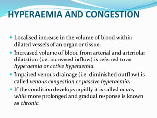
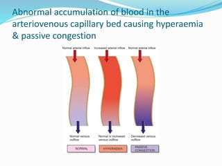
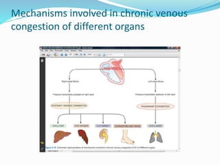
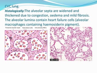
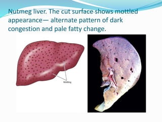
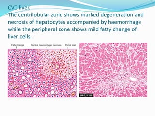
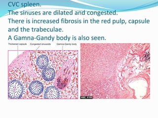
DR. ROOPAM JAIN
PROFESSOR & HEAD
DEPT. OF PATHOLOGY
HYPERAEMIA AND CONGESTION
 Localised increase in the volume of blood within
dilated vessels of an organ or tissue.
 ”
Increased volume of blood from arterial and arteriolar
dilatation (i.e. increased inflow) is referred to as
hyperaemia or active hyperaemia.
 ”
Impaired venous drainage (i.e. diminished outflow) is
called venous congestion or passive hyperaemia.
 If the condition develops rapidly it is called acute,
while more prolonged and gradual response is known
as chronic.
Abnormal accumulation of blood in the
arteriovenous capillary bed causing hyperaemia
& passive congestion
Mechanisms involved in chronic venous
congestion of different organs
MORPHOLOGY OF CVC OF ORGANS
CVC Lung
 Chronic venous congestion of the lung occurs in left
heart failure(e.g. in rheumatic mitral stenosis)
resulting in rise in pulmonary venous pressure.
 Grossly, the lungs are heavy and firm in
consistency.
 The sectioned surface is dark and rusty brown in
colour, referred to as brown induration of the lungs.
CVC lung.
Histologically:The alveolar septa are widened and
thickened due to congestion, oedema and mild fibrosis.
The alveolar lumina contain heart failure cells (alveolar
macrophages containing haemosiderin pigment).
CVC Liver
 Chronic venous congestion of the liver occurs in right heart
failure and sometimes due to occlusion of inferior vena cava and
hepatic vein.
 Grossly, the liver is enlarged and tender and the capsule is
tense.
 Cut surface shows characteristic nutmeg* appearance due to red
and yellow mottled appearance, corresponding to congested
centre of lobules and fatty peripheral zone respectively.
 Microscopically, the changes of passive congestion are more
marked in the centrilobular zone (zone 3) which is farthest from
blood supply (periportal zone, zone 1) and thus bears the brunt
of hypoxia the most.
Nutmeg liver. The cut surface shows mottled
appearance— alternate pattern of dark
congestion and pale fatty change.
CVC liver.
The centrilobular zone shows marked degeneration and
necrosis of hepatocytes accompanied by haemorrhage
while the peripheral zone shows mild fatty change of
liver cells.
CVC Spleen
 Chronic venous congestion of the spleen occurs in
right heart failure and in portal hypertension from
cirrhosis of liver.
 Grossly, the spleen in early stage is slightly to
moderately enlarged (up to 250 g as compared to
normal 150 g), while in long-standing cases there is
progressive enlargement and may weigh up to 500 to
1000 g.
CVC spleen (Congestive splenomegaly)
Sectioned surface shows
that the spleen is heavy
and enlarged in size.
The colour of sectioned
surface is grey-tan.
 Microscopically:
i) Red pulp is enlarged due to congestion and marked sinusoidal
dilatation and there are areas of recent and old haemorrhages.
ii) There is hyperplasia of reticuloendothelial cells in the red pulp of
the spleen (splenic macrophages).
iii) There is fibrous thickening of the capsule and of the trabeculae.
iv) Some of haemorrhages overlying fibrous tissue get deposits of
haemosiderin pigment and calcium salts; these organised
structures are termed as Gamna-Gandy bodies or siderofibrotic
nodules.
v) Firmness of the spleen in advanced stage is seen more
commonly in hepatic cirrhosis (congestive splenomegaly) and is
the commonest cause of hypersplenism.
CVC spleen.
The sinuses are dilated and congested.
There is increased fibrosis in the red pulp, capsule
and the trabeculae.
A Gamna-Gandy body is also seen.
CVC Kidney
 Grossly, the kidneys are slightly enlarged and the
medulla is congested.
 Microscopically, the changes are rather mild. The
tubules may show degenerative changes like cloudy
swelling and fatty change. The glomeruli may show
mesangial proliferation.
HAEMORRHAGE
 Haemorrhage is the escape of blood from a blood vessel.
 The bleeding may occur externally, or internally into the serous
cavities (e.g. haemothorax, haemoperitoneum,
haemopericardium), or into a hollow viscus.
 Extravasation of blood into the tissues with resultant swelling is
known as haematoma.
 Large extravasations of blood into the skin and mucous
membranes are called ecchymoses.
 Purpuras are small areas of haemorrhages (upto 1 cm) into the
skin and mucous membrane, whereas petechiae are minute
pinhead-sized haemorrhages.
 Microscopic escape of erythrocytes into loose tissues may occur
following marked congestion and is known as diapedesis.
ETIOLOGY
 The blood loss may be large and sudden (acute),
or small repeated bleeds may occur over a period of
time (chronic).
 The various causes of haemorrhage are as under:
1. Trauma to the vessel wall
2. Spontaneous haemorrhage
3. Inflammatory lesions of the vessel wall
4. Neoplastic invasion
5. Vascular diseases e.g. atherosclerosis
6. Elevated pressure within the vessels
EFFECTS
 The effects of blood loss depend upon 3 main factors:
i) the amount of blood loss
ii) the speed of blood loss
iii) the site of haemorrhage
 The loss up to 20% of blood volume suddenly or slowly
generally has little clinical effects because of compensatory
mechanisms.
 A sudden loss of 33% of blood volume may cause death,
while loss of up to 50% of blood volume gradually over a
period of 24 hours may not be necessarily fatal.

HYPERAEMIA & CONGESTION

  • 1.
    DR. ROOPAM JAIN PROFESSOR& HEAD DEPT. OF PATHOLOGY
  • 2.
    HYPERAEMIA AND CONGESTION Localised increase in the volume of blood within dilated vessels of an organ or tissue.  ” Increased volume of blood from arterial and arteriolar dilatation (i.e. increased inflow) is referred to as hyperaemia or active hyperaemia.  ” Impaired venous drainage (i.e. diminished outflow) is called venous congestion or passive hyperaemia.  If the condition develops rapidly it is called acute, while more prolonged and gradual response is known as chronic.
  • 3.
    Abnormal accumulation ofblood in the arteriovenous capillary bed causing hyperaemia & passive congestion
  • 4.
    Mechanisms involved inchronic venous congestion of different organs
  • 5.
    MORPHOLOGY OF CVCOF ORGANS CVC Lung  Chronic venous congestion of the lung occurs in left heart failure(e.g. in rheumatic mitral stenosis) resulting in rise in pulmonary venous pressure.  Grossly, the lungs are heavy and firm in consistency.  The sectioned surface is dark and rusty brown in colour, referred to as brown induration of the lungs.
  • 6.
    CVC lung. Histologically:The alveolarsepta are widened and thickened due to congestion, oedema and mild fibrosis. The alveolar lumina contain heart failure cells (alveolar macrophages containing haemosiderin pigment).
  • 7.
    CVC Liver  Chronicvenous congestion of the liver occurs in right heart failure and sometimes due to occlusion of inferior vena cava and hepatic vein.  Grossly, the liver is enlarged and tender and the capsule is tense.  Cut surface shows characteristic nutmeg* appearance due to red and yellow mottled appearance, corresponding to congested centre of lobules and fatty peripheral zone respectively.  Microscopically, the changes of passive congestion are more marked in the centrilobular zone (zone 3) which is farthest from blood supply (periportal zone, zone 1) and thus bears the brunt of hypoxia the most.
  • 8.
    Nutmeg liver. Thecut surface shows mottled appearance— alternate pattern of dark congestion and pale fatty change.
  • 9.
    CVC liver. The centrilobularzone shows marked degeneration and necrosis of hepatocytes accompanied by haemorrhage while the peripheral zone shows mild fatty change of liver cells.
  • 10.
    CVC Spleen  Chronicvenous congestion of the spleen occurs in right heart failure and in portal hypertension from cirrhosis of liver.  Grossly, the spleen in early stage is slightly to moderately enlarged (up to 250 g as compared to normal 150 g), while in long-standing cases there is progressive enlargement and may weigh up to 500 to 1000 g.
  • 11.
    CVC spleen (Congestivesplenomegaly) Sectioned surface shows that the spleen is heavy and enlarged in size. The colour of sectioned surface is grey-tan.
  • 12.
     Microscopically: i) Redpulp is enlarged due to congestion and marked sinusoidal dilatation and there are areas of recent and old haemorrhages. ii) There is hyperplasia of reticuloendothelial cells in the red pulp of the spleen (splenic macrophages). iii) There is fibrous thickening of the capsule and of the trabeculae. iv) Some of haemorrhages overlying fibrous tissue get deposits of haemosiderin pigment and calcium salts; these organised structures are termed as Gamna-Gandy bodies or siderofibrotic nodules. v) Firmness of the spleen in advanced stage is seen more commonly in hepatic cirrhosis (congestive splenomegaly) and is the commonest cause of hypersplenism.
  • 13.
    CVC spleen. The sinusesare dilated and congested. There is increased fibrosis in the red pulp, capsule and the trabeculae. A Gamna-Gandy body is also seen.
  • 14.
    CVC Kidney  Grossly,the kidneys are slightly enlarged and the medulla is congested.  Microscopically, the changes are rather mild. The tubules may show degenerative changes like cloudy swelling and fatty change. The glomeruli may show mesangial proliferation.
  • 15.
    HAEMORRHAGE  Haemorrhage isthe escape of blood from a blood vessel.  The bleeding may occur externally, or internally into the serous cavities (e.g. haemothorax, haemoperitoneum, haemopericardium), or into a hollow viscus.  Extravasation of blood into the tissues with resultant swelling is known as haematoma.  Large extravasations of blood into the skin and mucous membranes are called ecchymoses.  Purpuras are small areas of haemorrhages (upto 1 cm) into the skin and mucous membrane, whereas petechiae are minute pinhead-sized haemorrhages.  Microscopic escape of erythrocytes into loose tissues may occur following marked congestion and is known as diapedesis.
  • 16.
    ETIOLOGY  The bloodloss may be large and sudden (acute), or small repeated bleeds may occur over a period of time (chronic).  The various causes of haemorrhage are as under: 1. Trauma to the vessel wall 2. Spontaneous haemorrhage 3. Inflammatory lesions of the vessel wall 4. Neoplastic invasion 5. Vascular diseases e.g. atherosclerosis 6. Elevated pressure within the vessels
  • 17.
    EFFECTS  The effectsof blood loss depend upon 3 main factors: i) the amount of blood loss ii) the speed of blood loss iii) the site of haemorrhage  The loss up to 20% of blood volume suddenly or slowly generally has little clinical effects because of compensatory mechanisms.  A sudden loss of 33% of blood volume may cause death, while loss of up to 50% of blood volume gradually over a period of 24 hours may not be necessarily fatal.