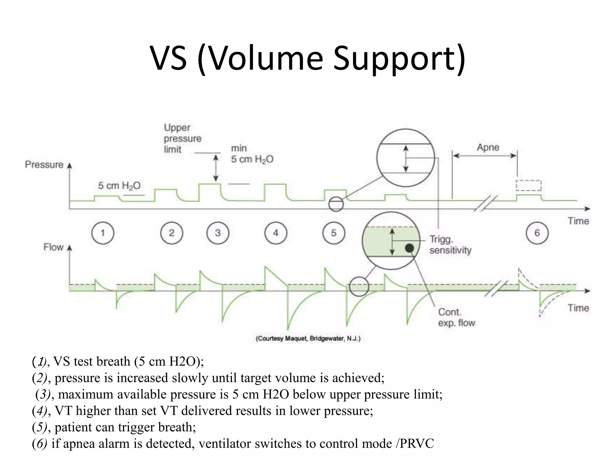
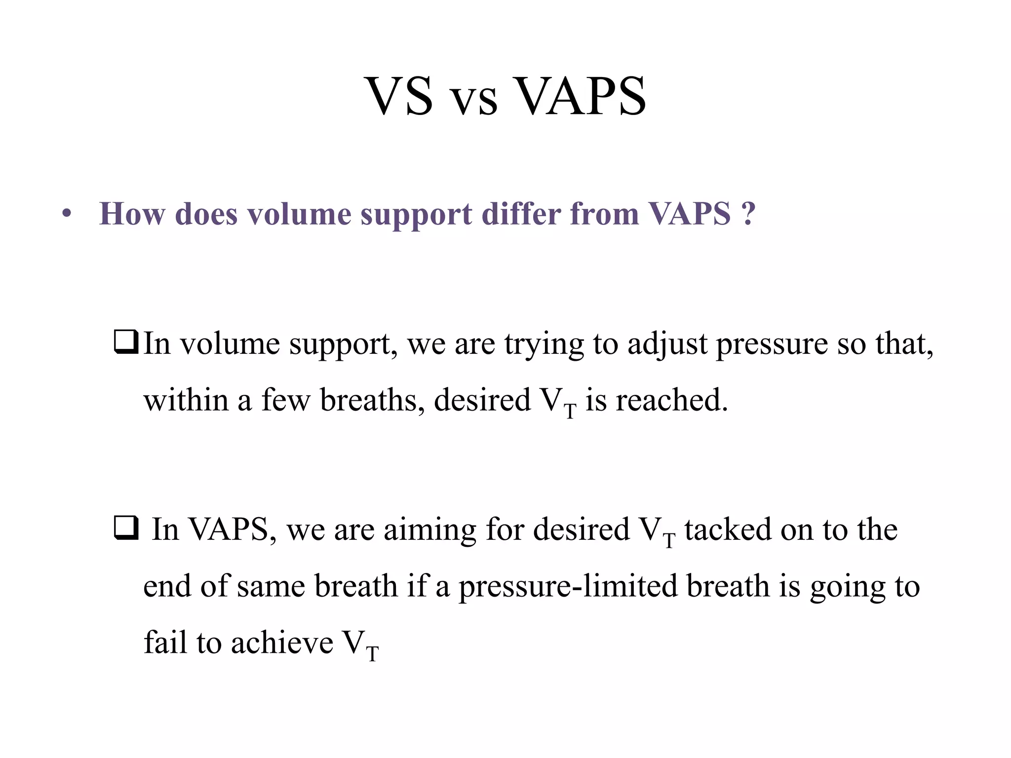
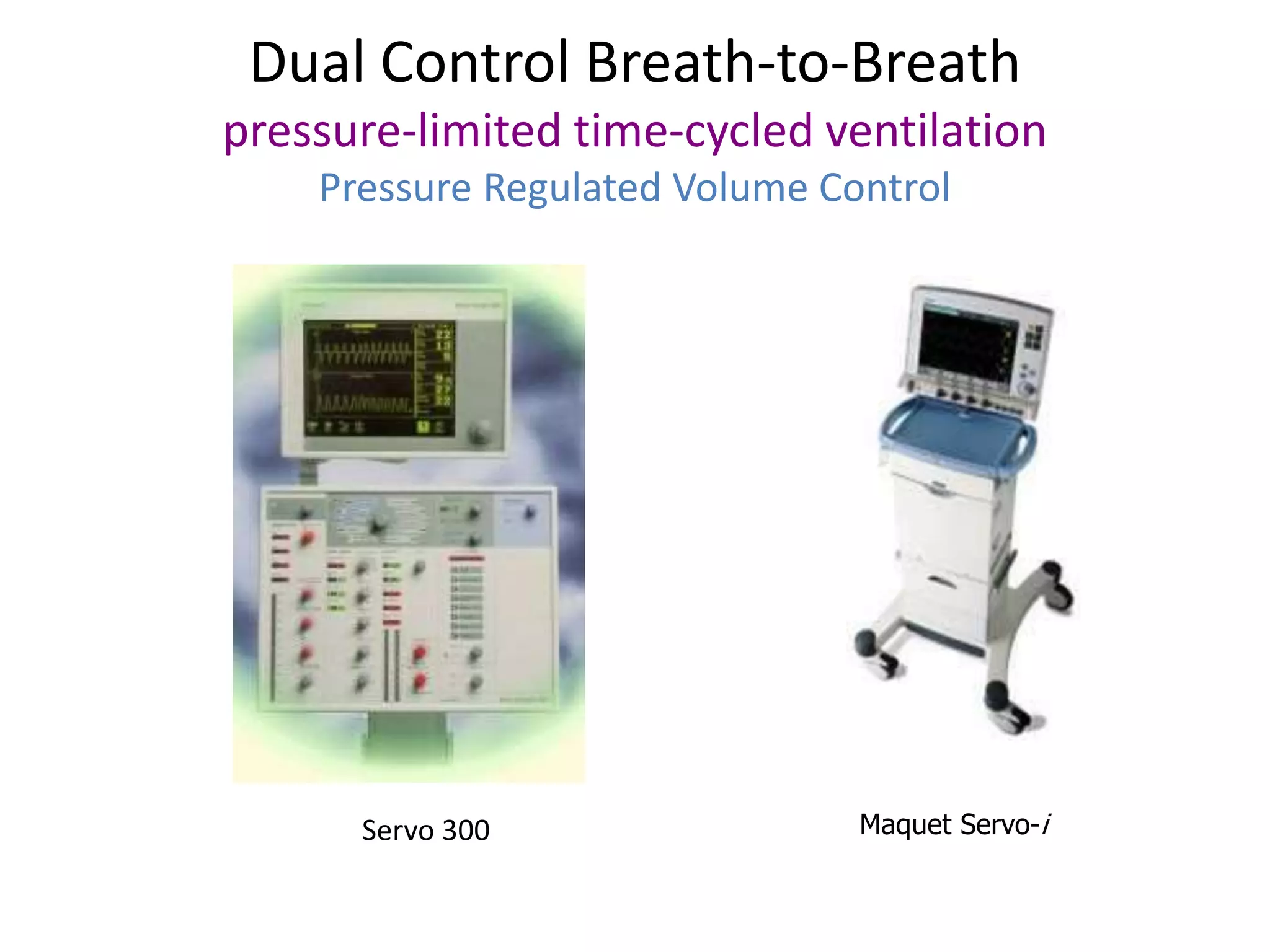
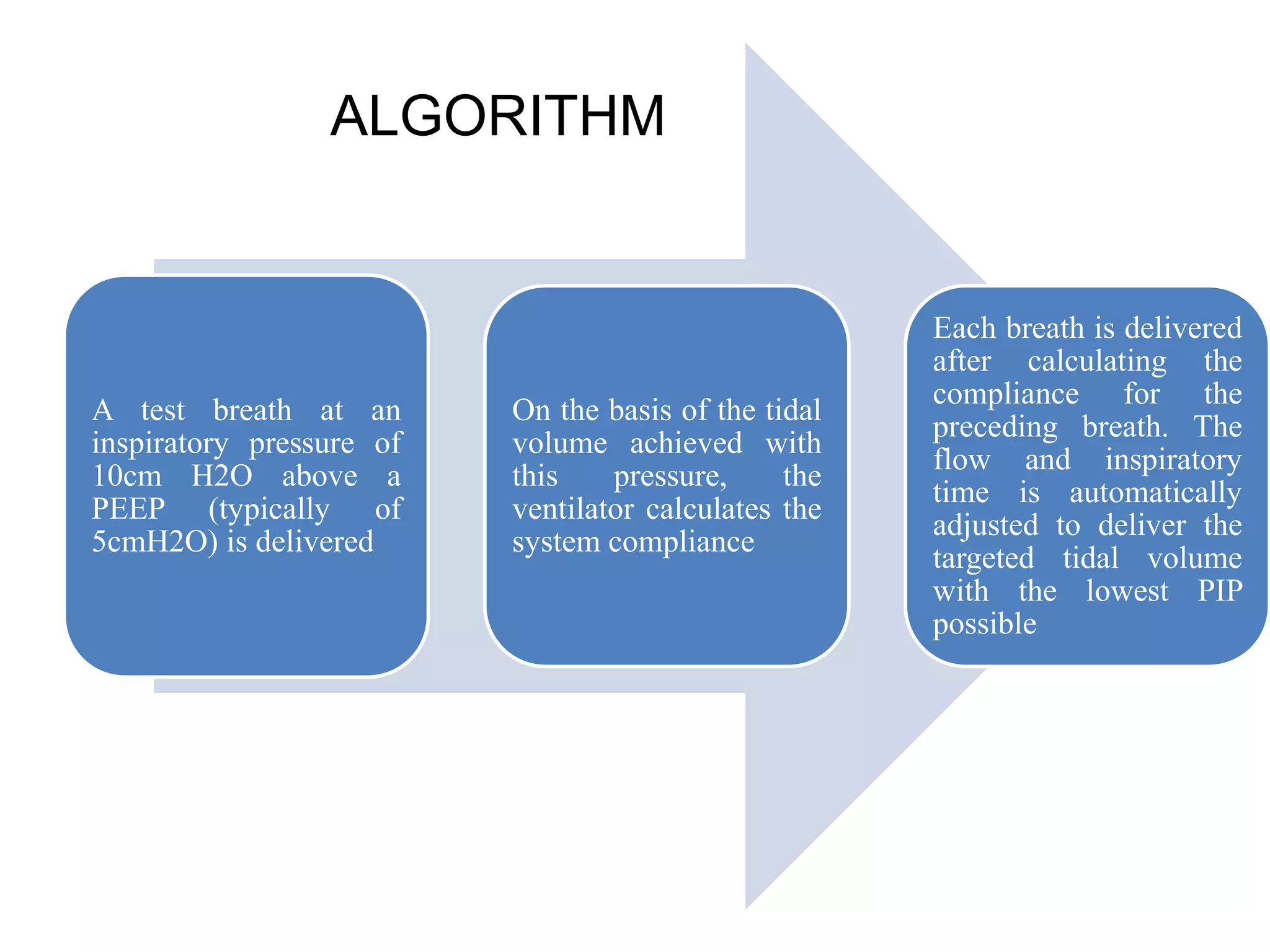
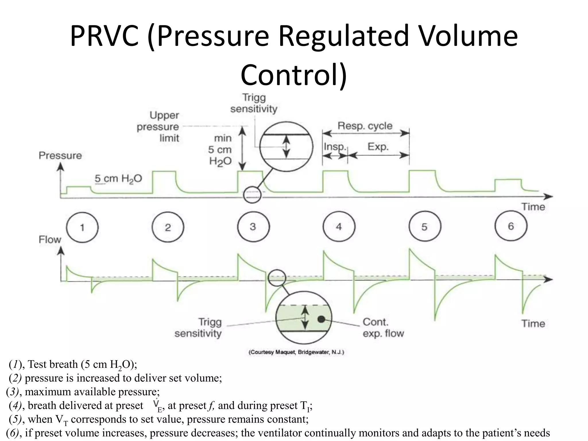
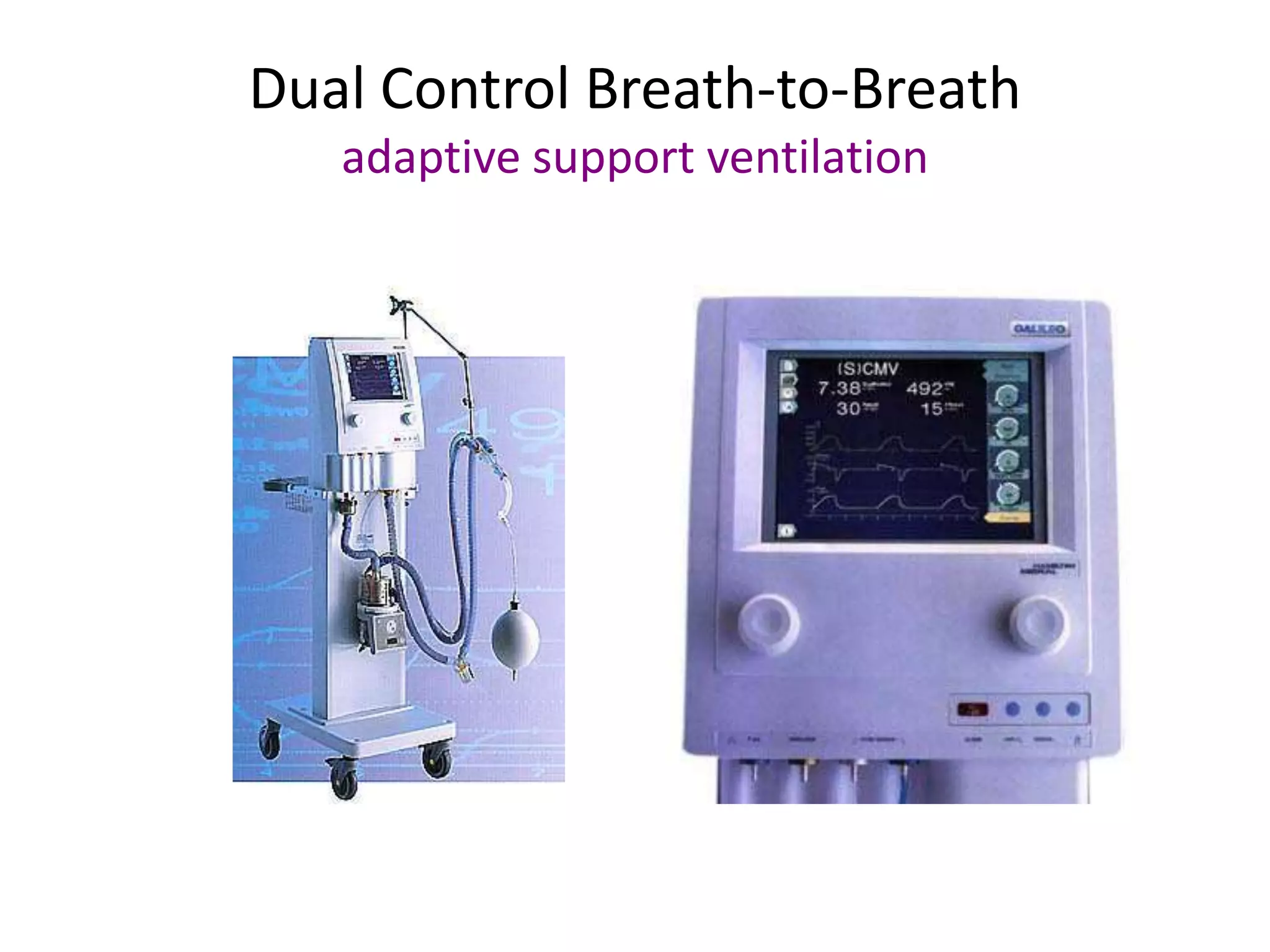
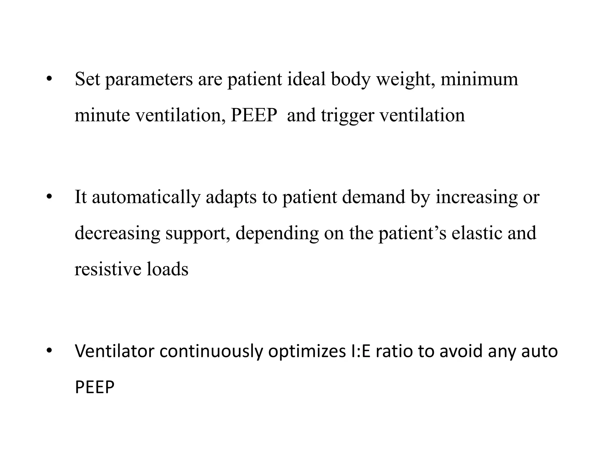
NEWER MODES OF
VENTILATION
PRESENTER- DR. RICHA KUMAR
MODERATOR- DR. AREFA JALIL
THE ORIGIN OF MECHANICAL
VENTILATION
• Andreas Vesalius is the first person to describe mechanical
ventilation in 1543
• “But the life may…be restored to the animal, an opening must be
attempted in the trunk of trachea, in which a tube of reed or cane
should be put; you will then blow into this, so that the lung may
rise again and the animal take in air.. And as I do this, and take
care that the lung is inflated in intervals, the motion of the heart
and arteries does not stop….”
• Mechanical ventilation is administered primarily in patients
unable to maintain adequate alveolar ventilation
• The role is supportive and is used to buy time while
addressing the condition that lead to respiratory failure.
• It should aim at “providing support , doing least harm”
• The ability of ventilator to initiate , maintain and terminate an
assisted breath derives its basis from “ equation of motion”
• Ventilator pressure to deliver a breath = pressure needed to
overcome the airway resistance + pressure needed to inflate
the chest wall and the lung
Equation of Motion
Ventilation
Pressure
(to deliver
tidal volume)
=
Elastic
Pressure
(to inflate lungs
and chest wall)
+
Resistive
Pressure
(to make air flow
through the
airways)
P = Resistance x Flow + Elastance x Volume
Pressure = Presistive + Pelastance
PROBLEMS WITH CONVENTIONAL MODES OF
VENTILATION
• IN critical care setting, all the parameters in equation of
motion change with time
• A ventilator setting appropriate for one point of time may not
be optimal with patient deterioration or improvement.
• Deliver the set parameters and take no feedback from the
patient
• Classical volume or pressure control modes are “OPEN
LOOPS” (feed back loop is absent )
• Newer modes target to make alterations with changing lung
and take feedback from patient parameters and are “closed
loop” type
Inspiration Expiration
0 1
20
0
0 1 2
3
-3
0
20
0
21
20
0
0 1 2
3
-3
0
20
0
2
Inspiration Expiration
Volume/Flow Control Pressure Control
Time (s) Time (s)
Paw
Paw
Pressure
Volume
Flow
Control Variables
Volume control
Tidal volumes are
guaranteed, even if
airway pressure
fluctuates
If lung mechanics are
poor, airway pressure
may rise to
unacceptable levels
Pressure control
Airway pressure level
is guaranteed
Tidal volumes are
not, and may change
if lung mechanics
vary
NEED OF NEWER MODES
• Conventional modes are uncomfortable
• More safely assist patient
• Less need for heavily sedation & paralysis
• More effectively ventilate/oxygenate
• Improve patient – ventilator synchrony
• Less haemodynamic compromise
• Lung protective ventilation : less likelihood of Ventilator Induced
Lung Injury
• More rapid weaning
DUAL MODES
PRESSURE
CONTROL
/ SUPPORT
VOLUME
CONTROL
EVOLUTION OF
MECHANICAL VENTILATORS
Modern ventilators now incorporate complex computer based algorithms, and are
capable of simultaneously controlling two variables.
 Intrabreath control (dual control WITHIN a single breath, DCWB):
During a part of an essentially pressure-targeted breath, flow is also controlled
Interbreath control (dual control from breath to breath, DCBB):
The configuration of a pressure-targeted breath is manipulated n SUBSEQUENT
breaths to deliver a targeted tidal volume
Dual-Controlled Modes
VOLUME ASSURED PRESSURE
SUPPORT
Dual Control within a Breath
volume-assured pressure support (VAPS)
• This is modification of pressure control mode
• This mode allows a feedback loop based on the volume
• It makes ventilator to switch from pressure control to volume
control if a minimum set TV is not achieved.
Bear 1000
Tbird
Bird 8400Sti
• operator adjustable parameters are same as in conventional PC mode
– pressure limit, peak flow rate, ventilator rate, and PEEP
• Additionally “minimum TV” is also defined
• This combination provides an optimal inspiratory flow during
assisted/controlled cycles, reducing the patient’s work of breathing
commonly seen during Ventilator Assisted Ventilation (VAV) and
causes lower intrinsic PEEP
• Unlike typical PSV, VAPS assures stable tidal volume along with
pressure support in patients with irregular breathing patterns
Benefits of VAPS
• Lower peak airway pressure
• Reduced patient work of breathing
• Improved gas distribution
• Less need for sedation
• Improved patient comfort
ADEQUATE PATIENT EFFORT
• If the delivered volume equals the preset volume, the
configuration of the breath is similar to that of a pressure
supported breath:
• the flow is decelerating
• the breath is flow-cycled
INADEQUATE PATIENT EFFORT
• If the flow fall below the set tidal volume within the assigned
inspiratory time, the machine delivers a volume controlled
breath :
•
• flow targeted
• volume cycled
Paw
cmH20
60
-20
60
Flow
L/min
Volume
Set flow limit
Set tidal volume cycle threshold
Set pressure limit
Tidal volume
met
Tidal volume
not met
Switch from Pressure control to
Volume/flow control
Inspiratory flow
greater than set flow
Flow cycle
Inspiratory flow
equals set flow
Pressure limit
overridden
L
0
0.6
40
• Set pressure limit should not be too high to cause
unwanted trauma to lung and generate higher volume
than minimal
• Set flow rates must not be very low as in situations
where minimal volume is not met it would cause a
delayed switch from pressure control to volume control
and would lead to unwanted prolongation of inspiratory
time
• Patients with airflow obstruction should be monitored
closely in order to prevent air trapping
DISADVANTAGES/ LIMITATIONS OF VAPS
Applications of VAPS
 A patient who requires a substantial level of ventilatory support and has
a vigorous ventilatory drive to improve gas distribution and synchrony
 A patient being weaned from the ventilator and having an unstable ventila
tory drive who may require backup tidal volume as a safety net
in case the patients effort or/and lung mechanics change
VOLUME SUPPORT
Dual control breath-to-breath
Pressure-limited flow-cycled ventilation
Volume Support
Servo 300 Maquet Servo-i
VS (Volume Support)
 Entirely a spontaneous mode
 Ventilator assesses initial breaths and steps up pressure support in
subsequent breaths if TV is low
 Tidal volume is used as feedback control to adjust the
pressure support level
 Intended to provide a control tidal volume and increased
patient comfort
 Delivers a patient triggered (pressure or flow), pressure
targeted, flow cycled breath
o Can also be timed cycled (if TI is extended for some reason) or pressure cycled
(if pressure rises too high).
 It adjusts pressure (up or down) to achieve the set volume (the
maximum pressure change is < 3 cm H2O and ranges from 0
cm H2O to 5 cm H2O below the high pressure alarm setting
INDICATIONS
 Spontaneous breathing patient who require minimum
ventilatory effort
 Patients who have inspiratory effort needing adaptive
support
 Patients who are asynchronous with the ventilator
 Used for patients ready to be “weaned” from the
ventilator
 Used for patients who cannot do all the WOB but who
are breathing spontaneously
The ventilator delivers a single spontaneous pressure support
type of breath and uses variable pressure support levels to
provide the target tidal volume
 During weaning or awakening from anesthesia, the patient
assumes a higher spontaneous tidal volume and the ventilator
decreases the pressure support level accordingly
 When the spontaneous tidal volume decreases, the ventilator
increases the pressure support level automatically to maintain
the target tidal volume.
 During VS, the ventilator frequency and minute ventilation are
determined by the triggering effort of the patient.
 The inspiratory time is determined by the patient respiratory
demand.
VS (Volume Support)
(1), VS test breath (5 cm H2O);
(2), pressure is increased slowly until target volume is achieved;
(3), maximum available pressure is 5 cm H2O below upper pressure limit;
(4), VT higher than set VT delivered results in lower pressure;
(5), patient can trigger breath;
(6) if apnea alarm is detected, ventilator switches to control mode /PRVC
VS vs VAPS
• How does volume support differ from VAPS ?
In volume support, we are trying to adjust pressure so that,
within a few breaths, desired VT is reached.
 In VAPS, we are aiming for desired VT tacked on to the
end of same breath if a pressure-limited breath is going to
fail to achieve VT
ADVANTAGES
• Guaranteed VT and VE
• Pressure supported breaths using the lowest required
pressure
• Decreases the patient’s spontaneous respiratory rate
• Decreases patient WOB
• Allows patient control of I:E time
• Breath by breath analysis
Disadvantages
• Spontaneous ventilation required
• VT selected may be too large or small for patient
• Varying mean airway pressure
• A sudden increase in respiratory rate and demand may
result in a decrease in ventilator support
PRESSURE REGULATED VOLUME
CONTROLLED (PRVC)
Dual Control Breath-to-Breath
pressure-limited time-cycled ventilation
Pressure Regulated Volume Control
Servo 300 Maquet Servo-i
PRESSURE REGULATED VOLUME CONTROL
• Ventilation that provides volume controlled breaths with
the lowest pressure possible by altering the flow and
inspiratory time
• Delivers patient or timed triggered, pressure-targeted
(controlled) and time-cycled breaths
• Ventilator measures VT delivered with VT set on the controls.
If delivered VT is less or more, ventilator increases or
decreases pressure delivered until set VT and delivered VT are
equal
• PRVC provides volume support while keeping the PIP at a
lowest level possible by altering the peak flow and inspiratory
time in response to changing airway or compliance
characteristics.
INDICATIONS
• Patient who require the lowest possible pressure and a
guaranteed consistent VT
• ALI/ARDS
• Patients requiring high and/or variable ventilatory effort
• Patient with the possibility of changes in compliance of
lung/ Resistance of airway
A test breath at an
inspiratory pressure of
10cm H2O above a
PEEP (typically of
5cmH2O) is delivered
On the basis of the tidal
volume achieved with
this pressure, the
ventilator calculates the
system compliance
Each breath is delivered
after calculating the
compliance for the
preceding breath. The
flow and inspiratory
time is automatically
adjusted to deliver the
targeted tidal volume
with the lowest PIP
possible
ALGORITHM
PRVC (Pressure Regulated Volume
Control)
(1), Test breath (5 cm H2O);
(2) pressure is increased to deliver set volume;
(3), maximum available pressure;
(4), breath delivered at preset E, at preset f, and during preset TI;
(5), when VT corresponds to set value, pressure remains constant;
(6), if preset volume increases, pressure decreases; the ventilator continually monitors and adapts to the patient’s needs
• The increasing airflow resistance may be due to increasing
airway resistance (nonelastic resistance) or decreasing lung
compliance (elastic resistance)
• At constant flow, the PIP is increased due to increasing
airflow resistance.
• Increased Airflow Resistance (nonelastic or elastic) :
Increase PIP / Flow
• PRVC lowers the flow to reduce the driving pressure.
• Increased Airflow Resistance (nonelastic or elastic) : PIP /
Decrease Flow
 To compensate for a lower inspiratory flow, PRVC prolongs the
inspiratory time to deliver the target volume
• (VT - Increased Constant Flow * decreased Inspiratory Time)
 The ventilator will not allow delivered pressure to rise higher
than 5 cm H2O below set upper pressure limit
 Example: If upper pressure limit is set to 35 cm H2O and
the ventilator requires more than 30 cm H2O to deliver a
targeted VT of 500 mL, an alarm will sound alerting the
clinician that too much pressure is being required to deliver
set volume
ADVANTAGES
• Maintains a minimum PIP
• Guaranteed VT and E
• Patient has very little WOB requirement
• Allows patient control of respiratory rate and E
• Variable E to meet patient demand
• Decelerating flow waveform for improved gas
distribution
• Breath by breath analysis
DISADVANTAGES
• Varying mean airway pressure
• May cause or worsen auto-PEEP
• When patient demand is increased, pressure level may
diminish when support is needed
• May be tolerated poorly in awake non-sedated patients
• A sudden increase in respiratory rate and demand may
result in a decrease in ventilator support
ADAPTIVE SUPPORT VENTILATION
(ASV)
Dual Control Breath-to-Breath
adaptive support ventilation
ASV (Adaptive Support Ventilation)
• A dual control mode that that PROVIDES A MANDATORY
MINUTE VENTILATION
• Unique: sets minimal work of breathing to deliver
desired minute ventilation
• Control variable is pressure
• Uses both pressure control and pressure support to
maintain a set minimum TV(volume target) using the least
required settings for minimal WOB depending on the
patient’s condition and effort
• Set parameters are patient ideal body weight, minimum
minute ventilation, PEEP and trigger ventilation
• It automatically adapts to patient demand by increasing or
decreasing support, depending on the patient’s elastic and
resistive loads
• Ventilator continuously optimizes I:E ratio to avoid any auto
PEEP
INDICATIONS
• Full or partial ventilatory support
• Patients requiring a lowest possible PIP and a
guaranteed VT
• ALI/ARDS
• Patients not breathing spontaneously and not triggering
the ventilator
• Patient with the possibility of work changes (CL and
Raw)
• Facilitates weaning
• Parameter input: patient’s body weight and desired percent
minute volume
• The body weight is used to estimate the dead space volume
and to calculate the alveolar volume
• For an estimated minute ventilation requirement for a patient,
the ventilator uses predetermined settings of 100 mL/min/kg
for adults and 200 mL/min/kg for children.
• The therapist may select the percent minute volume, ranging
from 20% to 200% of the predetermined adult or child setting
•
ASV WORKING PRINCIPLE
• For example, if 160% is selected for an adult, the minute
ventilation delivered by the ventilator will be about 160
mL/min/kg ( 100 X 160/100)
• Once the target minute ventilation is set, the ventilator uses test
breaths to measure the system compliance, airway resistance,
and any intrinsic PEEP.
• Following determination of these variables, the ventilator selects
and provides the frequency, inspiratory time, I:E ratio, and high
pressure limit for mandatory and spontaneous breaths
• If there is no spontaneous triggering effort, the ventilator
determines and provides the mandatory frequency, tidal
volume, and high pressure limit needed to deliver the
preselected tidal volume, inspiratory time, and I:E ratio
• As the patient begins to trigger the ventilator, the number of
mandatory breaths decreases and the pressure support level
increases until a calculated tidal volume is able to provide
adequate alveolar volume
• (i.e., tidal volume = alveolar volume + 2.2 mL/kg of
deadspace volume)
Mandatory breaths in
ASV mode
• TRIGGER
• machine
• LIMIT
• Pressure limited
• Cycle
• Time cycled
Assisted breaths in ASV
mode
• Trigger
• Pressure or flow ( by
the patient )
• Limit
• Pressure limited
• Cycle
• Flow , pressure or
time by the patient
• Clinician enters patient data & % support
• Ventilator calculates needed minute volume & best rate/TV to
produce least work
• Targeted TV’s given as pressure control or pressure support
breaths
• If pt.’s f > “set” by ventilator , MODE : PS
• Ifpt’s f < “set” by ventilator , MODE : PC SIMV/PS
• If patient is apneic, all breaths are PC
• Rate where WOB is minimal
• Pressure adjusts in +/‐2 cm H2O to achieve Tidal Volume
ADVANTAGES
• Guaranteed VT and E
• Minimal patient Work Of Breathing
• Ventilator adapts to the patient
• Weaning is done automatically and continuously
• Variable to meet patient demand
• Decelerating flow waveform for improved gas
distribution
• Breath by breath analysis
Disadvantages
• Inability to recognize and adjust to changes in
alveolar VD
• Possible respiratory muscle atrophy
• Varying mean airway pressure
• In patients with COPD, a longer TE may be
required
• A sudden increase in respiratory rate and demand
may result in a decrease in ventilator support
AUTOMODE
AUTOMODE
 The ventilator switch between mandatory and spontaneous breathing modes
 Combines volume support (VS) and pressure-regulated volume control
(PRVC)
 If patient is paralyzed; the ventilator will provide PRVC. All breaths are
mandatory that are ventilator triggered, pressure controlled and time cycled; the
pressure is adjusted to maintain the set tidal volume.
 If the patient breathes spontaneously for two consecutive breaths, the ventilator
switches to VS. All breaths are patient triggered, pressure limited, and flow
cycled.
 If the patient becomes apneic for 12 seconds; the ventilator switches back to
PRVC
Macquet Servo 300A
PROPOTIONAL ASSISST
VENTILATION (PAV)p
PROPOTIONAL ASSIST VENTILATION
• Advantage of improving ventilator patient synchrony
• Amplifies patient’s ventilatory effort giving patient freedom to
adopt his own breathing pattern
• Unloads respiratory muscles without imposing a fixed
breathing pattern thus allows synchrony
• Percentage of assistance to be delivered is set and other
parameters are adjusted automatically according to the patients
“air hunger”
• I:E ratio also decided by patient allowing more synchrony
• ADVANTAGES:
• Better synchrony
• More comfort in NIV with PAV compared to PSV
• Low airway pressures
• Optimal weaning
• Decreased work of breathing
• Early/late ALI/ARDS
• Hypercapnic ventilatory failure
DISADVANTAGE:
• If patient worsens or improves ,the proportion of assistance
needs to be readjusted according to patient’s clinical
condition
• This disadvantage has been adjusted in newer modification
“PAV + “ mode  capable of sensing patient respiratory
mechanics and adjusting accordingly
MANDATORY MINUTE VENTILATION
MANADATORY MINUTE VENTILATION
• Modification of PSV
• Ventilator takes feedback to alter both respiratory rate and
level of pressure support to achieve set minimum minute
ventilation
• Operator sets: minimum minute ventilation (70-90% of
current minute volume) ,which is readjusted according to
patients clinical condition
• Patient fails to achieve set volume then ventilator provides the
deficit
• Reliable weaning mode
• Apneic patient or central drive pathology, MMV sets safety by
providing a set value ventilation as mandatory ventilation
• Caution : MMV lower than current MV increased WOB
MMV more than required MV unloading of
muscles atrophy
o D/A Rapid shallow breathing if RR is set too high
BILEVEL VENTILATION MODES
BiLevels
 BiPaP
 ARPC ( airway pressure
release ventilation)
BiLevel Ventilation
 Is a spontaneous breathing mode in which two levels of pressure
i.e. high /low are set
 Substantial improvements for SPONTANEOUS BREATHING
Better synchronization,
More options for supporting spontaneous breathing
 Potential for improved monitoring
BiLevel Ventilation
Synchronized TransitionsSpontaneous Breaths
Spontaneous Breaths
Paw
cmH20
60
-20
1 2 3 4 5 6 7
Also called
Biphasic
Bivent
DuoPAP
These modes deliver
 pressure-controlled breaths
 time-triggered
 time-cycled breaths
using a set-point targeting scheme
This mode maintains a constant pressure
(set point) even in the face of spontaneous
breaths
There are two pressures to be set
Phigh
Plow
There are two time intervals to be set
Time spent on P high – Thigh
Time spent on P low - T low
Patient can breath spontaneously at both
these pressures
T high T low T high
BiPAP
CPAP
TIME
T high T low T high
PCV
BIPAP
Unrestricted spontaneous breathing
Allows reduced sedation and promote
weaning
Phigh
improves oxygenation
promotes alveolar recruitment
Plow
Allows exhalation
Maintains recruitment
P high & P low
The volume difference between the two
Two levels of functional FRC
Creates a driving pressure
Determines the VT
Permit gas to enter the lung units
Represents the difference between
airway pressure (Paw) and alveolar
pressure (Palv)
T high T low T high
PCV
Airway Pressure Release Ventilation
• Is a Bi-level form of ventilation with sudden short releases in
pressure to rapidly reduce FRC and allow for ventilation
• Can work in spontaneous or apneic patients
• APRV is similar but utilizes a very short expiratory time for
PRESSURE RELEASE
• this short time at low pressure allows for ventilation
 APRV always implies an inverse I:E ratio
 All spontaneous breathing is done at upper pressure level
INDICATIONS
• Partial to full ventilatory support
• Patients with ALI/ARDS
• Patients with refractory hypoxemia due to
collapsed alveoli
• Patients with massive atelectasis
• May use with mild or no lung disease
• Provides two levels of CPAP and allows spontaneous
breathing at both levels when spontaneous effort is present
• Both pressure levels are time triggered and time cycled
• The release phase ( expiratory phase) brings down mean airway
pressure and plays significant role in maintaining normocarbia.
• It has dual functionality:
• In spontaneously breathing pt patients breathes with supported
breaths thus reducing need for sedation
• In absence of spontaneous breathing bi-level pressure acts as
time cycled inverse ratio ventilation.
• Vt depends of upon respiratory compliance and difference between
two CPAP levels
Airway Pressure Release Ventilation
Paw
cmH20
60
-20
1 2 3 4 5 6 7 8
Spontaneous Breaths
Releases
ADVANTAGES
• Allows inverse ratio ventilation (IRV) with or without
spontaneous breathing (less need for sedation or paralysis)
• Improves patient-ventilator synchrony if spontaneous
breathing is present
• Improves mean airway pressure
• Improves oxygenation by stabilizing collapsed alveoli
• Allows patients to breath spontaneously while continuing
lung recruitment
• Lowers PIP
• May decrease physiologic deadspace
DISADVANTAGES AND RISKS
• Variable VT
• Could be harmful to patients with high expiratory resistance
(i.e., COPD or asthma)
• Auto-PEEP is usually present
• Caution should be used with hemodynamically unstable
patients
• Asynchrony can occur is spontaneous breaths are out of sync
with release time
• Requires the presence of an “active exhalation valve”
AUTOMATIC TUBE COMPRESSION
Automatic Tube Compensation
• Available in the Evita 4 ventilator (Dräger Medical)
• The PB840 ventilator has a similar feature which is
called tubing compensation (TC).
• Can be applied in all ventilation modes
• A mode of ventilation that offsets and compensates
for the airflow resistance imposed by the artificial
airway
• Overcome Work Of Breathing added by artificial airways
• It allows the patient to have a breathing pattern as if
breathing spontaneously without an artificial airway
• Improve patient/ventilator synchrony by providing variable
fast inspiratory flow
INDICATIONS
• patient who has compromised respiratory function
( COPD, malnutrition, respiratory muscle failure)
• Those who have failed previous extubation attempt
• The “difficult to wean” patient
Resistance due to ET Tube
• Due to varying inspiratory flow rates, no single level of pressure
support can actually fully compensate for WOB caused by the
ETT.
• ATC uses known static resistance for each size and type of
ETT/tracheal tube, and measures flow rates.
• Pressure is applied and continuously adjusted proportional to
resistance.
• With ATC, the pressure delivered by the ventilator to
compensate for the airflow resistance is active during
inspiration and expiration.
• It is dependent on the airflow characteristics and the flow
demand of the patient.
• For example, when the airway diameter decreases or flow
demand increases, the pressure is raised to overcome a higher
airflow resistance or increased flow demand.
ADVANTAGES
• Designed to Maintain Tracheal Pressure at Baseline
• Does not require ongoing assessment of resistance!
• Pressure Applied Based Upon Resistive Properties of the
Airway and Patients Inspiratory Flow
• Positive Pressure During Inspiration
• Negative Pressure During Exhalation
• Effectively unloads resistive effort imposed by ETT
• Improves patient – ventilator synchrony
• Reduces risk of lung injury
NEURALLY ADJUSTED VENTILATORY
ASSIST (NAVA)
Neurally adjusted ventilatory assist (NAVA)
• A mode of mechanical ventilation in which the patient’s electrical
activity of the diaphragm (EAdi or Edi) is used to guide the optimal
functions of the ventilator .
• The neural controls of respiration originated in the patient’s
respiratory center are sent to the diaphragm via the phrenic nerves.
• In turn, bipolar electrodes are used to pick up the electrical activity
• The electrodes are mounted on a disposable EAdi catheter and
positioned in the esophagus at the level of the diaphragm.
Neural adjusted ventilator assist
NAVA
Neuro-VentilatoryCoupling
Central Nervous System

Phrenic Nerve

Diaphragm Excitation

Diaphragm Contraction

Chest Wall and Lung Expansion

Airway Pressure, Flow and
Volume
New
Technology
Ideal
Technology
Current
Technology
Ventilator
Unit
INDICATIONS
• NAVA is available for adults, children, and neonates,
and it has been used successfully in the management
and weaning of mechanically ventilated patients with
spinal cord injury.
• Other uses and potential applications of NAVA include
patients with head injury, COPD, and history of
ventilator dependency.
•
• The ability to wean these patients rapidly reduces or
eliminates the incidence of disuse atrophy of the
diaphragm
Components
CATHETERS
SIGNAL CAPTURE
 All muscles (including the diaphragm
and other respiratory muscles) generate
electrical activity to excite muscle
contraction.
 The electrical activity of the diaphragm
is captured by an esophageal catheter
with an attached electrode array. The
signal is filtered in several steps and
provide the input for control of the
respiratory assist delivered by the
ventilator.
In a healthy subject only 5% of
maximum capacity of neuro
ventilatory coupling is used to
generate an adequate Vt
Benefits of NAVA
• Synchrony with least possible delay
• Safer and improved ventilation
• Leaks do not cause false initiation of breaths (unaffected by circuit)
• Eliminates man sleep disturbances
• Adapts to altered metabolic demand with consistent unloading
• Prevents disuse atrophy
• Improves NIV
• In other modes auto-PEEP increases work of ventilator
initiation in COPD and asthma , this does not effect ventilatory
cycle in NAVA thus overall WOB decreases in these patients
NEOGANESH (SMARTCARE)
NEOGANESH(SMARTCARE)
• Closed loop type modification of PSV WITH INREGRATED
ARTIFICIAL INTELLIGENCE
• Adjusts ventilator assistance depending on patient,s
respiratory pattern and literature based weaning protocols
• Based on 3 fundamental principles:
– Adapt PS to patient’s present clinical situation
– In case of stability wean off PS
– Initiate spontaneous breathing trials as per prerecorded clinical
guidelines
• Ventilator takes feedback from monitored RR, VT, etCO2
• Trials have shown that it reduces weaning failure and also
hastens weaning duration.
SUMMARY
Older modes & ventilators:
passive
operator‐dependant tools
New modes or new generation ventilators:
adaptively interactive
 goal oriented
patient centered
THANK YOU

Newer modes of ventilation

  • 1.
    NEWER MODES OF VENTILATION PRESENTER-DR. RICHA KUMAR MODERATOR- DR. AREFA JALIL
  • 2.
    THE ORIGIN OFMECHANICAL VENTILATION • Andreas Vesalius is the first person to describe mechanical ventilation in 1543 • “But the life may…be restored to the animal, an opening must be attempted in the trunk of trachea, in which a tube of reed or cane should be put; you will then blow into this, so that the lung may rise again and the animal take in air.. And as I do this, and take care that the lung is inflated in intervals, the motion of the heart and arteries does not stop….”
  • 3.
    • Mechanical ventilationis administered primarily in patients unable to maintain adequate alveolar ventilation • The role is supportive and is used to buy time while addressing the condition that lead to respiratory failure. • It should aim at “providing support , doing least harm”
  • 4.
    • The abilityof ventilator to initiate , maintain and terminate an assisted breath derives its basis from “ equation of motion” • Ventilator pressure to deliver a breath = pressure needed to overcome the airway resistance + pressure needed to inflate the chest wall and the lung
  • 5.
    Equation of Motion Ventilation Pressure (todeliver tidal volume) = Elastic Pressure (to inflate lungs and chest wall) + Resistive Pressure (to make air flow through the airways) P = Resistance x Flow + Elastance x Volume Pressure = Presistive + Pelastance
  • 6.
    PROBLEMS WITH CONVENTIONALMODES OF VENTILATION • IN critical care setting, all the parameters in equation of motion change with time • A ventilator setting appropriate for one point of time may not be optimal with patient deterioration or improvement. • Deliver the set parameters and take no feedback from the patient
  • 7.
    • Classical volumeor pressure control modes are “OPEN LOOPS” (feed back loop is absent ) • Newer modes target to make alterations with changing lung and take feedback from patient parameters and are “closed loop” type
  • 8.
    Inspiration Expiration 0 1 20 0 01 2 3 -3 0 20 0 21 20 0 0 1 2 3 -3 0 20 0 2 Inspiration Expiration Volume/Flow Control Pressure Control Time (s) Time (s) Paw Paw Pressure Volume Flow
  • 9.
    Control Variables Volume control Tidalvolumes are guaranteed, even if airway pressure fluctuates If lung mechanics are poor, airway pressure may rise to unacceptable levels Pressure control Airway pressure level is guaranteed Tidal volumes are not, and may change if lung mechanics vary
  • 10.
    NEED OF NEWERMODES • Conventional modes are uncomfortable • More safely assist patient • Less need for heavily sedation & paralysis • More effectively ventilate/oxygenate • Improve patient – ventilator synchrony • Less haemodynamic compromise • Lung protective ventilation : less likelihood of Ventilator Induced Lung Injury • More rapid weaning
  • 11.
  • 12.
    Modern ventilators nowincorporate complex computer based algorithms, and are capable of simultaneously controlling two variables.  Intrabreath control (dual control WITHIN a single breath, DCWB): During a part of an essentially pressure-targeted breath, flow is also controlled Interbreath control (dual control from breath to breath, DCBB): The configuration of a pressure-targeted breath is manipulated n SUBSEQUENT breaths to deliver a targeted tidal volume Dual-Controlled Modes
  • 13.
  • 14.
    Dual Control withina Breath volume-assured pressure support (VAPS) • This is modification of pressure control mode • This mode allows a feedback loop based on the volume • It makes ventilator to switch from pressure control to volume control if a minimum set TV is not achieved. Bear 1000 Tbird Bird 8400Sti
  • 15.
    • operator adjustableparameters are same as in conventional PC mode – pressure limit, peak flow rate, ventilator rate, and PEEP • Additionally “minimum TV” is also defined • This combination provides an optimal inspiratory flow during assisted/controlled cycles, reducing the patient’s work of breathing commonly seen during Ventilator Assisted Ventilation (VAV) and causes lower intrinsic PEEP • Unlike typical PSV, VAPS assures stable tidal volume along with pressure support in patients with irregular breathing patterns
  • 16.
    Benefits of VAPS •Lower peak airway pressure • Reduced patient work of breathing • Improved gas distribution • Less need for sedation • Improved patient comfort
  • 17.
    ADEQUATE PATIENT EFFORT •If the delivered volume equals the preset volume, the configuration of the breath is similar to that of a pressure supported breath: • the flow is decelerating • the breath is flow-cycled
  • 18.
    INADEQUATE PATIENT EFFORT •If the flow fall below the set tidal volume within the assigned inspiratory time, the machine delivers a volume controlled breath : • • flow targeted • volume cycled
  • 19.
    Paw cmH20 60 -20 60 Flow L/min Volume Set flow limit Settidal volume cycle threshold Set pressure limit Tidal volume met Tidal volume not met Switch from Pressure control to Volume/flow control Inspiratory flow greater than set flow Flow cycle Inspiratory flow equals set flow Pressure limit overridden L 0 0.6 40
  • 20.
    • Set pressurelimit should not be too high to cause unwanted trauma to lung and generate higher volume than minimal • Set flow rates must not be very low as in situations where minimal volume is not met it would cause a delayed switch from pressure control to volume control and would lead to unwanted prolongation of inspiratory time • Patients with airflow obstruction should be monitored closely in order to prevent air trapping DISADVANTAGES/ LIMITATIONS OF VAPS
  • 21.
    Applications of VAPS A patient who requires a substantial level of ventilatory support and has a vigorous ventilatory drive to improve gas distribution and synchrony  A patient being weaned from the ventilator and having an unstable ventila tory drive who may require backup tidal volume as a safety net in case the patients effort or/and lung mechanics change
  • 22.
  • 23.
    Dual control breath-to-breath Pressure-limitedflow-cycled ventilation Volume Support Servo 300 Maquet Servo-i
  • 24.
    VS (Volume Support) Entirely a spontaneous mode  Ventilator assesses initial breaths and steps up pressure support in subsequent breaths if TV is low
  • 25.
     Tidal volumeis used as feedback control to adjust the pressure support level  Intended to provide a control tidal volume and increased patient comfort  Delivers a patient triggered (pressure or flow), pressure targeted, flow cycled breath o Can also be timed cycled (if TI is extended for some reason) or pressure cycled (if pressure rises too high).  It adjusts pressure (up or down) to achieve the set volume (the maximum pressure change is < 3 cm H2O and ranges from 0 cm H2O to 5 cm H2O below the high pressure alarm setting
  • 26.
    INDICATIONS  Spontaneous breathingpatient who require minimum ventilatory effort  Patients who have inspiratory effort needing adaptive support  Patients who are asynchronous with the ventilator  Used for patients ready to be “weaned” from the ventilator  Used for patients who cannot do all the WOB but who are breathing spontaneously
  • 27.
    The ventilator deliversa single spontaneous pressure support type of breath and uses variable pressure support levels to provide the target tidal volume  During weaning or awakening from anesthesia, the patient assumes a higher spontaneous tidal volume and the ventilator decreases the pressure support level accordingly
  • 28.
     When thespontaneous tidal volume decreases, the ventilator increases the pressure support level automatically to maintain the target tidal volume.  During VS, the ventilator frequency and minute ventilation are determined by the triggering effort of the patient.  The inspiratory time is determined by the patient respiratory demand.
  • 29.
    VS (Volume Support) (1),VS test breath (5 cm H2O); (2), pressure is increased slowly until target volume is achieved; (3), maximum available pressure is 5 cm H2O below upper pressure limit; (4), VT higher than set VT delivered results in lower pressure; (5), patient can trigger breath; (6) if apnea alarm is detected, ventilator switches to control mode /PRVC
  • 30.
    VS vs VAPS •How does volume support differ from VAPS ? In volume support, we are trying to adjust pressure so that, within a few breaths, desired VT is reached.  In VAPS, we are aiming for desired VT tacked on to the end of same breath if a pressure-limited breath is going to fail to achieve VT
  • 31.
    ADVANTAGES • Guaranteed VTand VE • Pressure supported breaths using the lowest required pressure • Decreases the patient’s spontaneous respiratory rate • Decreases patient WOB • Allows patient control of I:E time • Breath by breath analysis
  • 32.
    Disadvantages • Spontaneous ventilationrequired • VT selected may be too large or small for patient • Varying mean airway pressure • A sudden increase in respiratory rate and demand may result in a decrease in ventilator support
  • 33.
  • 34.
    Dual Control Breath-to-Breath pressure-limitedtime-cycled ventilation Pressure Regulated Volume Control Servo 300 Maquet Servo-i
  • 35.
    PRESSURE REGULATED VOLUMECONTROL • Ventilation that provides volume controlled breaths with the lowest pressure possible by altering the flow and inspiratory time • Delivers patient or timed triggered, pressure-targeted (controlled) and time-cycled breaths
  • 36.
    • Ventilator measuresVT delivered with VT set on the controls. If delivered VT is less or more, ventilator increases or decreases pressure delivered until set VT and delivered VT are equal • PRVC provides volume support while keeping the PIP at a lowest level possible by altering the peak flow and inspiratory time in response to changing airway or compliance characteristics.
  • 37.
    INDICATIONS • Patient whorequire the lowest possible pressure and a guaranteed consistent VT • ALI/ARDS • Patients requiring high and/or variable ventilatory effort • Patient with the possibility of changes in compliance of lung/ Resistance of airway
  • 38.
    A test breathat an inspiratory pressure of 10cm H2O above a PEEP (typically of 5cmH2O) is delivered On the basis of the tidal volume achieved with this pressure, the ventilator calculates the system compliance Each breath is delivered after calculating the compliance for the preceding breath. The flow and inspiratory time is automatically adjusted to deliver the targeted tidal volume with the lowest PIP possible ALGORITHM
  • 39.
    PRVC (Pressure RegulatedVolume Control) (1), Test breath (5 cm H2O); (2) pressure is increased to deliver set volume; (3), maximum available pressure; (4), breath delivered at preset E, at preset f, and during preset TI; (5), when VT corresponds to set value, pressure remains constant; (6), if preset volume increases, pressure decreases; the ventilator continually monitors and adapts to the patient’s needs
  • 40.
    • The increasingairflow resistance may be due to increasing airway resistance (nonelastic resistance) or decreasing lung compliance (elastic resistance) • At constant flow, the PIP is increased due to increasing airflow resistance. • Increased Airflow Resistance (nonelastic or elastic) : Increase PIP / Flow • PRVC lowers the flow to reduce the driving pressure. • Increased Airflow Resistance (nonelastic or elastic) : PIP / Decrease Flow
  • 41.
     To compensatefor a lower inspiratory flow, PRVC prolongs the inspiratory time to deliver the target volume • (VT - Increased Constant Flow * decreased Inspiratory Time)  The ventilator will not allow delivered pressure to rise higher than 5 cm H2O below set upper pressure limit  Example: If upper pressure limit is set to 35 cm H2O and the ventilator requires more than 30 cm H2O to deliver a targeted VT of 500 mL, an alarm will sound alerting the clinician that too much pressure is being required to deliver set volume
  • 42.
    ADVANTAGES • Maintains aminimum PIP • Guaranteed VT and E • Patient has very little WOB requirement • Allows patient control of respiratory rate and E • Variable E to meet patient demand • Decelerating flow waveform for improved gas distribution • Breath by breath analysis
  • 43.
    DISADVANTAGES • Varying meanairway pressure • May cause or worsen auto-PEEP • When patient demand is increased, pressure level may diminish when support is needed • May be tolerated poorly in awake non-sedated patients • A sudden increase in respiratory rate and demand may result in a decrease in ventilator support
  • 44.
  • 45.
  • 46.
    ASV (Adaptive SupportVentilation) • A dual control mode that that PROVIDES A MANDATORY MINUTE VENTILATION • Unique: sets minimal work of breathing to deliver desired minute ventilation • Control variable is pressure • Uses both pressure control and pressure support to maintain a set minimum TV(volume target) using the least required settings for minimal WOB depending on the patient’s condition and effort
  • 47.
    • Set parametersare patient ideal body weight, minimum minute ventilation, PEEP and trigger ventilation • It automatically adapts to patient demand by increasing or decreasing support, depending on the patient’s elastic and resistive loads • Ventilator continuously optimizes I:E ratio to avoid any auto PEEP
  • 48.
    INDICATIONS • Full orpartial ventilatory support • Patients requiring a lowest possible PIP and a guaranteed VT • ALI/ARDS • Patients not breathing spontaneously and not triggering the ventilator • Patient with the possibility of work changes (CL and Raw) • Facilitates weaning
  • 49.
    • Parameter input:patient’s body weight and desired percent minute volume • The body weight is used to estimate the dead space volume and to calculate the alveolar volume • For an estimated minute ventilation requirement for a patient, the ventilator uses predetermined settings of 100 mL/min/kg for adults and 200 mL/min/kg for children. • The therapist may select the percent minute volume, ranging from 20% to 200% of the predetermined adult or child setting • ASV WORKING PRINCIPLE
  • 50.
    • For example,if 160% is selected for an adult, the minute ventilation delivered by the ventilator will be about 160 mL/min/kg ( 100 X 160/100) • Once the target minute ventilation is set, the ventilator uses test breaths to measure the system compliance, airway resistance, and any intrinsic PEEP. • Following determination of these variables, the ventilator selects and provides the frequency, inspiratory time, I:E ratio, and high pressure limit for mandatory and spontaneous breaths
  • 51.
    • If thereis no spontaneous triggering effort, the ventilator determines and provides the mandatory frequency, tidal volume, and high pressure limit needed to deliver the preselected tidal volume, inspiratory time, and I:E ratio • As the patient begins to trigger the ventilator, the number of mandatory breaths decreases and the pressure support level increases until a calculated tidal volume is able to provide adequate alveolar volume • (i.e., tidal volume = alveolar volume + 2.2 mL/kg of deadspace volume)
  • 52.
    Mandatory breaths in ASVmode • TRIGGER • machine • LIMIT • Pressure limited • Cycle • Time cycled Assisted breaths in ASV mode • Trigger • Pressure or flow ( by the patient ) • Limit • Pressure limited • Cycle • Flow , pressure or time by the patient
  • 53.
    • Clinician enterspatient data & % support • Ventilator calculates needed minute volume & best rate/TV to produce least work • Targeted TV’s given as pressure control or pressure support breaths • If pt.’s f > “set” by ventilator , MODE : PS • Ifpt’s f < “set” by ventilator , MODE : PC SIMV/PS • If patient is apneic, all breaths are PC • Rate where WOB is minimal • Pressure adjusts in +/‐2 cm H2O to achieve Tidal Volume
  • 54.
    ADVANTAGES • Guaranteed VTand E • Minimal patient Work Of Breathing • Ventilator adapts to the patient • Weaning is done automatically and continuously • Variable to meet patient demand • Decelerating flow waveform for improved gas distribution • Breath by breath analysis
  • 55.
    Disadvantages • Inability torecognize and adjust to changes in alveolar VD • Possible respiratory muscle atrophy • Varying mean airway pressure • In patients with COPD, a longer TE may be required • A sudden increase in respiratory rate and demand may result in a decrease in ventilator support
  • 56.
  • 57.
    AUTOMODE  The ventilatorswitch between mandatory and spontaneous breathing modes  Combines volume support (VS) and pressure-regulated volume control (PRVC)  If patient is paralyzed; the ventilator will provide PRVC. All breaths are mandatory that are ventilator triggered, pressure controlled and time cycled; the pressure is adjusted to maintain the set tidal volume.  If the patient breathes spontaneously for two consecutive breaths, the ventilator switches to VS. All breaths are patient triggered, pressure limited, and flow cycled.  If the patient becomes apneic for 12 seconds; the ventilator switches back to PRVC Macquet Servo 300A
  • 58.
  • 59.
    PROPOTIONAL ASSIST VENTILATION •Advantage of improving ventilator patient synchrony • Amplifies patient’s ventilatory effort giving patient freedom to adopt his own breathing pattern • Unloads respiratory muscles without imposing a fixed breathing pattern thus allows synchrony • Percentage of assistance to be delivered is set and other parameters are adjusted automatically according to the patients “air hunger” • I:E ratio also decided by patient allowing more synchrony
  • 60.
    • ADVANTAGES: • Bettersynchrony • More comfort in NIV with PAV compared to PSV • Low airway pressures • Optimal weaning • Decreased work of breathing • Early/late ALI/ARDS • Hypercapnic ventilatory failure
  • 61.
    DISADVANTAGE: • If patientworsens or improves ,the proportion of assistance needs to be readjusted according to patient’s clinical condition • This disadvantage has been adjusted in newer modification “PAV + “ mode  capable of sensing patient respiratory mechanics and adjusting accordingly
  • 62.
  • 63.
    MANADATORY MINUTE VENTILATION •Modification of PSV • Ventilator takes feedback to alter both respiratory rate and level of pressure support to achieve set minimum minute ventilation • Operator sets: minimum minute ventilation (70-90% of current minute volume) ,which is readjusted according to patients clinical condition
  • 64.
    • Patient failsto achieve set volume then ventilator provides the deficit • Reliable weaning mode • Apneic patient or central drive pathology, MMV sets safety by providing a set value ventilation as mandatory ventilation • Caution : MMV lower than current MV increased WOB MMV more than required MV unloading of muscles atrophy o D/A Rapid shallow breathing if RR is set too high
  • 65.
  • 66.
    BiLevels  BiPaP  ARPC( airway pressure release ventilation)
  • 67.
    BiLevel Ventilation  Isa spontaneous breathing mode in which two levels of pressure i.e. high /low are set  Substantial improvements for SPONTANEOUS BREATHING Better synchronization, More options for supporting spontaneous breathing  Potential for improved monitoring
  • 68.
    BiLevel Ventilation Synchronized TransitionsSpontaneousBreaths Spontaneous Breaths Paw cmH20 60 -20 1 2 3 4 5 6 7 Also called Biphasic Bivent DuoPAP These modes deliver  pressure-controlled breaths  time-triggered  time-cycled breaths using a set-point targeting scheme
  • 69.
    This mode maintainsa constant pressure (set point) even in the face of spontaneous breaths There are two pressures to be set Phigh Plow There are two time intervals to be set Time spent on P high – Thigh Time spent on P low - T low Patient can breath spontaneously at both these pressures T high T low T high BiPAP
  • 70.
    CPAP TIME T high Tlow T high PCV BIPAP Unrestricted spontaneous breathing Allows reduced sedation and promote weaning Phigh improves oxygenation promotes alveolar recruitment Plow Allows exhalation Maintains recruitment
  • 71.
    P high &P low The volume difference between the two Two levels of functional FRC Creates a driving pressure Determines the VT Permit gas to enter the lung units Represents the difference between airway pressure (Paw) and alveolar pressure (Palv) T high T low T high PCV
  • 72.
    Airway Pressure ReleaseVentilation • Is a Bi-level form of ventilation with sudden short releases in pressure to rapidly reduce FRC and allow for ventilation • Can work in spontaneous or apneic patients • APRV is similar but utilizes a very short expiratory time for PRESSURE RELEASE • this short time at low pressure allows for ventilation
  • 73.
     APRV alwaysimplies an inverse I:E ratio  All spontaneous breathing is done at upper pressure level
  • 74.
    INDICATIONS • Partial tofull ventilatory support • Patients with ALI/ARDS • Patients with refractory hypoxemia due to collapsed alveoli • Patients with massive atelectasis • May use with mild or no lung disease
  • 75.
    • Provides twolevels of CPAP and allows spontaneous breathing at both levels when spontaneous effort is present • Both pressure levels are time triggered and time cycled
  • 76.
    • The releasephase ( expiratory phase) brings down mean airway pressure and plays significant role in maintaining normocarbia. • It has dual functionality: • In spontaneously breathing pt patients breathes with supported breaths thus reducing need for sedation • In absence of spontaneous breathing bi-level pressure acts as time cycled inverse ratio ventilation.
  • 77.
    • Vt dependsof upon respiratory compliance and difference between two CPAP levels
  • 78.
    Airway Pressure ReleaseVentilation Paw cmH20 60 -20 1 2 3 4 5 6 7 8 Spontaneous Breaths Releases
  • 79.
    ADVANTAGES • Allows inverseratio ventilation (IRV) with or without spontaneous breathing (less need for sedation or paralysis) • Improves patient-ventilator synchrony if spontaneous breathing is present • Improves mean airway pressure
  • 80.
    • Improves oxygenationby stabilizing collapsed alveoli • Allows patients to breath spontaneously while continuing lung recruitment • Lowers PIP • May decrease physiologic deadspace
  • 81.
    DISADVANTAGES AND RISKS •Variable VT • Could be harmful to patients with high expiratory resistance (i.e., COPD or asthma) • Auto-PEEP is usually present • Caution should be used with hemodynamically unstable patients • Asynchrony can occur is spontaneous breaths are out of sync with release time • Requires the presence of an “active exhalation valve”
  • 82.
  • 83.
    Automatic Tube Compensation •Available in the Evita 4 ventilator (Dräger Medical) • The PB840 ventilator has a similar feature which is called tubing compensation (TC). • Can be applied in all ventilation modes • A mode of ventilation that offsets and compensates for the airflow resistance imposed by the artificial airway
  • 84.
    • Overcome WorkOf Breathing added by artificial airways • It allows the patient to have a breathing pattern as if breathing spontaneously without an artificial airway • Improve patient/ventilator synchrony by providing variable fast inspiratory flow
  • 85.
    INDICATIONS • patient whohas compromised respiratory function ( COPD, malnutrition, respiratory muscle failure) • Those who have failed previous extubation attempt • The “difficult to wean” patient
  • 86.
  • 87.
    • Due tovarying inspiratory flow rates, no single level of pressure support can actually fully compensate for WOB caused by the ETT. • ATC uses known static resistance for each size and type of ETT/tracheal tube, and measures flow rates. • Pressure is applied and continuously adjusted proportional to resistance.
  • 88.
    • With ATC,the pressure delivered by the ventilator to compensate for the airflow resistance is active during inspiration and expiration. • It is dependent on the airflow characteristics and the flow demand of the patient. • For example, when the airway diameter decreases or flow demand increases, the pressure is raised to overcome a higher airflow resistance or increased flow demand.
  • 89.
    ADVANTAGES • Designed toMaintain Tracheal Pressure at Baseline • Does not require ongoing assessment of resistance! • Pressure Applied Based Upon Resistive Properties of the Airway and Patients Inspiratory Flow • Positive Pressure During Inspiration • Negative Pressure During Exhalation • Effectively unloads resistive effort imposed by ETT • Improves patient – ventilator synchrony • Reduces risk of lung injury
  • 90.
  • 91.
    Neurally adjusted ventilatoryassist (NAVA) • A mode of mechanical ventilation in which the patient’s electrical activity of the diaphragm (EAdi or Edi) is used to guide the optimal functions of the ventilator . • The neural controls of respiration originated in the patient’s respiratory center are sent to the diaphragm via the phrenic nerves. • In turn, bipolar electrodes are used to pick up the electrical activity • The electrodes are mounted on a disposable EAdi catheter and positioned in the esophagus at the level of the diaphragm.
  • 92.
    Neural adjusted ventilatorassist NAVA Neuro-VentilatoryCoupling Central Nervous System  Phrenic Nerve  Diaphragm Excitation  Diaphragm Contraction  Chest Wall and Lung Expansion  Airway Pressure, Flow and Volume New Technology Ideal Technology Current Technology Ventilator Unit
  • 93.
    INDICATIONS • NAVA isavailable for adults, children, and neonates, and it has been used successfully in the management and weaning of mechanically ventilated patients with spinal cord injury. • Other uses and potential applications of NAVA include patients with head injury, COPD, and history of ventilator dependency. • • The ability to wean these patients rapidly reduces or eliminates the incidence of disuse atrophy of the diaphragm
  • 94.
  • 95.
  • 96.
    SIGNAL CAPTURE  Allmuscles (including the diaphragm and other respiratory muscles) generate electrical activity to excite muscle contraction.  The electrical activity of the diaphragm is captured by an esophageal catheter with an attached electrode array. The signal is filtered in several steps and provide the input for control of the respiratory assist delivered by the ventilator.
  • 97.
    In a healthysubject only 5% of maximum capacity of neuro ventilatory coupling is used to generate an adequate Vt
  • 98.
    Benefits of NAVA •Synchrony with least possible delay • Safer and improved ventilation • Leaks do not cause false initiation of breaths (unaffected by circuit) • Eliminates man sleep disturbances • Adapts to altered metabolic demand with consistent unloading • Prevents disuse atrophy • Improves NIV
  • 99.
    • In othermodes auto-PEEP increases work of ventilator initiation in COPD and asthma , this does not effect ventilatory cycle in NAVA thus overall WOB decreases in these patients
  • 100.
  • 101.
    NEOGANESH(SMARTCARE) • Closed looptype modification of PSV WITH INREGRATED ARTIFICIAL INTELLIGENCE • Adjusts ventilator assistance depending on patient,s respiratory pattern and literature based weaning protocols • Based on 3 fundamental principles: – Adapt PS to patient’s present clinical situation – In case of stability wean off PS – Initiate spontaneous breathing trials as per prerecorded clinical guidelines
  • 102.
    • Ventilator takesfeedback from monitored RR, VT, etCO2 • Trials have shown that it reduces weaning failure and also hastens weaning duration.
  • 103.
    SUMMARY Older modes &ventilators: passive operator‐dependant tools New modes or new generation ventilators: adaptively interactive  goal oriented patient centered
  • 104.