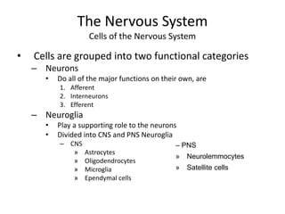
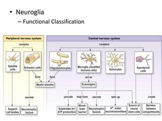
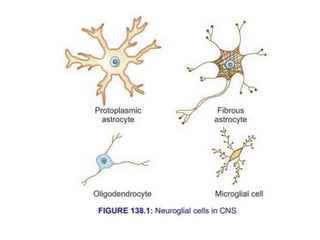
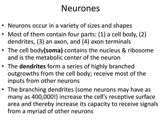
Nervous System & Neuron
kamran
The Nervous System
General Overview
The
integration
of
all
three
major
functions
• What are the major functional components of
the Nervous System?
– Brain
– Spinal Cord
– Afferent Tracts
– Efferent Tracts
– Ganglia
The Nervous System
Functional Components
Central Nervous
System
Peripheral Nervous
System
The Nervous System
Organization of the CNS
• The Brain
– Functional organization
• Neuronal circuits organized into
– Gray matter & White Matter
Gray Matter
Consists of
– Non myelinated axon terminals
– Dendrites
– Cell Bodies
Functions in
– signal processing (integration)
– Neurocrine / neurohormone
secretion
White Matter
Consists of
– Myelinated axons
Functions in
– Formation of tracts
• ascending or descending (SC)
• commissural, association,
projection (B)
The Nervous System
Cells of the Nervous System
• Cells are grouped into two functional categories
– Neurons
• Do all of the major functions on their own, are
1. Afferent
2. Interneurons
3. Efferent
– Neuroglia
• Play a supporting role to the neurons
• Divided into CNS and PNS Neuroglia
– CNS
» Astrocytes
» Oligodendrocytes
» Microglia
» Ependymal cells
– PNS
» Neurolemmocytes
» Satellite cells
• The human CNS contains about 1011(100 billion)
neurons
• Also contains 2–10 times this number of glial cells
• About 40% of the human genes participate, at least
to a degree, in formation of CNS
• The neurons are the basic building blocks of the
nervous system
• Neurons perform the specialized function of
integration & transmission of nerve impulse
• Neurons and glial cells along with brain capillaries
form a functional unit that is required for normal
brain function, including synaptic activity, ECL fluid
homeostasis, energy metabolism, and neural
protection
• Disturbances in the interaction of these elements
are the pathophysiological basis for many
neurological disorders (eg, cerebral ischemia,
seizures, neurodegenerative diseases, and cerebral
edema)
GLIAL CELLS
• Previously, glial cells (or glia) were viewed as CNS
connective/ supporting tissue; also called neuroglia
or glia (glia = glue)
• Today theses cells are recognized for their role in
communication within the CNS in partnership
with neurons
• non-excitable and do not transmit nerve impulse
(action potential)
• Unlike neurons, glial cells continue to undergo
cell division in adulthood and their ability to
proliferate is particularly noticeable after brain
injury (eg, stroke)
• Neuroglia
– Functional Classification
• Most commonly, neuroglial cells constitute the
site of tumors in nervous system
• Two major types of glial cells in the nervous system;
microglia and macroglia
• Microglia; smallest neuroglial cells; derived from
macrophages outside of the nervous system; often
called the macrophages of CNS
• Scavenger cells that remove debris resulting from
injury, infection, and disease (eg, multiple sclerosis,
AIDS-related dementia, Parkinson disease, and
Alzheimer disease)
• Three types of macroglia: oligodendrocytes,
Schwann cells, and astrocytes
• Oligodendrocytes and Schwann cells are involved in
myelin formation around axons in the CNS and
peripheral nervous system, respectively
• Astrocytes are star-shaped neuroglial cells present
in all the parts of the brain; Two types of astrocytes
are found in human brain:
• i. Fibrous astrocytes- occupy mainly the white
matter
ii. Protoplasmic astrocytes- found in gray matter
and have a granular cytoplasm
• Both send processes to blood vessels of brain,
particularly, the capillaries, forming tight junction
with capillary membrane; Tight junction in turn
forms the blood-brain barrier
• Also send processes that envelop synapses and the
surface of nerve cells
• Protoplasmic astrocytes have a membrane potential
that varies with the external K+ concentration but
do not generate propagated potentials
• Produce substances that are tropic to neurons, and
help maintain the appropriate conc. of ions and
neurotransmitters by taking up K+ and the
neurotransmitters glutamate and γ-aminobutyrate
Neurones
• Neurons occur in a variety of sizes and shapes
• Most of them contain four parts: (1) a cell body, (2)
dendrites, (3) an axon, and (4) axon terminals
• The cell body(soma) contains the nucleus & ribosome
and is the metabolic center of the neuron
• The dendrites form a series of highly branched
outgrowths from the cell body; receive most of the
inputs from other neurons
• The branching dendrites (some neurons may have as
many as 400,000!) increase the cell’s receptive surface
area and thereby increase its capacity to receive signals
from a myriad of other neurons
• Particularly in the cerebral and cerebellar cortex,
the dendrites have small knobby projections called
dendritic spines
• The axon, sometimes also called a nerve fiber, is
a single long process that extends from the cell
body to its target cells (from μm to m in length)
• The part of the cell body where the axon is joined is
known as the initial segment, or axon hillock
• The initial segment is the “trigger zone” where, in
most neurons, the electric signals are generated &
are then propagated away from the cell body along
the axon or, sometimes, back along the dendrites
• The main axon may have branches, called
collaterals, along its course
• Near the ends both the main axon and its collaterals
undergo further branching; The greater the degree
of branching of the axon and axon collaterals, the
greater the cell’s sphere of influence
• Each branch ends in an axon presynaptic terminal;
composed of a number of synaptic knobs which are
also called terminal buttons or boutons
• They contain granules or vesicles in which the
synaptic transmitters secreted by the nerves are
stored
• The axons of many neurons are myelinated, that is,
they acquire a sheath of myelin, a protein–lipid
complex that is wrapped around the axon
• In the PNS, myelin forms when a Schwann cell wraps its
membrane around an axon up to 100 times
• The myelin is compacted when the extracellular
portions of a membrane protein called protein zero (P0)
lock to the extracellular portions of P0 in the apposing
membrane
• Various mutations in the gene for P0 cause peripheral
neuropathies; 29 different mutations have been
described that cause symptoms ranging from mild to
severe
• Myelin sheath is not a continuous sheath; absent at
regular intervals (1-μm constrictions that are about 1
mm apart); the area where myelin sheath is absent is
called node of Ranvier
• Segment of the nerve fiber between two nodes
is called internode
• Myelin sheath is responsible for white color of nerve
fibers
• Unmyelinated neurones are simply surrounded by
Schwann cells without the wrapping of the Schwann
cell membrane that produces myelin around the axon
• In the CNS, most neurons are myelinated; the myelin-
forming cells are the oligodendroglia
• Here oligodendrocytes emit multiple processes that
form myelin on many neighboring axons
• In multiple sclerosis, a crippling autoimmune
disease, patchy destruction of myelin occurs in the
CNS
• The loss of myelin is associated with delayed or
blocked conduction in the demyelinated axons
• Neurons are classified by three different methods.
A. Depending upon the number of poles
B. Depending upon the function
C. Depending upon the length of axon
• Depending Upon The Number Of Poles:
• Based on the number of poles from which the nerve
fibers arise, neurons are divided into three types:
• 1. Unipolar Neurons: Neurons that have only one
pole; from a single pole, both axon and dendrite
arise; this type of nerve cells is present only in
embryonic stage in human beings
• 2. Bipolar Neurons: Neurons with two poles are
known as bipolar neurons; Axon arises from one
pole and dendrites arise from the other pole
• 3. Multipolar Neurons: Neurons which have many
poles; one of the poles gives rise to axon and all
other poles give rise to dendrites
• Depending Upon The Function
• On the basis of function, nerve cells are classified into
two types:
1. Motor or efferent neurons
2. Sensory or afferent neurons
• 1. Motor or Efferent Neurons: Neurons which carry
the motor impulses from CNS to peripheral effector
organs like muscles, glands, blood vessels, etc.
Generally, each motor neuron has a long axon and
short dendrites
• 2. Sensory or Afferent Neurons: Neurons which
carry the sensory impulses from periphery to CNS;
generally, each sensory neuron has a short axon and
long dendrites
• Depending Upon The Length Of Axon
• Depending upon the length of axon, neurons are
divided into two types:
1. Golgi type I neurons
2. Golgi type II neurons
• 1. Golgi Type I Neurons: They have long axons; cell
body of these neurons is in different parts of CNS
and their axons reach the remote peripheral organs
• 2. Golgi Type II Neurons: Such neurons have short
axons; present in cerebral cortex and spinal cord
AXONAL TRANSPORT
• Neurons-secretory cell; differ from other secretory
cells in that the secretory zone is generally at the
end of the axon, far away from the cell body
• Protein synthesis occurs in cell body & transported
to the axonal ending by axoplasmic flow
• The functional and anatomic integrity of the axon is
very important; if the axon is cut, the part distal to
the cut degenerates (wallerian degeneration)
• Orthograde transport occurs along microtubules
that run along the length of the axon and requires
two molecular motors, dynein and kinesin
• Orthograde transport moves from the cell body toward
the axon terminals
• Has both fast and slow components; fast axonal
transport occurs at about 400 mm/day, and slow
axonal transport occurs at 0.5 to 10 mm/day
• Retrograde transport, which is in the opposite
direction (from the nerve ending to the cell body),
occurs along microtubules at about 200 mm/day
• Synaptic vesicles recycle in the membrane, but some
used vesicles are carried back to the cell body and
deposited in lysosomes
• Some materials taken up at the ending by endocytosis,
including nerve growth factor (NGF) and some viruses,
are also transported back to the cell body
Neurons.pptx...............................

Neurons.pptx...............................

  • 1.
    Nervous System &Neuron kamran
  • 2.
    The Nervous System GeneralOverview The integration of all three major functions
  • 3.
    • What arethe major functional components of the Nervous System? – Brain – Spinal Cord – Afferent Tracts – Efferent Tracts – Ganglia The Nervous System Functional Components Central Nervous System Peripheral Nervous System
  • 4.
    The Nervous System Organizationof the CNS • The Brain – Functional organization • Neuronal circuits organized into – Gray matter & White Matter Gray Matter Consists of – Non myelinated axon terminals – Dendrites – Cell Bodies Functions in – signal processing (integration) – Neurocrine / neurohormone secretion White Matter Consists of – Myelinated axons Functions in – Formation of tracts • ascending or descending (SC) • commissural, association, projection (B)
  • 5.
    The Nervous System Cellsof the Nervous System • Cells are grouped into two functional categories – Neurons • Do all of the major functions on their own, are 1. Afferent 2. Interneurons 3. Efferent – Neuroglia • Play a supporting role to the neurons • Divided into CNS and PNS Neuroglia – CNS » Astrocytes » Oligodendrocytes » Microglia » Ependymal cells – PNS » Neurolemmocytes » Satellite cells
  • 6.
    • The humanCNS contains about 1011(100 billion) neurons • Also contains 2–10 times this number of glial cells • About 40% of the human genes participate, at least to a degree, in formation of CNS • The neurons are the basic building blocks of the nervous system • Neurons perform the specialized function of integration & transmission of nerve impulse
  • 7.
    • Neurons andglial cells along with brain capillaries form a functional unit that is required for normal brain function, including synaptic activity, ECL fluid homeostasis, energy metabolism, and neural protection • Disturbances in the interaction of these elements are the pathophysiological basis for many neurological disorders (eg, cerebral ischemia, seizures, neurodegenerative diseases, and cerebral edema)
  • 8.
    GLIAL CELLS • Previously,glial cells (or glia) were viewed as CNS connective/ supporting tissue; also called neuroglia or glia (glia = glue) • Today theses cells are recognized for their role in communication within the CNS in partnership with neurons • non-excitable and do not transmit nerve impulse (action potential) • Unlike neurons, glial cells continue to undergo cell division in adulthood and their ability to proliferate is particularly noticeable after brain injury (eg, stroke)
  • 9.
  • 10.
    • Most commonly,neuroglial cells constitute the site of tumors in nervous system • Two major types of glial cells in the nervous system; microglia and macroglia • Microglia; smallest neuroglial cells; derived from macrophages outside of the nervous system; often called the macrophages of CNS • Scavenger cells that remove debris resulting from injury, infection, and disease (eg, multiple sclerosis, AIDS-related dementia, Parkinson disease, and Alzheimer disease)
  • 11.
    • Three typesof macroglia: oligodendrocytes, Schwann cells, and astrocytes • Oligodendrocytes and Schwann cells are involved in myelin formation around axons in the CNS and peripheral nervous system, respectively • Astrocytes are star-shaped neuroglial cells present in all the parts of the brain; Two types of astrocytes are found in human brain: • i. Fibrous astrocytes- occupy mainly the white matter ii. Protoplasmic astrocytes- found in gray matter and have a granular cytoplasm
  • 13.
    • Both sendprocesses to blood vessels of brain, particularly, the capillaries, forming tight junction with capillary membrane; Tight junction in turn forms the blood-brain barrier • Also send processes that envelop synapses and the surface of nerve cells • Protoplasmic astrocytes have a membrane potential that varies with the external K+ concentration but do not generate propagated potentials • Produce substances that are tropic to neurons, and help maintain the appropriate conc. of ions and neurotransmitters by taking up K+ and the neurotransmitters glutamate and γ-aminobutyrate
  • 16.
    Neurones • Neurons occurin a variety of sizes and shapes • Most of them contain four parts: (1) a cell body, (2) dendrites, (3) an axon, and (4) axon terminals • The cell body(soma) contains the nucleus & ribosome and is the metabolic center of the neuron • The dendrites form a series of highly branched outgrowths from the cell body; receive most of the inputs from other neurons • The branching dendrites (some neurons may have as many as 400,000!) increase the cell’s receptive surface area and thereby increase its capacity to receive signals from a myriad of other neurons
  • 18.
    • Particularly inthe cerebral and cerebellar cortex, the dendrites have small knobby projections called dendritic spines • The axon, sometimes also called a nerve fiber, is a single long process that extends from the cell body to its target cells (from μm to m in length) • The part of the cell body where the axon is joined is known as the initial segment, or axon hillock • The initial segment is the “trigger zone” where, in most neurons, the electric signals are generated & are then propagated away from the cell body along the axon or, sometimes, back along the dendrites
  • 20.
    • The mainaxon may have branches, called collaterals, along its course • Near the ends both the main axon and its collaterals undergo further branching; The greater the degree of branching of the axon and axon collaterals, the greater the cell’s sphere of influence • Each branch ends in an axon presynaptic terminal; composed of a number of synaptic knobs which are also called terminal buttons or boutons • They contain granules or vesicles in which the synaptic transmitters secreted by the nerves are stored
  • 21.
    • The axonsof many neurons are myelinated, that is, they acquire a sheath of myelin, a protein–lipid complex that is wrapped around the axon • In the PNS, myelin forms when a Schwann cell wraps its membrane around an axon up to 100 times • The myelin is compacted when the extracellular portions of a membrane protein called protein zero (P0) lock to the extracellular portions of P0 in the apposing membrane • Various mutations in the gene for P0 cause peripheral neuropathies; 29 different mutations have been described that cause symptoms ranging from mild to severe
  • 22.
    • Myelin sheathis not a continuous sheath; absent at regular intervals (1-μm constrictions that are about 1 mm apart); the area where myelin sheath is absent is called node of Ranvier • Segment of the nerve fiber between two nodes is called internode • Myelin sheath is responsible for white color of nerve fibers • Unmyelinated neurones are simply surrounded by Schwann cells without the wrapping of the Schwann cell membrane that produces myelin around the axon • In the CNS, most neurons are myelinated; the myelin- forming cells are the oligodendroglia
  • 23.
    • Here oligodendrocytesemit multiple processes that form myelin on many neighboring axons • In multiple sclerosis, a crippling autoimmune disease, patchy destruction of myelin occurs in the CNS • The loss of myelin is associated with delayed or blocked conduction in the demyelinated axons • Neurons are classified by three different methods. A. Depending upon the number of poles B. Depending upon the function C. Depending upon the length of axon
  • 24.
    • Depending UponThe Number Of Poles: • Based on the number of poles from which the nerve fibers arise, neurons are divided into three types: • 1. Unipolar Neurons: Neurons that have only one pole; from a single pole, both axon and dendrite arise; this type of nerve cells is present only in embryonic stage in human beings • 2. Bipolar Neurons: Neurons with two poles are known as bipolar neurons; Axon arises from one pole and dendrites arise from the other pole • 3. Multipolar Neurons: Neurons which have many poles; one of the poles gives rise to axon and all other poles give rise to dendrites
  • 27.
    • Depending UponThe Function • On the basis of function, nerve cells are classified into two types: 1. Motor or efferent neurons 2. Sensory or afferent neurons • 1. Motor or Efferent Neurons: Neurons which carry the motor impulses from CNS to peripheral effector organs like muscles, glands, blood vessels, etc. Generally, each motor neuron has a long axon and short dendrites • 2. Sensory or Afferent Neurons: Neurons which carry the sensory impulses from periphery to CNS; generally, each sensory neuron has a short axon and long dendrites
  • 28.
    • Depending UponThe Length Of Axon • Depending upon the length of axon, neurons are divided into two types: 1. Golgi type I neurons 2. Golgi type II neurons • 1. Golgi Type I Neurons: They have long axons; cell body of these neurons is in different parts of CNS and their axons reach the remote peripheral organs • 2. Golgi Type II Neurons: Such neurons have short axons; present in cerebral cortex and spinal cord
  • 29.
    AXONAL TRANSPORT • Neurons-secretorycell; differ from other secretory cells in that the secretory zone is generally at the end of the axon, far away from the cell body • Protein synthesis occurs in cell body & transported to the axonal ending by axoplasmic flow • The functional and anatomic integrity of the axon is very important; if the axon is cut, the part distal to the cut degenerates (wallerian degeneration) • Orthograde transport occurs along microtubules that run along the length of the axon and requires two molecular motors, dynein and kinesin
  • 31.
    • Orthograde transportmoves from the cell body toward the axon terminals • Has both fast and slow components; fast axonal transport occurs at about 400 mm/day, and slow axonal transport occurs at 0.5 to 10 mm/day • Retrograde transport, which is in the opposite direction (from the nerve ending to the cell body), occurs along microtubules at about 200 mm/day • Synaptic vesicles recycle in the membrane, but some used vesicles are carried back to the cell body and deposited in lysosomes • Some materials taken up at the ending by endocytosis, including nerve growth factor (NGF) and some viruses, are also transported back to the cell body