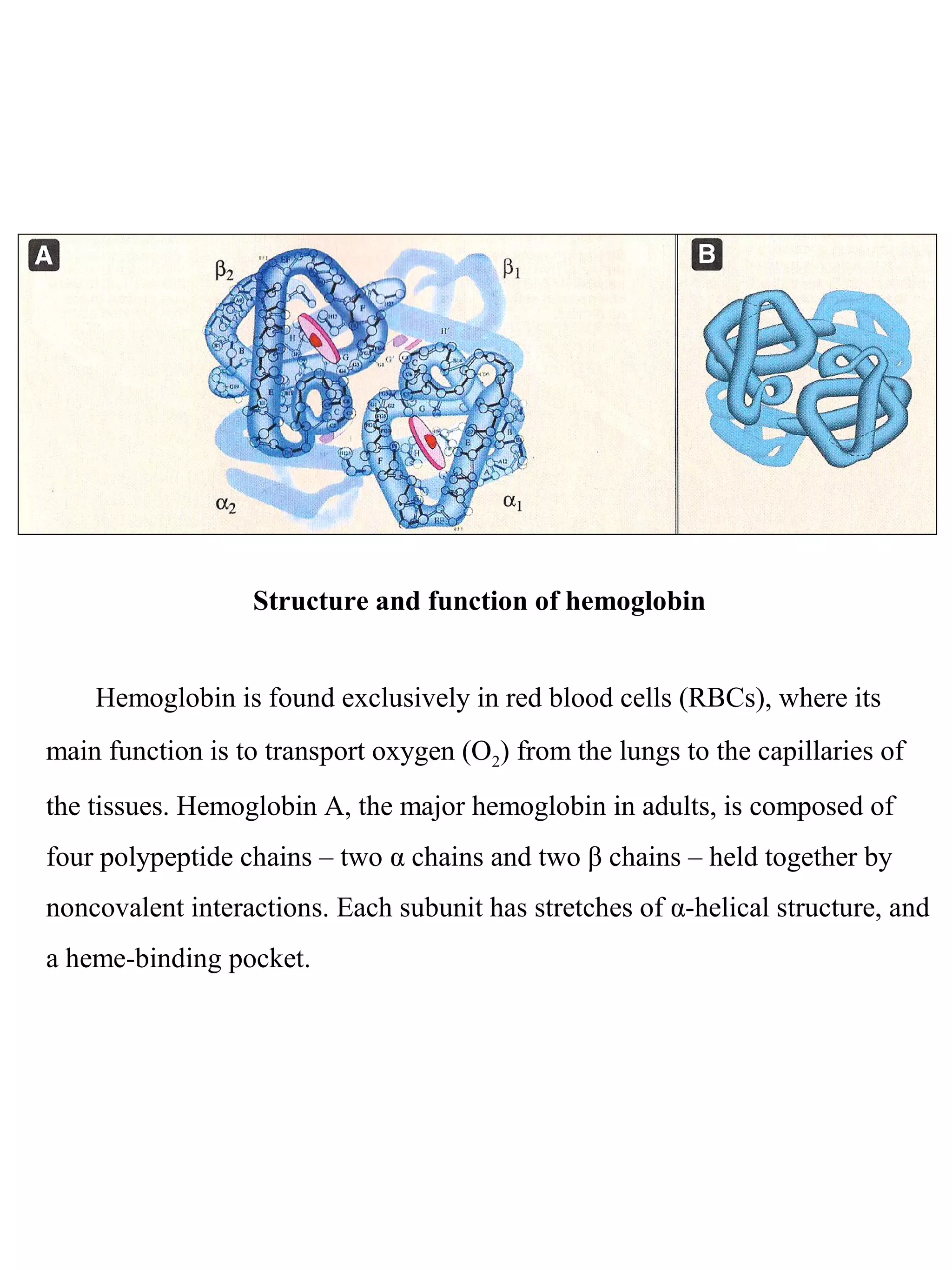
1. Hemoglobin transports oxygen in red blood cells through a cooperative binding mechanism between its four subunits. Each subunit contains a heme group that reversibly binds oxygen.
2. In tissues, higher carbon dioxide and hydrogen ion concentrations cause hemoglobin to release oxygen. However, in the lungs where oxygen levels are high, hemoglobin becomes saturated with oxygen.
3. Sickle cell anemia results from a mutation where glutamate is replaced by valine in the beta chain of hemoglobin. This causes deoxygenated hemoglobin to polymerize and distort red blood cells into a sickle shape, blocking blood flow.
A living organism is a highly complex and unstable system. Its existence is precarious because of its complexity. Metabolism is the sum
total of all biochemical reactions designed to maintain and replicate
the structure of the organism and to counteract a continuous drive
towards an increase in the disorder, or entropy, of the system.
To build complex structures requires energy, i.e., a system needs
energy for synthesis and maintenance. Human metabolism derives
energy from degradation of organized structures (food) from plants,
and, therefore, indirectly from the sun. In more affluent societies of
the world, a significant amount of energy is obtained from animals
which themselves have survived by utilizing plants and other animals.
For the most part, food represents complex organic molecules which
require energy to be synthesized and therefore can yield energy by
being destroyed. (Food also contains some constituents which are
not utilized for energy production, but have important roles in maintaining life, e.g., minerals and vitamins.)
Introduction
Definition
Classification of polysaccharides
1- Homopolysaccharides
2-Heteropolysaccharides
What is heteropolysaccarides?
Type of heteropolysaccharides
Function of heteropolysaccharides
Conclusion
References
Polysaccharide introduction, example, structure, starch, cellulose, chitin those structure and important functions and their presence in plants and animals, polysaccharide types based on functions and their composition , functions of polysaccharides , important images for relevant polysaccharides types, polysaccharide role in plants and animal cells. Starch - structure and functions, cellulose structure and functions, chitin - structure and functions
This Presentation is made for S.Y.Bsc. Students. This presentation includes the structure of nucliec acids DNA, RNA and biological significance of nucliec acids.
This ppt explains the properties of monosaccharides, polysaccharides. the properties like mutarotation, reduction, optical activity, caramerlization, osazone is given in the ppt. Also the determination of ring size of the monosaccharide is explained/
Protein structures, Detail about protein dystrophin DMD and BMD primary structures, secondary structures, tertiary structures, Quaternary structures, functions of proteins ,
different sub types of protein structures, dystropins proteins structures , locations of it in chromosomes, chromosomal abnormalities, facts of Duchenne Muscular Dystrophy
Proteins are polypeptide structures made up of one or more extended chains of residues from the amino acid. They provide a wide range of organism tasks, including as DNA replication, molecule transport, metabolic process catalysis, and cell structural support.
The albumins seen in vast quantities in egg whites typically have a distinct 3D structure as a result of bonds that form between the protein’s various amino acids. These bonds are broken by heating, exposing the hydrophobic (water-hating) amino acids that are typically maintained on the inside of the protein 1, 1 comma, 2 end superscript, 2, start superscript. In an effort to escape the water that surrounds them in the egg white, the hydrophobic amino acids will bind to one another, creating a protein network that gives the egg white structure and makes it white and opaque. Ta-da! Protein denaturation, thank you for another wonderful breakfast
Similar to Proteins and their biological structures (20)
HOT NEW PRODUCT! BIG SALES FAST SHIPPING NOW FROM CHINA!! EU KU DB BK substit...GL Anaacs
Contact us if you are interested:
Email / Skype : kefaya1771@gmail.com
Threema: PXHY5PDH
New BATCH Ku !!! MUCH IN DEMAND FAST SALE EVERY BATCH HAPPY GOOD EFFECT BIG BATCH !
Contact me on Threema or skype to start big business!!
Hot-sale products:
NEW HOT EUTYLONE WHITE CRYSTAL!!
5cl-adba precursor (semi finished )
5cl-adba raw materials
ADBB precursor (semi finished )
ADBB raw materials
APVP powder
5fadb/4f-adb
Jwh018 / Jwh210
Eutylone crystal
Protonitazene (hydrochloride) CAS: 119276-01-6
Flubrotizolam CAS: 57801-95-3
Metonitazene CAS: 14680-51-4
Payment terms: Western Union,MoneyGram,Bitcoin or USDT.
Deliver Time: Usually 7-15days
Shipping method: FedEx, TNT, DHL,UPS etc.Our deliveries are 100% safe, fast, reliable and discreet.
Samples will be sent for your evaluation!If you are interested in, please contact me, let's talk details.
We specializes in exporting high quality Research chemical, medical intermediate, Pharmaceutical chemicals and so on. Products are exported to USA, Canada, France, Korea, Japan,Russia, Southeast Asia and other countries.
Prix Galien International 2024 Forum ProgramLevi Shapiro
June 20, 2024, Prix Galien International and Jerusalem Ethics Forum in ROME. Detailed agenda including panels:
- ADVANCES IN CARDIOLOGY: A NEW PARADIGM IS COMING
- WOMEN’S HEALTH: FERTILITY PRESERVATION
- WHAT’S NEW IN THE TREATMENT OF INFECTIOUS,
ONCOLOGICAL AND INFLAMMATORY SKIN DISEASES?
- ARTIFICIAL INTELLIGENCE AND ETHICS
- GENE THERAPY
- BEYOND BORDERS: GLOBAL INITIATIVES FOR DEMOCRATIZING LIFE SCIENCE TECHNOLOGIES AND PROMOTING ACCESS TO HEALTHCARE
- ETHICAL CHALLENGES IN LIFE SCIENCES
- Prix Galien International Awards Ceremony
Ethanol (CH3CH2OH), or beverage alcohol, is a two-carbon alcohol
that is rapidly distributed in the body and brain. Ethanol alters many
neurochemical systems and has rewarding and addictive properties. It
is the oldest recreational drug and likely contributes to more morbidity,
mortality, and public health costs than all illicit drugs combined. The
5th edition of the Diagnostic and Statistical Manual of Mental Disorders
(DSM-5) integrates alcohol abuse and alcohol dependence into a single
disorder called alcohol use disorder (AUD), with mild, moderate,
and severe subclassifications (American Psychiatric Association, 2013).
In the DSM-5, all types of substance abuse and dependence have been
combined into a single substance use disorder (SUD) on a continuum
from mild to severe. A diagnosis of AUD requires that at least two of
the 11 DSM-5 behaviors be present within a 12-month period (mild
AUD: 2–3 criteria; moderate AUD: 4–5 criteria; severe AUD: 6–11 criteria).
The four main behavioral effects of AUD are impaired control over
drinking, negative social consequences, risky use, and altered physiological
effects (tolerance, withdrawal). This chapter presents an overview
of the prevalence and harmful consequences of AUD in the U.S.,
the systemic nature of the disease, neurocircuitry and stages of AUD,
comorbidities, fetal alcohol spectrum disorders, genetic risk factors, and
pharmacotherapies for AUD.
Tom Selleck Health: A Comprehensive Look at the Iconic Actor’s Wellness Journeygreendigital
Tom Selleck, an enduring figure in Hollywood. has captivated audiences for decades with his rugged charm, iconic moustache. and memorable roles in television and film. From his breakout role as Thomas Magnum in Magnum P.I. to his current portrayal of Frank Reagan in Blue Bloods. Selleck's career has spanned over 50 years. But beyond his professional achievements. fans have often been curious about Tom Selleck Health. especially as he has aged in the public eye.
Follow us on: Pinterest
Introduction
Many have been interested in Tom Selleck health. not only because of his enduring presence on screen but also because of the challenges. and lifestyle choices he has faced and made over the years. This article delves into the various aspects of Tom Selleck health. exploring his fitness regimen, diet, mental health. and the challenges he has encountered as he ages. We'll look at how he maintains his well-being. the health issues he has faced, and his approach to ageing .
Early Life and Career
Childhood and Athletic Beginnings
Tom Selleck was born on January 29, 1945, in Detroit, Michigan, and grew up in Sherman Oaks, California. From an early age, he was involved in sports, particularly basketball. which played a significant role in his physical development. His athletic pursuits continued into college. where he attended the University of Southern California (USC) on a basketball scholarship. This early involvement in sports laid a strong foundation for his physical health and disciplined lifestyle.
Transition to Acting
Selleck's transition from an athlete to an actor came with its physical demands. His first significant role in "Magnum P.I." required him to perform various stunts and maintain a fit appearance. This role, which he played from 1980 to 1988. necessitated a rigorous fitness routine to meet the show's demands. setting the stage for his long-term commitment to health and wellness.
Fitness Regimen
Workout Routine
Tom Selleck health and fitness regimen has evolved. adapting to his changing roles and age. During his "Magnum, P.I." days. Selleck's workouts were intense and focused on building and maintaining muscle mass. His routine included weightlifting, cardiovascular exercises. and specific training for the stunts he performed on the show.
Selleck adjusted his fitness routine as he aged to suit his body's needs. Today, his workouts focus on maintaining flexibility, strength, and cardiovascular health. He incorporates low-impact exercises such as swimming, walking, and light weightlifting. This balanced approach helps him stay fit without putting undue strain on his joints and muscles.
Importance of Flexibility and Mobility
In recent years, Selleck has emphasized the importance of flexibility and mobility in his fitness regimen. Understanding the natural decline in muscle mass and joint flexibility with age. he includes stretching and yoga in his routine. These practices help prevent injuries, improve posture, and maintain mobilit
Title: Sense of Taste
Presenter: Dr. Faiza, Assistant Professor of Physiology
Qualifications:
MBBS (Best Graduate, AIMC Lahore)
FCPS Physiology
ICMT, CHPE, DHPE (STMU)
MPH (GC University, Faisalabad)
MBA (Virtual University of Pakistan)
Learning Objectives:
Describe the structure and function of taste buds.
Describe the relationship between the taste threshold and taste index of common substances.
Explain the chemical basis and signal transduction of taste perception for each type of primary taste sensation.
Recognize different abnormalities of taste perception and their causes.
Key Topics:
Significance of Taste Sensation:
Differentiation between pleasant and harmful food
Influence on behavior
Selection of food based on metabolic needs
Receptors of Taste:
Taste buds on the tongue
Influence of sense of smell, texture of food, and pain stimulation (e.g., by pepper)
Primary and Secondary Taste Sensations:
Primary taste sensations: Sweet, Sour, Salty, Bitter, Umami
Chemical basis and signal transduction mechanisms for each taste
Taste Threshold and Index:
Taste threshold values for Sweet (sucrose), Salty (NaCl), Sour (HCl), and Bitter (Quinine)
Taste index relationship: Inversely proportional to taste threshold
Taste Blindness:
Inability to taste certain substances, particularly thiourea compounds
Example: Phenylthiocarbamide
Structure and Function of Taste Buds:
Composition: Epithelial cells, Sustentacular/Supporting cells, Taste cells, Basal cells
Features: Taste pores, Taste hairs/microvilli, and Taste nerve fibers
Location of Taste Buds:
Found in papillae of the tongue (Fungiform, Circumvallate, Foliate)
Also present on the palate, tonsillar pillars, epiglottis, and proximal esophagus
Mechanism of Taste Stimulation:
Interaction of taste substances with receptors on microvilli
Signal transduction pathways for Umami, Sweet, Bitter, Sour, and Salty tastes
Taste Sensitivity and Adaptation:
Decrease in sensitivity with age
Rapid adaptation of taste sensation
Role of Saliva in Taste:
Dissolution of tastants to reach receptors
Washing away the stimulus
Taste Preferences and Aversions:
Mechanisms behind taste preference and aversion
Influence of receptors and neural pathways
Impact of Sensory Nerve Damage:
Degeneration of taste buds if the sensory nerve fiber is cut
Abnormalities of Taste Detection:
Conditions: Ageusia, Hypogeusia, Dysgeusia (parageusia)
Causes: Nerve damage, neurological disorders, infections, poor oral hygiene, adverse drug effects, deficiencies, aging, tobacco use, altered neurotransmitter levels
Neurotransmitters and Taste Threshold:
Effects of serotonin (5-HT) and norepinephrine (NE) on taste sensitivity
Supertasters:
25% of the population with heightened sensitivity to taste, especially bitterness
Increased number of fungiform papillae
MANAGEMENT OF ATRIOVENTRICULAR CONDUCTION BLOCK.pdfJim Jacob Roy
Cardiac conduction defects can occur due to various causes.
Atrioventricular conduction blocks ( AV blocks ) are classified into 3 types.
This document describes the acute management of AV block.
The prostate is an exocrine gland of the male mammalian reproductive system
It is a walnut-sized gland that forms part of the male reproductive system and is located in front of the rectum and just below the urinary bladder
Function is to store and secrete a clear, slightly alkaline fluid that constitutes 10-30% of the volume of the seminal fluid that along with the spermatozoa, constitutes semen
A healthy human prostate measures (4cm-vertical, by 3cm-horizontal, 2cm ant-post ).
It surrounds the urethra just below the urinary bladder. It has anterior, median, posterior and two lateral lobes
It’s work is regulated by androgens which are responsible for male sex characteristics
Generalised disease of the prostate due to hormonal derangement which leads to non malignant enlargement of the gland (increase in the number of epithelial cells and stromal tissue)to cause compression of the urethra leading to symptoms (LUTS
micro teaching on communication m.sc nursing.pdfAnurag Sharma
Microteaching is a unique model of practice teaching. It is a viable instrument for the. desired change in the teaching behavior or the behavior potential which, in specified types of real. classroom situations, tends to facilitate the achievement of specified types of objectives.
Ozempic: Preoperative Management of Patients on GLP-1 Receptor Agonists Saeid Safari
Preoperative Management of Patients on GLP-1 Receptor Agonists like Ozempic and Semiglutide
ASA GUIDELINE
NYSORA Guideline
2 Case Reports of Gastric Ultrasound
1. Catalyzing substrates
1. Chemical evolution, in
which biopolymers were
formed from small molecules
Proteins and
nucleic acids
2. Self organization, in which
biopolymers developed the
capacity for self-replication
3. Biological evolution, in which
primitive living cells generated
sophisticated metabolic systems
and eventually the ability to form
multicellular organisms.
2.
3.
4. 1. Enzymes (more 2000 proteins) - very specific catalysis up to 1020
times
2. Regulatory proteins (hormones, gene expression)
3. Transport proteins (Hb, HSA, membrane proteins)
4. Storage proteins (ovalbumin, casein, ferritin - 460kD -4500 atoms of
iron)
5. Contractile and motile proteins (actin, myosin, tubulin, dynein,
kinesin)
6. Structural proteins (keratins, collagen -1/3 of total)
7. Protective and exploitive proteins (immunoglobulins, thrombin,
fibrinogen)
8. Exotic proteins (antifreeze proteins, monellin – sweetener,
resilin – elastics, glue proteins)
BIOLOGICAL FUNCTIONS OF PROTEINS
5. Although more than 300 different
amino acids have been described in nature,
only 20 are commonly found as constituents
of mammalian proteins. These are the only
amino acids that are coded for by DNA, the
genetic material in the cell. At physiologic
pH, the carboxyl group is dissociated,
forming the negatively charged carboxylate
ion (–COO-
), and the amino group is
protonated (–NH3
+
). In proteins, almost all
carboxyl and amino groups are combined
through peptide linkage and, in general, are
not available for chemical reaction except
for hydrogen bond formation.
8. Protein function can be understood only in terms of protein structure
1. Primary structure is the amino acid sequence of its polypeptide chain
2. Secondary structure in the local spatial arrangement of a polypeptide’s
backbone atoms without regards to the conformations f its side chain
3. Tertiary structure refers to the three-dimensional structure of an entire
polypeptide
4. Quaternary structure refers to the spatial arrangement of the subunits of the
protein associated through noncovalent interactions and disulfide bonds.
(a) – Lys – Ala – His – Gly – Lys – Lys – Val – Leu – Gly – Ala –
Primary structure (amino acid sequence in a polypeptide chain)
(b)
Secondar
y
structure
(helix)
(c) (d) β2
β
Tertiary structure: one
complete protein chain
(β chain of
hemoglobin)
β1
α2 α1
Quatemary structure:
the four separate chains
of hemoglobin
assembled into an
oligomeric protein
The structural hierarchy in proteins: (a) primary structure, (b) secondary structure, (c) tertiary
structure, and (d) quaternary structure.
9. Interactions between the side chains of amino acid residues in proteins.
1- electrostatic interactions; 2 – hydrogen bonds; 3 – hydrophobic
interactions; 4 – disulfide bonds.
10. SECONDARY STRUCTURE OF PROTEINS
The polypeptide backbone forms regular
arrangements of amino acids that are located
near to each other in the linear sequence.
These arrangements are termed the secondary
structure of the polypeptide. The α-helix, β-
sheet, and β-bend (β-turn) are examples of
secondary structures frequently encountered
in proteins.
11. The 20 amino acids commonly
found in proteins are joined together by
peptide bonds. The linear sequence of
the linked amino acids contains the
information necessary to generate a
protein molecule with a unique three-
dimensional shape. The complexity of
protein structure is best analyzed by
considering the molecule in terms of
four organizational levels, namely,
primary, secondary, tertiary, and
quaternary.
12. Interactions stabilizing tertiary structure
The unique three-dimensional structure
of each polypeptide is determined by its
amino acid sequence. Interactions between
the amino acid side chains guide the folding
of the polypeptide to form a compact
structure.
1. Disulfide bonds
2. Hydrophobic interactions
3. Hydrogen bonds
4. Ionic interactions
14. PROTEIN FOLDING
Protein folding, which occurs within the cell in seconds to minutes, employs
a shortcut through the maze of all folding possibilities. For example, positively
and negatively charged side chains attract each other, conversely, similarly
charged side chains repel each other. In addition, interactions involving hydrogen
bonds, hydrophobic interactions, and disulfide bonds all exert an influence on the
folding process. This results in a correctly folded protein with a low-energy state.
DENATURATION OF PROTEINS
Protein denaturation results in the unfolding and disorganization of the
protein’s secondary and tertiary structures, which are not accompanied by
hydrolysis of peptide bonds. Denaturing agents include heat, organic solvents,
mechanical mixing, strong acids or bases, detergents, and ions of heavy metals
such as leas and mercury. Most proteins, once denatured, remain permanently
disordered. Denatured proteins are often insoluble and, therefore, precipitate from
solution.
15. Structure and function of hemoglobin
Hemoglobin is found exclusively in red blood cells (RBCs), where its
main function is to transport oxygen (O2) from the lungs to the capillaries of
the tissues. Hemoglobin A, the major hemoglobin in adults, is composed of
four polypeptide chains – two α chains and two β chains – held together by
noncovalent interactions. Each subunit has stretches of α-helical structure, and
a heme-binding pocket.
16. Amino acid substitution in Hb S β chains:
A molecule of Hb S contains two normal α-
globin chains and two mutant β-globin chains
(βS
), in which glutamate at position six has
been replaced with valine.
The replacement of the charged
glutamate with the nonpolar valine forms a
protrusion on the β-globin that fits into a
complementary site on the β chain of another
hemoglobin molecule in the cell. At low
oxygen tension, deoxyhemoglobin S
polymerizes inside the RBC, forming a
network of fibrous polymers that stiffen and
distort the cell, producing rigid, misshapen
erythrocytes. Such sickled cells frequently
block the flow of blood in the narrow
capillaries. This interruption in the supply of
oxygen leads to localized anoxia (oxygen
deprivation) in the tissue, causing pain and
eventually death (infarction) of cells in the
vicinity of the blockage.
17. 1. Amino acid substitution in Hb S β chains:
A molecule of Hb S contains two normal α-globin chains
and two mutant β globin chains (βS
), in which glutamate
at position six has been replaced with valine.
2. Sickling and tissue anoxia: The replacement
of the charged glutamate with the nonpolar valine
forms a protrusion on the β-globin that fits into a
complementary site on the β chain of another
hemoglobin molecule in the cell. At low oxygen tension,
deoxyhemoglobin S polymerizes inside the RBC,
forming a network of fibrous polymers that stiffen and
distort the cell, producing rigid, misshapen
erythrocytes. Such sickled cells frequently block the
flow of blood in the narrow capillaries. This
interruption in the supply of oxygen leads to localized
anoxia (oxygen deprivation) in the tissue, causing pain
and eventually death (infarction) of cells in the vicinity
of the blockage.
18.
19. Hemeproteins are a group of
specialized proteins that contain heme as a
tightly bound prosthetic group. The role of
the heme group is dictated by the
environment created by the three-
dimensional structure of the protein. In
hemoglobin and myoglobin, the two most
abundant heme-proteins in humans, the
heme group serves to reversibly bind
oxygen.
Heme is a complex of protoporphyrin
IX and ferrous iron (Fe2+
). The iron is held
in the center of the heme molecule by bonds
to the four nitrogens of the porphyrin ring.
The heme Fe2+
can form two additional
bonds, one on each side of the planar
porphyrin ring. In myoglobin and
hemoglobin, one of these positions is
coordinated to the side chain of a histidine
residue of the globin molecule, whereas the
other position is available to bind oxygen.
20. Structure and function of myoglobin
Myoglobin, a hemeprotein present in heart and skeletal
muscle, functions both as a reservoir for oxygen, and as an
oxygen carrier. Myoglobin consists of a single polypeptide
chain that is structurally similar to the individual subunit
polypeptide chains of the hemoglobin molecule.
Myoglobin is a compact molecule, with approximately 80
% of its polypeptide chain folded into eight stretches of α-
helix.
21. An artist's rendering of sperm whale myoglobin. One of the heme group's propionic
acid side chains has been displaced for clarity. The amino acid residues are
consecutively numbered, starting from the N-terminus, and the eight helices are
likewise designated A through H.
22. Sickle cell hemoglobin (HbS) Glu (β-chain) replaced by Val
DeoxyHbS undergoes self association and forms a liquid
crystalline phase, which distorts the erythrocytes into a sickle
shape. Sickling: leads to their aggregation and to a decrease in
blood circulation (anemia, tissue infarction and chronic failure of
organ function)
HEMOGLOBIN: colored component of the blood 34% solution
in erythrocytes
Mr 64500 - tetramer composed of 2 polypeptide chain pairs
With a Fe2+ content of 0.334%, the total of 950g Hb in a human
represent 3.5 g or 80% of the total body iron
Several forms of human Hb (α, β, γ, ε, δ)
Presently known 153 abnormal Hb (87 are variations of β-chain)
β-Thalassemia – no β-chain – patients contains HbF instead HbA1
23. Quaternary structure of hemoglobin: The hemoglobin tetramer
can be envisioned as being composed of two identical dimers. The two
polypeptide chains within each dimer are held tightly together, primarily
by hydrophobic interactions. Ionic and hydrogen bonds also occur
between the members of the dimer.
• T form: The deoxy form of hemoglobin is called the “T” (tense)
form. In the T form, the two αβ dimers interact through a network
of ionic bonds and hydrogen bonds that constrain the movement of
the polypeptide chains. The T form is the low-oxygen-affinity form
of hemoglobin.
• R form: The binding of oxygen to hemoglobin causes the rupture
of some of the ionic bonds and hydrogen bonds between the αβ
dimers. This leads to a structure called the “R”, or relaxed form, in
which the polypeptide chains have more freedom of movement. The
R form is the high-oxygen-affinity form of hemoglobin.
24. Aggregation or association of two or more identical or
different polypeptide chains by noncovalent interaction
leading to stable oligomeric (or multimeric) structure.
650 proteins (including 500 enzymes) with subunit
structure had been described by 1974. Most of the known
multimeric proteins contain either 2 or 4 similar sized
subunits.
Far less common are proteins with uneven numbers of
subunits (by 1973, 28 trimeric proteins, including 23
enzymes; 5 pentameric, 2 heptameric and 1 nonameric).
Possession of quarternary structure appear to confer a
flexibility of shape and activity, which is necessary for the
physiological role of the proteins. Monomers derived from
multimeric enzymes are usually inactive.
The subunit composition can be determined by
dissociation of aggregate and investigation of the separate
subunits by:ultracentrifugation, polyaerylamide disc
electrophoresis, gel-filtration, ion-exchange chromatography
The structure of the intact aggregate can be studied by:
electron microscopy, low angle X-ray or neutron diffraction.
QUARTERNARY STRUCTURE OF PROTEINS
25. The binding of a hypothetical pair of proteins by two ionic
bonds, one hydrogen bond, and one large combination of
hydrophobic and van der Waals interactions. The structural
complementarity of the surfaces of surfaces of the two molecules
gives rise to this particular combination of weak bonds and hence to
the specificity of binding between the molecules.
26. Binding of oxygen to myoglobin and
hemoglobin
Myoglobin can bind only one molecule of
oxygen. Hemoglobin can bind four oxygen molecules
– one at each of its four heme groups.
Oxygen dissociation curve: A plot of Y
measured at different partial pressures of oxygen
(pO2) is called the oxygen dissociation curve. This
graph illustrates that myoglobin has a higher oxygen
affinity at all pO2 values than does hemoglobin.
• Myoglobin (Mb): The oxygen dissociation
curve for myoglobin has a hyperbolic shape. The
equilibrium is shifted to the right or to the left as
oxygen is added to or removed from the system.
Hemoglobin (Hb): The oxygen dissociation
curve for hemoglobin is sigmoidal in shape,
indicating that the subunits cooperate in binding
oxygen. Cooperative binding of oxygen by the
four subunits of hemoglobin means that the
binding of an oxygen molecule at one heme
group increases the oxygen affinity of the
remaining heme groups in the same hemoglobin
molecule. Allosteric effects
The ability of hemoglobin to reversibly
bind oxygen is affected by the pO2, the pH of
the environment, the partial pressure of
carbon dioxide, pCO2, and the availability of
2,3-bisphosphoglycerate. These are
collectively called allosteric (“other site”)
effectors.
27. Heme-heme interactions
The net effect is that the affinity of
hemoglobin for the last oxygen bound is
approximately 300 times greater than its affinity
for the first oxygen bound.
Loading and unloading oxygen
For example, in the lung, the concentration of
oxygen is high and hemoglobin becomes virtually
saturated (or “loaded”) with oxygen. In contrast,
in the peripheral tissues, oxyhemoglobin releases
(or “unloads”) much of its oxygen for use in the
oxidative metabolism of the tissues.
The concentration of both CO2 and H+
in the
capillaries of metabolically active tissues is higher
than that observed in alveolar capillaries of the
lungs. In the tissues, CO2 is converted by carbonic
anhydrase to carbonic acid:
CO2 + H2O H2CO3
H2CO3 HCO3
–
+ H+
which spontaneously loses a proton, becoming
bicarbonate:
The H+
contributes to the lowering of pH.
This differential pH gradient (lungs having a
higher pH, tissues a lower pH) favors the
unloading of oxygen in the peripheral tissues, and
the loading of oxygen in the lung. Thus, the oxygen
affinity of the hemoglobin molecule responds to
small shifts in pH between the lungs and oxygen-
consuming tissues, making hemoglobin a more
efficient transporter of oxygen.
Source of the protons that lower the pH
28. Effect of 2,3-bisphosphoglycerate on oxygen affinity
It is the most abundant organic phosphate in the
RBC, where its concentration is approximately that of
hemoglobin. 2,3-BPG is synthesized from an intermediate
of the glycolytic pathway.
• Binding of 2,3-BPG to deoxyhemoglobin: 2,3-BPG
decreases the oxygen affinity of hemoglobin by binding to
deoxyhemoglobin. This binding stabilizes the taut
conformation of deoxyhemoglobin.
HbO2 + 2,3-BPG Hb–2,3-BPG + O
oxyhemoglobin deoxyhemoglobin
• One molecule of 2,3-BPG binds to a pocket, formed by
the two β-globin chains, in the center of the
deoxyhemoglobin tetramer. This pocket contains several
positively charged amino acids that form ionic bonds with
the negatively charged phosphate groups of 2,3-BPG.
• Shift of the oxygen dissociation curve: The presence of
2,3-BPG significantly reduces the affinity of hemoglobin
for oxygen, shifting the oxygen dissociation curve to the
right. This reduced affinity enables hemoglobin to release
oxygen efficiently found in the tissues.
• Response of 2,3-BPG levels to chronic hypoxia or
anemia: The concentration of 2,3-BPG in the RBC
increases in response to chronic hypoxia, such as that
observed in chronic obstructive pulmonary disease
(COPD) like emphysema, or at high altitudes, where
circulating hemoglobin may have difficulty receiving
sufficient oxygen.
29. 1. Amino acid substitution in Hb S β chains:
A molecule of Hb S contains two normal α-globin chains
and two mutant β globin chains (βS
), in which glutamate
at position six has been replaced with valine.
2. Sickling and tissue anoxia: The replacement
of the charged glutamate with the nonpolar valine
forms a protrusion on the β-globin that fits into a
complementary site on the β chain of another
hemoglobin molecule in the cell. At low oxygen tension,
deoxyhemoglobin S polymerizes inside the RBC,
forming a network of fibrous polymers that stiffen and
distort the cell, producing rigid, misshapen
erythrocytes. Such sickled cells frequently block the
flow of blood in the narrow capillaries. This
interruption in the supply of oxygen leads to localized
anoxia (oxygen deprivation) in the tissue, causing pain
and eventually death (infarction) of cells in the vicinity
of the blockage.
30.
31. 1. H+
decreases affinity to O2
2. Blood to tissues → acidification
3. Binding of H+
to Hb conformation
4. Lung O2 to Hb, H+
release
5. Enhancement of pH
6. Blood – O2 binding, tissue – O2
release
BOHR'S EFFECT
Effect of H+
on oxygen binding by hemoglobin (Hb). A. In the tissues,
CO2 is released. In the red blood cell, this CO2 forms carbonic acid, which
releases protons. The protons bind to Hb, causing it to release O2 to the
tissues. B. In the lungs, the reactions are reversed. O2 binds to protonated
Hb, causing the release of protons. They bind to bicarbonate (HCO3
-
),
forming carbonic acid which is cleaved to H2O and CO2, which is exhaled.
32. 1. Selection of protein source (tissues, cells, E.coli, yeasts, cloning)
2. Solubilization
a. cytosol - osmotic lysis of cells by hypotonic solution + centrifugation
b. membrane - homogenization + centrifugation
3. Stabilization - isolation in buffers, temperature, inhibitors of proteases
PROTEIN ISOLATION
PROTEIN PURIFICATION
CHARACTERISTIC PROCEDURE
1. CHARGE 1.Ion-exchange chromatography
2. Electrophoresis
3. Isoelectric focusing
2. POLARITY 1. Adsorption chromatography
2. Paper chromatography
3.Reverse-phase chromatography
4.Hydrophobic chromatography
3. SIZE 1. Dialysis and ultrafiltration
2. Gel electrophoresis
3. Gel filtration chromatography
4. Ultracentrifugation
4. SPECIFICITY 1. Affinity chromatography
33. In the process of ion exchange, ions that electrostatically bound to an insoluble and chemically inert
matrix are reversibly replaced by ions in solution:
R + A-
+ = R + B-
+ A-
A schematic diagram illustrating the separation of several
proteins by ion exchange chromatography using stepwise elution.
A device for generating a
linear concentration gradient. Two
connected open chambers, which
have identical cross-sectional
areas, are initially filled with
equal volumes of solutions of
different concentrations.
As the column is washed, a process known as elution, those proteins with
relatively low affinities to ion exchanger move through the column faster than the
proteins that bind to the ion exchanger with higher affinities.
ION-EXCHANGE CHROMATOGRAPHY
34. The molecules are separated according to their size and shape
Molecules with molecular masses ranging below the exclusion limit
(the molecular mass of the smallest molecule unable to penetrate the
pores) of a gel will elute from the gel in the order of their molecular
masses, with the largest eluting first.
Dialysis is a process that
separates molecules
according to size through
the use of semipermeable
membranes containing
pores of less then
macromolecular
dimensions
GEL FILTRATION CHROMATOGRAPHY
At start of
dialysis
(a) (b) At equilibrium
Dialysis
bad
Buffer
Concentrated
solution
35. Molecule known as a ligand, specifically binds to the protein of interest is
covalently attached to an inert and porous matrix
The separation of
macromolecules by affinity
chromatography.
The formation of cyanogen bromide-
activated agarose (top) and its reaction
with a primary amine to form a covalently
attached ligand for affinity
-chromatography (bottom).
Examples of the various types of nucleophilic groups that can be covalently
attached to epoxy-activated agarose via reation with its epoxide groups.
AFFINITY CHROMATOGRAPHY
36. ELECTROPHORESIS
Migration of ions in an electric field
Polyacrylamide and agarose
Paper electrophoresis, (a) A schematic diagram of
the apparatus used. The sample is applied to a point on
the buffer-moistened paper. The ends of the paper are
dipped into reservoirs of buffer in which the electrodes
are immersed and an electric field is applied. Uncharged
molecules remain the point of sample application.
Visualization with amido black, Coomassie brilliant blue, silver stain, radioactivity
–
+
–
+
Cathode Cathode
Plastic
frame Plastic
frame
Anode
Anode
Buffer
Buffer
Sample
wells
Sample
wells
Sample
Stacking
gel
Running
gel
37. [CH3-(CH2)10-CH2-O-SO3
-
]Na+
– sodium dodecyl sulfate SDS-treated
protein stend to have identical charge-to-mass ratios and similar shapes.
SDS-PAG ELECTROPHORESIS
The SDS – polyacrylamide
electrophoresis pattern of the
supernatant (left) and membrane
fractions (right) of various strains of
the Salmonella typhimurium. Samples
of 200-µg of protein each were run in
parallel lanes on a 35-cm - long x 0.8-
mm-thick slab gel containing 10%
polyacrylamide. The lane marked
MW contains molecular weight
standards.
A logarithmic plot of the
molecular masses of 37 different
polypeptide chains ranging from
11 to 70 kD versus their relative
electrophoretic mobilities on an
SDS-polyacrylamide gel.
Many proteins contain more than one polypeptide chain → SDS
treatment disrupt the noncovalent interactions between these subunits
38. If a mixture of proteins is electrophoresed through a
solution having stable pH gradient in which the pH smoothly
increases from anode to catode, each protein will migrate to
the position in the pH gradient corresponding to its isoelectric
point.
IMMUNOBLOTTING
ISOELECTRIC FOCUSING
The detection of proteins by immunoblotting