SlideShare a Scribd company logo
NERVOUS SYSTEM
PRESENTED BY:- PANDIT ROHIT
A
B.SC. NURSING 3RD YR
2019-20
Presentation
INTRODUCTION
• Nervous system is the chief controlling and coordinating
system of the body.
• It is the body’s most organised, complex, structural and
functional system.
• It is responsible for the judgement, intellegence and
memory.
• It detects and responds to changes occur inside and
outside the body.
• It consists :- brain, spinal cord and peripheral nerves.
• With a mass of only 2kg, the nervous system is 3% of
total body weight (almost).
• The size of the nervous system ranges from a few
hundred cells in the simplest worms, to around 300 billion
cells in African elephants.
• Neural signaling is capable of a much higher level. The
fastest nerve signals travel at speeds that exceed 100
meters per second.
• According to the U.S. National Library of Medicine
there are more than 600 neurologic diseases.
• Most neurons send signals via their axons, although
some types are capable of dendrite-to-dendrite
communication. (In fact, the types of neurons called
amacrine cells have no axons, and communicate only
via their dendrites).
• Most amacrine cells are inhibitory neurons in the
vertebrate retina, containing the common inhibitory
neurotransmitters GABA.
• The nervous system is made up of nervous tissues and
nervous tissues made up of Neurons and Neuroglia(Neuroglial
cells).
• At the cellular level, the nervous system is defined by the
presence of two special type of cells, called the NEURON and
GLIAL cells
• Neurology :- deals with normal functioning and disorders
of the nervous system.
• Neurologist :- physician who diagnose and treat
disorders related to nervous system through various
diagnostic tests like :- EEG, MRI, CAT Scan.
• Neurosurgeon :- A neurosurgeon is a doctor having a
degree of MS(master of surgery) and treat patients
through surgery.
• “In India, there is one neurosurgeon available per 10 lakh
people. The ideal ratio should be at least one
neurosurgeon for every one lakh population
NERVE
• A nerve is an enclosed, cable-like bundle of nerve fibers
(called axons) in the peripheral nervous system.
• A nerve transmits electrical impulses.
• A nerve provides a common pathway for the
electrochemical nerve impulses called action potentials
that are transmitted along each of the axons.
• Within a nerve, each axon is surrounded by a layer of
connective tissue called the endoneurium.
ENDONEURIUM
• Also called Henle’s sheath.
• It is a layer of delicate connective tissue around the
myelin sheath of each myelinated nerve fiber in the PNS.
• Its component cells are called endoneurial cells.
• The endoneurium contains a liquid known as endoneurial
fluid.
• Peripheral nerve injuries commonly release increased
amounts of endoneurial fluid into surrounding tissues;
these can be detected by MRI.
NERVE FASCICLE
• A nerve fascicle, is a bundle of nerve fibers.
• A nerve fascicle is enclosed by perineurium.
• Fascicles enclose many thousands of axons.
PERINEURIUM
• The perineurium is a protective sheath that
surrounds a nerve fascicle.
• The perineurium is composed from fibroblasts.
EPINEURIUM
• The epineurium is the outermost layer of dense irregular
connective tissue surrounding a peripheral nerve.
• It usually surrounds multiple nerve fascicles as well as blood
vessels which supply the nerve.
• Epineurium is primarily made from collagen.
• The epineurium is usually most abundant around joints, as its
function is to protect the nerves from stretching.
1. NEURON
• Neurons can be distinguished from other cells in a
number of ways, but their most fundamental property is
that they communicate with other cells via synapses.
• Some nerve cells can be comparatively smaller by 0.1
millimeters or can be longer by 1 meter. The size of
nerve cells is usually based on their functions i.e how
long electrical impulse is transmitted within our body.
• The length of a nerve cell can vary from nanometers to
meters.
• Each neuron is made up of
1. Cell body/ Cyton/ Soma/ Perikaryon
2. Dendrites
3. Axon
• Neurotrophins: These are proteins which are required for
the growth and survival of neurons. These are produced
by astrocytes
PROTEINS
• Neurotrophins are a family of proteins that induce the survival,
development, and function of neurons.
• Glial cells(Astrocyte) that exist in the peripheral nervous system,
which secrete NGF (Neurotrophins).
• Neurotrophins are chemicals that help to stimulate and control
neurogenesis.
A)Cell body :-
• It contains uni-nucleated cytoplasm.
• Centriole is absent and thus cell division doesn’t take
place.
• Nissls’s granules are responsible for protein synthesis.
• Collectively they form gray matter and are found at
periphery of the brain and spinal cord.
• The cell body contains genetic information, maintains the
neuron's structure, and provides energy to drive
activities.
B) Dendrites :-
Dendrites are many short richely branched and oftenly
swallen branches of neuron, that recieve and carry
incoming impulses towards cell body.They resemble a
tree-like structure
Conduct the electrochemical charge to the cell body
C) Axon :-
Axon, also called nerve fibre, portion of a nerve cell
(neuron) that carries nerve impulses away from the cell
body.
Mostly(normally) a neuron has one axon that connects it
with other neurons
The longest axons of the human body are those that make
up the sciatic nerve where the length can exceed one
meter.
MYELINOGENESIS
1. PNS
• Axon is covered by a layer of phospholipids which is called
myelin sheath
• This myelin sheath is covered by thin membrane called
neurilemma.
• Where the sheath is discontious around axon, are called
nodes of ranvier
• This myelin sheath prevents leakage of ions.
• The plasma membrane layer of Schwann cells is called the neurilemma.
2. CNS
Neurilemma/Schwann cell are absent in CNS. Therefore,
myelination occurs by Neuroglia (Oligodendrocytes).
✓Multiple sclerosis disease is caused by the demyelination
of cranial nerves
FUNCTIONS OF MYELIN SHEATH
1. Faster conduction (saltatory conduction)
Myelin sheath is responsible for faster conduction of
impulse through the nerve fibers. In myelinated nerve
fibers, the impulses jump from one node to another node.
Conduction of impulse is 50 times faster than
unmyelinated neurons.
2. Insulating capacity
Myelin sheath has a high insulating capacity. Because of
this quality, myelin sheath restricts the nerve impulse
within single nerve fiber and prevents the stimulation of
Myelinated Neuron Unmyelinated Neuron
1. Large diameter of axon 1. Small
2. Nodes of Ranveir are present 2. Absent
3. Prevents loss of impulse 3. Can lose nerve impulse
4. Nerve fibres are white in colour 4. Gray in colour
5. Nerve impulse is faster 5. Slower
6. Salutatory conduction - present 6. Absent
7. Found in Spinal and cranial
nerves
7. Found in ANS and SNS
TYPES OF NEURONS
A)Acc. to the number of their process
1.Multipolar neuron :- most neurons in humans are
these. Commonly found in the cortex of the brain and
the spinal cord.
2.Bipolar neuron :- Bipolar neurons are found in the
retina of the eye, roof of the nasal cavity, and inner ear.
3. Pseudounipolar neuron :- Pseudounipolar neurons
have one projection from the cell body, which splits into
two axons: one that extends into the periphery and one
that extends into the central nervous system. Found in
dorsal nerve root ganglia and sensory ganglia of cranial
nerve.
4. Unipolar neuron :- A unipolar neuron only has one
axon that extends into dendrites. And are present in
mesencephalic nucleus of trigeminal nerve.
B) acc. to the length of axon(nerve fibre)
1.Golgi type 1 :- These neurons have long axons and
short dendrites. These are seen in pyramidal cells of
vertebral cortex, purkinje cells of cerebellum and
anterior horn cells of spinal cord
2.Golgi type 2 :- neurons with short axons and establish
synapse with neighbouring neurons. These are also
seen in cerebral cortex.
3.Amacrine neurons :- These are one without axons(only
dendrites are present). They are present in retina.
CLASSIFICATION OF NERVE FIBRES
1. Depending upon structure
2. Depending upon diameter
3. Depending upon origin
4. Depending upon function
5. Depending upon secretion of neurotransmitter
6. Depending upon diameter and conduction of impulse
1. Depending upon structure
a) myelinated , b) unmyelinated
2. Depending upon distribution
a) somatic , b) visceral
3. Depending upon origin
a) cranial , b) spinal
4. Depending upon function
a) sensory , b) motor
5. Depending upon secretion of neurotransmitters
a) adrenergic , b) cholinergic
4. Depending upon function
a) sensory b)motor
5. Depending upon secretion
of neurotransmitters
a) adrenergic , b) cholinergic
6. Depending upon diameter and conduction of impulse
Velocity of impulse through a nerve fiber is directly
proportional to the thickness of the fiber.
2. NEUROGLIA
• These are the cells that provide metabolic support and
immune protection for neurons.
• Ratio of neuron and neuroglia is 10:1
• Neuroglia don’t generate nerve impulses
• Neuroglia can regenerate if required
TYPES OF NEUROGLIA
1.Astrocyte :- • Provide energy and metabolic needs for
neurons
• Give structural support
• When neurons are injured/destroyed, they
are
replaced with scar tissue made by
astrocytes. This
process is called gliosis.
2. Microglia :- Phagocytic cell ( similar to macrophages ).
3. Ependymal cells :- production of CSF.
SYNAPSE ( NEURONAL JUNCTION )
• A synapse is the junction b/w two neurons.
• Here a special activity occurs :- a weak signal may be
allowed to die-out or amplified into a strong signal.
Similarly, at synapse a signal can be both convergent as
well as divergent.
• The neurons are connected to one another by their
processes forming long chains along which the impulses
are conducted.
• The site of contact b/w the nerve cells is known as
synapse.
• The impulse is transmitted across synapse through
The synapse consists of :-
1.Presynaptic ending :- contains neurotransmitter
2.Postsynaptic ending :- contains receptors
3.Synaptic Cleft :- space b/w Presynaptic and Postsynaptic
endings.
• Name Synapse was given by Charles Sherrington.
• Synapse = Presynaptic knob + synaptic cleft +
Postsynaptic membrane
• When action potential develops in Presynaptic
membrane, it become permeable for calcium ions, that
enter Presynaptic membrane and neurotransmitter
comes out of synaptic vesicles.
TYPES OF SYNAPSE
A)Synapse with another neuron
• Axodendritic :- most prominent kind of synapses that one
neuron makes onto the dendrite of another neuron.
• Axosomatic :- synapse b/w body and axon
• Axoaxonic :- impulse carried by an axon onto axon of
another neuron. It can induce inhibitory or excitatory
effects in the postsynaptic neuron.
B) Neuromuscular :- synapse of a motor neuron and a
muscle
C) Neuroglandular :- synapse of a neuron and a
endo/exocrine gland.
SYNAPTIC TRANSMISSION
• It is the process where nerve cells communicate between
them/muscle/glands
TYPES OF TRANSMISSION
1.Electrical transmission :- In this, the two cells actually
touch each other and they share proteins. Found only in
heart and eye.
2. Chemical transmission :- This include the release of
chemical neurotransmitters to propagate signals from one
neuron (presynaptic) to another (postsynaptic). The two
cells are separated by the synaptic cleft, a gap of
approximately 40 nm between the presynaptic and
CHEMICAL TRANSMISSION
• A synaptic cleft is about 30-50nm( 1nm=10-9 ) which separates
synaptic knob to the Postsynaptic cell.
• There are three kinds of synaptic vesicles :-
a) Small clear synaptic vesicles that contain neurotransmitters.
b) Small vesicles with dense core that contain catecholamines.
c) Large vesicles with dense core that contain neuropeptide. That
are released after depolarization of the cell.
• As the nerve reaches the axon terminal, it opens voltage gated
calcium channels. This increased calcium concentration causes
rupture of the synaptic vesicles.
• In this, electrical activity in the presynaptic neuron is converted
into the release of chemical called neurotransmitter, that bind to
the receptors located in the plasma membrane of the postsynaptic
cell.
Reuptake of neurotransmitters :-
In case of many neurotransmitters, most of the amount released
into the synaptic cleft is actively transported back into the
presynaptic nerve terminals and restored in the microvesicles.
NEUROTRANSMITTERS
• These are chemical messangers that transmits signals
from a neuron to a target cell across a synapse.
• Target cell may be a neuron/muscle cell/gland cells.
Types
Inhibitory Excitatory
•Excitatory neurotransmitters have excitatory
effects on the neuron. This means they increase
the likelihood that the neuron will fire an action
potential.
•Inhibitory neurotransmitters have inhibitory effects
on the neuron. This means they decrease the
likelihood that the neuron will fire an action.
*Some neurotransmitters, such as dopamine,
depending on the receptors present, create both
excitatory and inhibitory effects.
TYPES OF NEUROTRANSMITTERS
1. INHIBITORY
• GABA
• Serotonin
• Dopamine
2. EXCITATORY
• Ach
• Epinephrine
• Norepinephrine
• Histamine
• Glutamate
• Dopamine
DISORDERS LINKED TO
NEUROTRANSMITTERS
1. Alzheimer’s :- Lack of Ach and glutamte.
2. Schizophrenia :- Excessive Dopamine.
3. Parkinson’s disease :- lack of Dopamine.
4. Epilepsy :- lowered GABA.
5. Anxiety :- low serotonin.
GENERATION AND CONDUCTION OF
NERVE IMPULSE
1. RESTING POTENTIAL :- resting potential may be
defined as the difference in voltage due to ion
exchange between inside and outside of cell. ( In
simple terms :- when a neron is at rest). Avg. Potential
range ( -40mV to -90mV).
2. A resting neuron’s cell membrane is
polarized(charged).
3. Outside of the membrane is
more +vely charged.
4. Normal resting membrane has a potential
• Depolarization occur when the Na+ channels open and
they moved inside the cell.
• The exit of K+ causes the membrane to once again
become more +vely charged.
• Action potential causes calcium to enter the synaptic
knob.
• Calcium entering the synaptic knob triggers the release
of neurotransmitters.
• Atomic mass of calcium is :- 22.98u
and potassium is :- 39.09u
• Depolarization : Na+ influx
• Repolarization :- K+ efflux
ACTION POTENTIAL
• Action potential is basically the way of neurons to transport
electrical signals from one cell to next.
• The electrical signals are very rapid. Being measured in
milliseconds and potential changes measured in miliVolts(mV).
• Action potentials are generated by special types of voltage-
gated ion channels
• Voltage gated Sodium-ion channels are highly concentrated in
the nodes of ranvier.
RMP DEPOLARIZATION + REPOLARIZATION =
ACTION
POTENTIAL
WHY THE IMPULSE JUMPS FROM NODE
TO NODE ( SALTATORY CONDUCTION )
• The myelin sheath is wrapped around an axon in such a
fashion, that there are a few gaps(node of ranvier).
• The nodes of Ranvier have 2,000 to 12,000 voltage-
gated channels per square micrometer (μm²) of
membrane.
• Now, there are alot of ion channels on the cell membrane
(neurilemma) of the nerve cells. These ion channels
selectively allow some ions to pass through them, and
prevent some of the ions.
• If the membrane is preventing certain positively charged
ions to come inside the cell, there will be more positive
charge outside the cell.
• Now when a signal reaches the cell body through its
dendrites, this potential is disturbed.
• This disturbance goes in a linear fashion in an
unmyelinated neuron, so should pass the entire neuron.
• In a myelinated neuron, the disturbance does not have to
pass along the entire length.
• The signal jumps from one node to another.
• Hence myelinated neurons are much much faster than
unmyelinated neurons in terms of conduction.
REFRACTORY PERIOD
• Refractory period is the period at which the nerve does
not give any response to a stimulus.
Types of Refractory period
1. Absolute Refractory Period
2. Relative Refractory Period
• Absolute Refractory Period is the period during which the
nerve does not show any response at all, whatever may
be the strength of stimulus come.
• Relative Refractory Period is the period during which the
nerve fiber shows response, if/when strength of stimulus
is increased.
• If a stimulus is applied, of any strength, the muscle
responds to the max. or it doesn’t respond at all. This is
the law.
ALL OR NONE LAW
SUMMATION
• When one subliminal stimulus is applied ( when we are aware ), it
doesn’t produce any response in the nerve fiber.
• However, if two or more stimuli are applied within a short period
(~0.5 msec.) the response is produced.
• This is because the subliminal stimuli are summed up together.
This is called summation.
DALE’S PRINCIPLE
Henry Hallett Dale
• Dale’s principle states that one type of neuron contains
and releases only one type of neurotransmitter but this
does not mean that every neuron exerts same effects on
all of its targets, because the effect of a synapse depends
not on the neurotransmitter but on the receptors that it
activates.
• Dale’s principle ensures that biological neurons are either
exclusively excitatory or inhibitory.
• Because different targets can use different types of receptors.
Example:- motor neuron contains Ach
sensory neuron contains glutamate
• Some modern writers have understood the principle to state that
neurons release one and only one transmitter at all of their
synapses, which is false.
• In later publications, have taken it to mean that neurons release
the same set of transmitters at all of their synapses.
CENTRAL NERVOUS SYSTEM
• It includes brain(Cranial cavity) and spinal cord.
• These structures are protected by rigid bony
encasement.
• Spinal cord is connected to the brain through ‘Foramen
Magnum’ and is encircled by the bones of vertebral
column.
• Brain is covered by meninges :- Dura mater, Arachnoid
mater and Pia mater.
• The blood supply to the brain originates from the first
major arterial branches from the heart, ensuring that over
20% of the entire supply of oxygenated blood flows
directly into the brain.
• Thee structures of brain and spinal cord are arranged in
two layers
(gray matter and white matter).
• Gray matter is formed by cell bodies.
• White matter is formed by nerve fibres.
• In Brain, the white matter is centrally placed and gray
matter is in the outer part. But in spinal cord it is vice-
• Brain and Spinal cord are surrounded by three layers of
meninges(Dura+Arachnoid+Pia mater).
• The space b/w Dura mater and Arachnoid Mater is called
‘Subdural Space’, between Arachnoid mater and Pia
mater is called Sub-Arachnoid space.
• The Sub-arachnoid space is filled with
CSF(Cerebrospinal fluid).
1. BRAIN
• Brain is the one of the largest and most complex part of the
nervous system.
• Brain weighs around 1300-1400g in an adult and lies within the
cranial cavity.
• Circulus Arteriosus and contributing arteries play vital role in
maintaining supply of blood to brain.
• Venous blood from brain drain into the Dural venous sinuses,
and then downwards into the jugular veins.
• The brain itself contains more than 100 billion neurons.
✓ Isotropic Fractionator: A Simple, Rapid Method for the
Quantification of Total Cell and Neuron Numbers in the Brain.
FOREBRAIN(PROSENCEPHALON)
• The forebrain plays a central role in the processing
activities:- sensory and associative functions, and
voluntary motor activities.
1. TELENCEPHALON
A)Cerebral hemispheres make up the uppermost portion
of the brain and are involved in sensory integration, control
of voluntary movement, and higher intellectual functions,
such as speech and abstract thought.
B)Basal Ganglia are a group of structures near the center
of the brain that allow different areas of the brain to work
together. The basal ganglia manage the signals the brain
BASAL GANGLIA
• Ganglia is a group of neurons, so we can call it as Basal
Nuclei.
• The basal ganglia are situated at the base of the
forebrain and top of the midbrain.
• The main components of the basal ganglia :- striatum,
globus pallidus, ventral pallidum, substantia nigra, and
the sub-thalamic nucleus.
C) LIMBIC SYSTEM
• Is a set of brain structures located on both sides of the thalamus.
• The structures and interacting areas of the limbic system are
involved in motivation, emotion, learning, and memory.
• It is highly interconnected with the nucleus accumbens, which
plays a role in sexual arousal.
• It has two major parts :-
a) Hippocampus
b) Amygdala
Hippocampus is a complex brain structure
embedded deep into temporal lobe. It has a major
role in learning and memory.
Amygdala, region of the brain primarily associated
with emotional processes. The name amygdala is
derived from the Greek word amygdale, meaning
“almond, leads to almond like shape.
FUNCTIONS OF LIMBIC SYSTEM
1. Olfaction
2. Regulation of endocrine glands
3. Regulation of autonomic functions :- heart rate, bp, body temp.
4. Regulation of food intake.
5. Control of circadian rhythm.
6. Regulation of sexual function.
7. Role in memory and motivation.
8. Role in emotional state.
2. DIENCIPHALON
A)THALAMUS
• Is a large mass of gray matter located in the dorsal part of the
diencephalon.
• It has several functions, such as the relaying of sensory signals,
including motor signals to the cerebral cortex and the regulation of
consciousness, sleep, and alertness.
• Anatomically, it is a paramedian symmetrical structure of two
halves (left and right),situated between the cerebral cortex and the
midbrain.
• Damage to the thalamus can lead to permanent coma.
Blood Supply To The Thalamus
• Mainly basilar artery supplies to the thalamus.
• The thalamus derives its blood supply from a number of
arteries.
a) The polar(posterior communicating) artery
b) Paramedian thalamic-subthalamic arteries.
c) Inferolateral(thalamogeniculate) arteries
d) Posterior choroidal arteries
FUNCTIONS OF THALAMUS
• Act as a relay station
• Plays an important role in regulating states of sleep and
wakefulness.
• This is the case for many of the sensory systems :- auditory,
somatic, visceral, gustatory and visual system
THALAMOTOMY
• Is a surgical procedure in which an opening is made into the
thalamus.
• It is primarily effective for patients with severe Parkinson’s
disease, where a selected portion of the thalamus is surgically
destroyed.
B) HYPOTHALAMUS
• One of the most important functions is to link the nervous
system to the endocrine system via the pituitary gland.
• The hypothalamus is located below the thalamus and is
part of the limbic system.
• In humans, it is the size of an almond.
• It synthesizes and secretes certain neurohormones,
called releasing hormone.
• The hypothalamus is divided into 3 regionsevents.praoptic, tuberal
and mammillary.
A) Supraoptic Hypothalamus
Produce and secrete the peptide hormone vasopressin, also known
as antidiuretic hormone (ADH) and oxytocin.
B) Tuberal Hypothalamus
Is mainly involved in feeding behavior and is said to be a “satiety
center”.
C) Mammillary Hypothalamus
Important for episodic memory. It is the memory of everyday events.
MID-BRAIN(MESENCEPHALON)
• It is 2cm long and connects b/w forebrain and hindbrai.
• It is divided into two parts :-
a) The tectum, b) Cerebral Peduncle
A)The Tectum is the posterior part to aqueduct.
*Aqueduct = allows CSF to flow between the 3rd and 4th
ventricle.
B) Cerebral Peduncle are the two structures located each
half of the mid-brain anterior to the aqueduct.
a) Crus cerbri(crura), b) Substantia nigra, c)
Tegmentum
a)Crus Cerebri :- The crura transmit axons of the primary
motor pathway descending from the cerebral cortex to
the spinal cord.
b)Substantia Nigra :- The substantia nigra is a critical
brain region for the production of dopamine. The
substantia nigra consists of two parts, the pars
reticulata and the pars compacta. Cells of the pars
compacta contain the dark pigment melanin; these cells
synthesize dopamine.
c) Tegmentum :- It is a motor center that relays inhibitory
signals to the thalamus and prevent unwanted body
movement.
HIND-BRAIN(RHOMBENCEPHALON)
1. METENCEPHALON
a) Pons
b) Cerebellum
A)Pons :- A structure that links brain to the spinal cord. It
handles unconscious processes and jobs, such as your
sleep-wake cycle and breathing.
B)Cerebellum :- Composed of gray and white matter.
Maintaimg balance, co-ordimating movements, Vision,
Motor learning, Ect.
2. Myelencephalon( Medulla Oblongata)
The medulla oblongata is divided into two parts:
•Superior section :- connects to brain’s 4th ventricle
•Inferior section :- connects to the spinal cord.
✓Also helps control vital processes like heartbeat,
breathing and blood pressure.
✓ Out of 12 cranial nerves, 9 to 12 Cranial nerves starts
from Medulla Oblongata.
BLOOD SUPPLY TO THE BRAIN
• The entire blood supply to the brain, depends on two branches of
Aorta.
a) Internal Carotid Arteries
b) Vertebral Arteries
• The brain recieves about 15-20% blood supply of total body and
receives 20-25% oxygen of the total inhaled oxygen.
• 10 seconds of brain ischemia leads to unconciousness.
• 20 seconds brain ischemia leads to cessation of electrical activity
of the brain.
• 3-4min of brain ischemia leads to permanent brain damage.
1. ARTERIAL BLOOD SUPPLY TO THE
BRAIN
• The arterial blood reaches to the brain through the :-
a) Internal Carotid Arteries (80% to Forebrain).
b) Vertebral Arteries(20% to brainstem and cerebellum).
2. VENOUS DRAINAGE OF THE BRAIN
• Runs in Sub-arachnoid space.
• It includes dural sinus and cerebral sinus(Superficial and Deep).
A) Cerebral Sinus :-
• Superficial/External veins :- Drains the cortex of the brain.
• Deep :- This system drains the thalamus, hypothalamus, etc.
B) Dural Sinus :- subsequently drain to the internal jugular vein.
BRAINSTEM
•Brainstem is the part of brain formed by
Medulla Oblongata, Pons, And mid-brain.
•It is the pathway between brain and spinal
cord.
•It is the location of many important centers for
the regulation of vital functions in the body.
MEDULLA OBLONGATA
• Medulla Oblongata or Medulla is the lowermost part of
the brain.
• It is situated below pons.
• It contains centres for heartbeat, breathing, and blood
pressure
✓ Controls vital functions
1. Respiratory center🫁
2. Vasomotor center
3. Deglutition center
4. Vomiting center 🫁
5. Salivation center🫁
PONS
• Pons means “bridge”, it is the bridge between Medulla
and Mid-brain.
• The pons can be broadly divided into two parts : ventral
and dorsal
• The pons in humans measures about 2.5 centimetres
(0.98 in) in length.
• Four cranial nerves are present here. ( 5th-8th )
• It handles unconscious processes and jobs, such as our
sleep-wake cycle and breathing.
2. SPINAL CORD
✓ INTRODUCTION
• The spinal cord is the long cylindrical lower part of central
nervous system.
• It is the main pathway for information between brain and
PNS.
• Spinal cord is 17-18inch long ( in an adult normally ).
• Though spinal cord is surrounded by three meninges. But
below the level of conus( L1 starts), only Pia mater is
continued.
• The spinal cord is enclosed by Dura mater and the space
between dura mater and vertebral canal is called
• Spinal cord is made up of 31 segments :
cervical = 8
thoracic = 12
lumbar = 5
sacral = 5
coccyx = 1
• It gives rise to 31 pairs of spinal nerves.
• Golgi type 1(in Anterior horns) and Golgi 2(in Posterior horns)
are found in Gray matter of spinal cord. Both are multipolar
neurons.
• Length of spinal cord :- 45cm
• Weight of spinal cord :- 35cm
DEVELOPMENT OF SPINAL CORD IN
EMBRYO
• During the initial third month of embryonic development,
the spinal cord extends the entire length of the vertebral
canal and both grow at about the same rate.
• As development continues, the body and the vertebral
column continue to grow at a much greater rate than the
spinal cord. This results in displacement of the lower
parts of the spinal cord with relation to the vertebrae
column.
FUNCTIONS OF SPINAL CORD
• Protection of spinal cord, nerves and internal organs.
• Flexibility of motion
• Pass information from and to brain
ASCENDING TRACTS OF SPINAL CORD
• Carries the impulses to the brain.
• The pathway is formed by three types of neurons :-
a) first order neurons
b) second order neurons
c) third order neurons
• 1st order neurons receive sensory impulses from the
receptors and send them to the sensory neurons(2nd
order) present in the posterior gray horn of spinal cord.
• 2nd order neurons present in posterior gray horn carry
impulses to the subcortical area (thalamus),where 3rd
order neurons are present.
• 3rd order neurons carry impulses from subcortical
area(thalamus) to the cerebral cortex.
Diastematomyelia
• Diastematomyelia (occasionally diastomyelia) is a
congenital disorder in which a part of the spinal cord is
split
• Usually at the level of the upper lumbar vertebra in the
longitudinal (sagittal) direction.
• Diastematomyelia, a type of spina bifida, resulting in a
split cord malformation.
• The spinal cord is longitudinally divided into two
‘hemicords,’ each surrounded by its own dural tube and
separated by a midline bony spur or cartilaginous or
CEREBRO-SPINAL FLUID(CSF)
• The CSF is a colorless fluid.
• Amount :- 100-200ml.
• Lies within the Sub-arachnoid space.
• Rate of formation :- 0.3-0.4 ml/min.
• Slightly alkaline in nature.
• Composition :- water (99.13%), solids (0.87%)
• CSF also contains lymphocytes.
• CSF is formed by Epithelium and Ependymal cells in
Coroid Plexus.
FORMATION OF CSF
• Inside the Ependymal cell sodium efflux from the cell by using ATP
due to sodium-potassium pump.
• With the efflux of sodium, chlorine also efflux from the cell.
• This creates the osmotic gradient, which drives the secretion of
H2O(water).
• Glucose channel is also present and glucose is secreted from the
cells of choroid capillaries.
CIRCULATION OF CSF
Lateral ventricles
Third ventricle
Fourth ventricle
Sub-Arachnoid space
Cerebral hemisphere Spinal cord
✓ FUNCTIONS OF CSF
• Protects brain and spinal cord from shock.
• Prevents the movements of brain against the bones of skull.
• Medium of exchange through which substances
• Excretion of waste products.
• It is the procedure of removing CSF through a needle to examine
CSF in a lab.
• Needle is inserted into Sub-arachnoid space.
• Spinal cord ends at L1-L2, so the ideal site for sample collection
is L2-L4
• Lateral recumbent position
✓ LUMBAR PUNCTURE
MENINGES OF BRAIN AND SPINAL CORD
1. Dura mater ( Pachymeninx )
2. Arachnoid mater
3. Pia mater
1. Dura mater
• It is made up of two layers :- an outer endosteal layer and an
inner meningeal layer
• The periosteal or endosteal layer of the dura mater is
simply a layer of periosteum that covers the inner surface of
the skull.
• A deep layer called the meningeal layer. When it covers the
spinal cord it is known as the dural sac or thecal sac.
• Unlike cranial dura mater, spinal dura mater only has one
layer, known as the meningeal layer. The potential space
between these two layers is known as the epidural space.
• The dura mater is the outermost, thickest and toughest
membrane covering the brain.
DURAL FOLDS
• The function of the dural folds is to limit the rotational
displacement of the brain.
• The dura separates into four layers at dural reflections
(also known as dural folds).
1. Tentorium cerebri
2. Falx cerebri
3. Cerebellar Falx
4. Diaphragm Sellar
Falx
cerebri
Tentoriu
m
cerebelli
HEMMORHAGE IN DURA MATER
• Epidural hematoma is when bleeding occurs between the
tough outer membrane covering the brain (dura mater)
and the skull.
• Often there is loss of consciousness following a head
injury, a brief regaining of consciousness, and then loss
of consciousness again.
✓ Symptoms :- headache, confusion, vomiting, and an
inability to move parts of body, seizures.
Causes :-
Head injury, bleeding disorder, blood vessel malformation
•Diagnosis :- CT-Scan, MRI
•Treatment :
A) It may not require surgery when :-
• If glasgow coma score is above 8.
• If the volume of the epidural hematoma is less than 30
ml.
• The clot diameter less than 15 mm.
B) Epidural hematoma is a surgical emergency. Delayed
surgery can result in permanent brain damage or death.
The hematoma is evacuated through a burr hole or
Epidural hematoma
as seen on a CT
scan with overlying
skull fracture. Note
the biconvex shaped
collection of blood.
LUCID INTERVAL
• Lucid interval is the period between initial unconsciousness, which
occurs immediately after accident and after some time of
consciousness then again unconciousness at later stage.
Unconciousness Consciousness
Unconsciousness
2. Arachnoid mater
• The Arachnoid mater is a thin transparent membrane that
loosely surrounds the brain without dipping into sulci.
• The arachnoid mater makes arachnoid villi, small
protrusions through the dura mater into the venous
system of the brain, which allow CSF to exit the
subarachnoid space and enter the blood stream.
• They may, however, play an important role in the
absorption of CSF’s waste products of neuronal
metabolism (mainly CSF proteins) under normal
physiological conditions.
3. Pia Mater
• Thin vascular membrane which closely invests the brain,
dipping into various sulci and other irregularities of it’s
surface.
• Pia mater is the delicate innermost layer of the
meninges, the membranes surrounding the brain and
spinal cord.
• The pia mater is a thin fibrous tissue that is permeable to
water and small solutes.
• A network of blood vessels travels to the brain and spinal
cord by interlacing through the pia membrane. These
capillaries are responsible for nourishing the brain.
• At the point where the pia mater reaches the conus
medullaris or medullary cone at the end of the spinal
cord, the membrane extends as a thin filament called the
filum terminale or terminal filum(Figure on next slide).
• The pia mater attaches to the dura mater through 21
pairs of denticulate ligaments that pass through the
arachnoid mater and dura mater of the spinal cord.
BLOOD BRAIN BARRIER
• As the name suggests, this is a barrier between the brain
and blood vessels
• To maintain normal brain functions, the neural
environment must be in homeostasis.
• This requires a tight regulation and transportation of
cells, molecules and ions between the blood and the
brain.
• There are three barrier layers contribute to the
separation of blood and neural tissues.
• BBB is the highly selective semipermeable membrane
barrier that separates the circulating blood from the brain
and extracellular fluid
• A few water soluble substances such as glucose croas
BBB by active transport.
• The purpose of the blood–brain barrier is to protect
against circulating toxins or pathogens that could cause
brain infections, while at the same time allowing vital
nutrients to reach the brain.
• When a nerve fiber is injured, various changes occur in
the nerve fibers and nerve cell body(Soma).
• All these changes are together called the degenerative
changes.
• The injury occurs due to the obstruction of blood flow,
local injection of toxins, crushing/transection of nerve
fiber.
DEGENERATION AND
REGENERATION OF NERVE FIBRES
DEGREES OF INJURY
1. First degree
• Most common type of injury
• Caused by applying pressure over nerves for a short period of
time
• Leads to hypoxia
• But the axon is not destroyed
• The normal functioning returns within few hours.
• Also called Seddon’s Neuropraxia.
2. Second degree
• Due to prolonged severe pressure
• Repair and restoration take about 18 months.
• Also called Axonotmesis.
3. Third degree
• After degeneration, the recovery is slow, poor and incomplete.
4. Fourth degree
• Injury is so severe.
• Regeneration is poor/incomplete.
5. Fifth degree
• Involves complete transaction of the nerve with loss of continuity.
• Regeneration is not possible unless the cutends are rearranged
by surgery.
DEGENERATION
• Degeneration refers to impairment or pathological changes of an
injured tissue.
• The degeneraive changes are classified into three types.
1. Wallarian degeneration
2. Retrograde degeneration
3. Transneural degeneration
1.Wallarian/Orthograde Degeneration :-
• Wallarian degeneration is the pathological change that occurs
in the distal cut end of nerve fiber(axon).
• Wallarian degenration starts within 24hrs. of injury.
• Axon cylinder swells and breaks up into small pieces and then
appear as debris into the space.
• The myelin sheath is slowly disintegrated into fat droplets.
These changes occur from 8th to 35th day.
• The macrophages invade from outside and remove the debris
of axon.
• Later the void(empty space) is filled by the cytoplasm of
Schwann cell.
2. Retrograde degeneration :-
• Retrograde degenration is the pathological changes
which occur in the nerve cell body and axon.
• Retrograde degeneration starts within 48hrs.
• First, the Nissl’s granules disintegrate into fragments.
• Then the Golgi apparatus is disintegrated.
• The cell body(Soma) swells due to accumulation of fluid
and becomes round.
• *Sometimes, the nucleus is extruded out of the cell
resulting permanent death of neuron and thus
regeneration is impossible.
3. Transneuronal degenration :-
• If an Afferent nerve fiber is cut, the degenrative changes
occur in the Neuron with which the Afferent nerve fiber
synapses.
• Some researchers believe that transneuronal
degeneration leads to degeneration and atrophy of
retinal cells and photoreceptors, resulting in damage to
vision loss.
REGENERATION
• Regeneration means regrowth of lost or destroyed part of
tissue.
• The injured and degenrated nerve fiber can regenerate.
• It starts as earliest as 4th day after injury but becomes
more effective only after 30 days and is completed in
about 80 days.
CRITERIA FOR REGENERATION
Regeneration can only be possible if :-
• Gap between cut-ends shouln’t exceed 3mm.
• Neurilemma should be present (As neurilemma is absent
in CNS, the regeneration of nerve doest not occur in
CNS).
• Nucleus must be intact(unbroken).
• Two ends should remain in the same line.
STAGES OF REGENERATION
• Schwann cell synthesize nerve growth factors and myelin
sheath It takes about a year.
• The newly formed internodes are shorter than original
ones.
• In the nerve cell body, first the Nissl’s granules appear
and then Golgi body.
• The cell loose excess fluid and nucleus occupies the
central portion.
PERIPHERAL NERVOUS SYSTEM
• The PNS consists of nerves and ganglia, which lie outside the
brain and the spinal cord.
• The main function of the PNS is to connect the CNS to the limbs
and organs
• Unlike the CNS, the PNS is not protected by the vertebral column
and skull
• The peripheral nervous system is divided into the somatic
nervous system and the autonomic nervous system
AUTONOMIC (SPLENCHNIC) NERVOUS
SYSTEM
• ANS is concerned with the regulation of visceral functions of the
body.
• Involuntary in control.
• Connected with organs that have smooth muscle, such as the
heart, bladder, and other cardiac, exocrine, and endocrine related
organs
• Divisions of ANS :-
a) Sympathetic division
b) Parasympathetic division
• There are 31 pair of spinal nerves :- 8 cervical, 12
thoracic, 5 lumbar, 5 sacral, 1 coccygeal
1. SYMPATHETIC NERVOUS SYSTEM
• When the body requires sympathy to help the body to face and
overcome critical situations, it is called SNS of body.
• The sympathetic system is activated during a “fight or flight”
situation in which mental stress or physical danger is
encountered.[
• There are two kinds of neurons involved in the transmission of any
signal through the sympathetic system: pre-ganglionic and post-
ganglionic.
2. PARASYMPATHETIC NERVOUS SYSTEM
• The parasympathetic system allows the body to function in a “rest
and digest” state.
• When the parasympathetic system dominates the body, there are
increases in salivation and activities in digestion, while heart rate
and other sympathetic response decrease.
SYMPATHETIC NERVOUS
SYSTEM
PARA-SYMPATHETIC
NERVOUS SYSTEM
1. Pupil dilates 1. Constricts
2. Heart rate increase 2. Decrease
3. Inhibits salivary secretion 3. Stimulation
4. Dilated lungs 4. Constrict lungs
5. Peristaltic movt. inhibited 5. Stimulation of peristaltic movt.
6. Sweat secretion 6. Inhibits sweat secretion
7. Inhibits micturition 7. Micturition
8. Closes anus by contracting anal sphincters 8. Relaxes anal sphincters and opens the anus.
9. Stimulation of adrenal hormones 9. Inhibits adrenal secretion
10. Increased activity of digestive system 10. Decreased activity
SOMATIC NERVOUS SYSTEM
• The somatic nervous system (SNS), or voluntary
nervous system is the part of the peripheral nervous
system associated with the voluntary control of body
movements.
• The SNS has further two divisions :- a) motor division
b) sensory division
Sound, smell, Vision, Taste and Touch
1.Motor division
• Motor neurins are those who recieve signals from CNS
and send them to tissues, glands, or organs.
• The dendrites and cell body of the motor neuron
originate in the spinal cord, once the signal is picked up
by the dendrites it sends it to the axon and axon to axon
terminal upto the site.
2. Sensory division
• Receptors pick up stimulus from the environment and
that stimulus is transformed into an electrical signal by
sensory neurons.
• These neurons carry signals upto the spinal cord and to
REFLEX ACTIVITY
• Reflex activity is the response to a peripheral nervous
stimulation that occurs without our consciousness.
• It is a type of protective mechanism that protects us from
damages.
✓ Reflex Arc
• Reflex Arc is the anatomical nervous pathway for a reflex
action
• A simple reflex Arc includes five components
Receptors, Afferent Nerve, Center(Spinal cord), Efferent
CLASSIFICATION OF REFLEXES
• Reflexes are classified by five different methods
depending upon various factors :-
1. Whether inborn or acquired
2. Situation of the center
3. Purpose
4. Number of synapse
5. Clinical basis
1. Depending upon whether inborn or acquired
A)Unconditional Reflex/Inborn reflex
B)Conditioned Reflex/Acquired Reflex
2. Depending upon situation of the center
A)Cerebellar Reflexes(Cerebellum)
B)Cortical Reflexes(Cerebral cortex)
C)Mid-brain Reflexes
D)Bulbar Reflexes(Center in Medulla Oblongata)
E)Spinal reflexes
3. Depending upon purpose
A)Protective Reflexes( to protect body from harmful
stimuli )
B)Antigravity Reflexes( protect the body against gravity,
muscles contraction )
4. Depending upon the no. of synapse
A)Monosynaptic( Reflexes having one synapse in reflex
Arc )
B)Polysynaptic( Reflexes having more than one synapse
in reflex arc )
5. Depending upon clinical basis
• Superficial Reflexes ; Eye blinking
• Deep Reflexes ; Closure of the mouth
• Visceral Reflexes ; Brady/Tachy-cardia, Dilation of pupil
• Pathological Reflexes ; Babinski’s sign
GLASGOW COMA SCALE
• The Glasgow Coma Scale(GCS) is a clinical scale used to reliably
measure a person’s level of consciousness after a brain injury.
• A patient is accessed against the criteria of the scale, and the
resulting points give a patient score between 3(min) to 15(max).
• GSC check three components
1. Eye response (1-4)
2. Verbal response (1-5)
3. Motor response (1-6)
EYE RESPONSE (1-4)
1. No eye opening
2. Eyes opening in response to pain
3. Eye opening in response to a speech
4. Eyes opening spontaneously
VERBAL RESPONSE (1-5)
1. No verbal response
2. Incomprehensible sound(moaning only)
3. Speaks but inappropriate and random words
4. The patient responds to questions but the patient is little bit
confused
5. Patient responds correctly and appropriately
MOTOR RESPONSE (1-6)
1. No motor response
2. Decerebrate posture ( Abnormal extension )
3. Decorticate posture ( Abnormal flexion )
4. Withdrwal from painful stimuli
5. Purposeful movement towards painful stimuli
6. Obeys commands (the patient does the things simply as asked
to do)
✓ Generally brain injury is classified as :-
• Severe ; GCS ≤ 8
• Moderate ; GCS (9-12)
• Mild ; GCS ≥ 13
BIBLIOGRAPHY
• Brunner Siddarth’s textbook of medical surgical nursing
14th edition
• Gerard J. Tortora, Principles of Anatomy And Physiology
• Bd Chaurasia, Human Anatomy Vol.3
• Textbook of medical physiology, GK Pal
• Dr. Najeeb Lectures on Youtube
• Ross and Wilson, Ross & Wilson Anatomy and Physiology in
Health and Illness
• Google Wikipedia
• SlideShare
• Essentials Of Medical Physiology Fifth Edition, K sembulingam
and Prema Sembulingam
• Guyton and Hall textbook of medical physiology
Thank you
|| जय श्री राम ||

More Related Content

What's hot

Đồ án 1_Thiết kế mạch đo nhiệt độ
Đồ án 1_Thiết kế mạch đo nhiệt độĐồ án 1_Thiết kế mạch đo nhiệt độ
Đồ án 1_Thiết kế mạch đo nhiệt độ
Lê Công
 
Neuromuscular junction and synapses by DR.IRUM
Neuromuscular junction and synapses by DR.IRUMNeuromuscular junction and synapses by DR.IRUM
Neuromuscular junction and synapses by DR.IRUMSMS_2015
 
Sensory and motor pathways
Sensory and motor pathwaysSensory and motor pathways
Sensory and motor pathways
Subramani Parasuraman
 
Anatomy of the nervous system
Anatomy of the nervous systemAnatomy of the nervous system
Anatomy of the nervous system
frankkangesa
 
Posterior cerebral artery-Brain blood supply
Posterior cerebral artery-Brain blood supplyPosterior cerebral artery-Brain blood supply
Posterior cerebral artery-Brain blood supply
Muhammad Jamil
 
Corpus callosum
Corpus callosumCorpus callosum
Corpus callosum
Chaitra Chinmayee M K
 
Blood supply of the brain
Blood supply of the brainBlood supply of the brain
Blood supply of the brain
Pawan Maurya
 
Ascending tracts
Ascending tractsAscending tracts
Ascending tracts
aditya romadhon
 
Synapse
SynapseSynapse
Synapse
bigboss716
 
Brodmann’s areas 3, 1, 2
Brodmann’s areas 3, 1, 2Brodmann’s areas 3, 1, 2
Brodmann’s areas 3, 1, 2
Deepak Chinagi
 
Lập trình PLC S7 1200 tiếng Việt-Chuong 3 cấu hình thiết bị
Lập trình PLC S7 1200 tiếng Việt-Chuong 3 cấu hình thiết bịLập trình PLC S7 1200 tiếng Việt-Chuong 3 cấu hình thiết bị
Lập trình PLC S7 1200 tiếng Việt-Chuong 3 cấu hình thiết bị
Xuân Thủy Nguyễn
 
PROPERTIES OF NERVE FIBRES.pptx
PROPERTIES OF NERVE FIBRES.pptxPROPERTIES OF NERVE FIBRES.pptx
PROPERTIES OF NERVE FIBRES.pptx
Reena Gollapalli
 
Central nervous system (brain and spinal cord) CNS
Central nervous system (brain and spinal cord) CNSCentral nervous system (brain and spinal cord) CNS
Central nervous system (brain and spinal cord) CNS
Ravish Yadav
 
Anatomy & functions of the Brainstem & Cerebellum
Anatomy & functions of the Brainstem & CerebellumAnatomy & functions of the Brainstem & Cerebellum
Anatomy & functions of the Brainstem & Cerebellum
Rafid Rashid
 
Spinal cord
Spinal cord Spinal cord
Spinal cord
laraib jameel
 
Ứng Dụng PLC Điều Khiển Và Ổn Định Lò Nhiệt (Kèm Bản Vẽ, Datasheet, Plc)
Ứng Dụng PLC Điều Khiển Và Ổn Định Lò Nhiệt (Kèm Bản Vẽ, Datasheet, Plc) Ứng Dụng PLC Điều Khiển Và Ổn Định Lò Nhiệt (Kèm Bản Vẽ, Datasheet, Plc)
Ứng Dụng PLC Điều Khiển Và Ổn Định Lò Nhiệt (Kèm Bản Vẽ, Datasheet, Plc)
nataliej4
 
Anatomy Lect 4 Neuroanatomy
Anatomy Lect 4 NeuroanatomyAnatomy Lect 4 Neuroanatomy
Anatomy Lect 4 NeuroanatomyMiami Dade
 
Mid brain part 1
Mid brain part 1Mid brain part 1
Mid brain part 1
DHABHAI
 
Lumbosacral plexus
Lumbosacral plexusLumbosacral plexus
Lumbosacral plexus
AbdulMohsin143
 
Đồ án tốt nghiệp - Ứng dụng xử lý ảnh trong hệ thống phân loại sản phẩm.doc
Đồ án tốt nghiệp - Ứng dụng xử lý ảnh trong hệ thống phân loại sản phẩm.docĐồ án tốt nghiệp - Ứng dụng xử lý ảnh trong hệ thống phân loại sản phẩm.doc
Đồ án tốt nghiệp - Ứng dụng xử lý ảnh trong hệ thống phân loại sản phẩm.doc
Dịch vụ viết thuê đề tài trọn gói 🥰🥰 Liên hệ ZALO/TELE: 0917.193.864 ❤❤
 

What's hot (20)

Đồ án 1_Thiết kế mạch đo nhiệt độ
Đồ án 1_Thiết kế mạch đo nhiệt độĐồ án 1_Thiết kế mạch đo nhiệt độ
Đồ án 1_Thiết kế mạch đo nhiệt độ
 
Neuromuscular junction and synapses by DR.IRUM
Neuromuscular junction and synapses by DR.IRUMNeuromuscular junction and synapses by DR.IRUM
Neuromuscular junction and synapses by DR.IRUM
 
Sensory and motor pathways
Sensory and motor pathwaysSensory and motor pathways
Sensory and motor pathways
 
Anatomy of the nervous system
Anatomy of the nervous systemAnatomy of the nervous system
Anatomy of the nervous system
 
Posterior cerebral artery-Brain blood supply
Posterior cerebral artery-Brain blood supplyPosterior cerebral artery-Brain blood supply
Posterior cerebral artery-Brain blood supply
 
Corpus callosum
Corpus callosumCorpus callosum
Corpus callosum
 
Blood supply of the brain
Blood supply of the brainBlood supply of the brain
Blood supply of the brain
 
Ascending tracts
Ascending tractsAscending tracts
Ascending tracts
 
Synapse
SynapseSynapse
Synapse
 
Brodmann’s areas 3, 1, 2
Brodmann’s areas 3, 1, 2Brodmann’s areas 3, 1, 2
Brodmann’s areas 3, 1, 2
 
Lập trình PLC S7 1200 tiếng Việt-Chuong 3 cấu hình thiết bị
Lập trình PLC S7 1200 tiếng Việt-Chuong 3 cấu hình thiết bịLập trình PLC S7 1200 tiếng Việt-Chuong 3 cấu hình thiết bị
Lập trình PLC S7 1200 tiếng Việt-Chuong 3 cấu hình thiết bị
 
PROPERTIES OF NERVE FIBRES.pptx
PROPERTIES OF NERVE FIBRES.pptxPROPERTIES OF NERVE FIBRES.pptx
PROPERTIES OF NERVE FIBRES.pptx
 
Central nervous system (brain and spinal cord) CNS
Central nervous system (brain and spinal cord) CNSCentral nervous system (brain and spinal cord) CNS
Central nervous system (brain and spinal cord) CNS
 
Anatomy & functions of the Brainstem & Cerebellum
Anatomy & functions of the Brainstem & CerebellumAnatomy & functions of the Brainstem & Cerebellum
Anatomy & functions of the Brainstem & Cerebellum
 
Spinal cord
Spinal cord Spinal cord
Spinal cord
 
Ứng Dụng PLC Điều Khiển Và Ổn Định Lò Nhiệt (Kèm Bản Vẽ, Datasheet, Plc)
Ứng Dụng PLC Điều Khiển Và Ổn Định Lò Nhiệt (Kèm Bản Vẽ, Datasheet, Plc) Ứng Dụng PLC Điều Khiển Và Ổn Định Lò Nhiệt (Kèm Bản Vẽ, Datasheet, Plc)
Ứng Dụng PLC Điều Khiển Và Ổn Định Lò Nhiệt (Kèm Bản Vẽ, Datasheet, Plc)
 
Anatomy Lect 4 Neuroanatomy
Anatomy Lect 4 NeuroanatomyAnatomy Lect 4 Neuroanatomy
Anatomy Lect 4 Neuroanatomy
 
Mid brain part 1
Mid brain part 1Mid brain part 1
Mid brain part 1
 
Lumbosacral plexus
Lumbosacral plexusLumbosacral plexus
Lumbosacral plexus
 
Đồ án tốt nghiệp - Ứng dụng xử lý ảnh trong hệ thống phân loại sản phẩm.doc
Đồ án tốt nghiệp - Ứng dụng xử lý ảnh trong hệ thống phân loại sản phẩm.docĐồ án tốt nghiệp - Ứng dụng xử lý ảnh trong hệ thống phân loại sản phẩm.doc
Đồ án tốt nghiệp - Ứng dụng xử lý ảnh trong hệ thống phân loại sản phẩm.doc
 

Similar to Nervous system.pptx

Introduction to nervous system
Introduction to nervous systemIntroduction to nervous system
Introduction to nervous system
Kanwal Abbasi Ph.D
 
Lect 1 introduction to nerve
Lect 1 introduction to nerveLect 1 introduction to nerve
Lect 1 introduction to nerve
Ayub Abdi
 
Anatomy-Nervous-System Anatomy and Physiology updated.pptx
Anatomy-Nervous-System Anatomy and Physiology updated.pptxAnatomy-Nervous-System Anatomy and Physiology updated.pptx
Anatomy-Nervous-System Anatomy and Physiology updated.pptx
JRRolfNeuqelet
 
Neural control n coordination by BNP.pdf
Neural control n coordination by BNP.pdfNeural control n coordination by BNP.pdf
Neural control n coordination by BNP.pdf
College of pharmaceutical sciences
 
NERVE CELLS FINAL( NEURON AND GLIAL CELLS.pptx FOR NURSING STUDENTS
NERVE CELLS FINAL( NEURON AND GLIAL CELLS.pptx FOR NURSING STUDENTSNERVE CELLS FINAL( NEURON AND GLIAL CELLS.pptx FOR NURSING STUDENTS
NERVE CELLS FINAL( NEURON AND GLIAL CELLS.pptx FOR NURSING STUDENTS
WINCY THIRUMURUGAN
 
Report in biology (nervous system)
Report in biology (nervous system)Report in biology (nervous system)
Report in biology (nervous system)Mary Tuazon
 
central nervous system
central nervous systemcentral nervous system
central nervous system
VenkeyReddy
 
Anatomy and physiology of brain ppt, its organization
Anatomy and physiology of brain ppt,  its organizationAnatomy and physiology of brain ppt,  its organization
Anatomy and physiology of brain ppt, its organization
Jyotirmayee Mandal
 
Nervous system.pdf
Nervous system.pdfNervous system.pdf
Nervous system.pdf
WdEaAlBoNy
 
Nervous tissue
Nervous tissueNervous tissue
Nervous tissue
BithikaBaidya
 
6. Nerves tissue-1.pptx
6. Nerves tissue-1.pptx6. Nerves tissue-1.pptx
6. Nerves tissue-1.pptx
FranciKaySichu
 
nervous system
 nervous system nervous system
nervous system
ishikachoudhary6
 
Central nervous system 1
Central nervous system 1Central nervous system 1
Central nervous system 1
Dr. Arpit Gohel
 
central nervous system
central nervous systemcentral nervous system
central nervous system
laraib jameel
 
Nervous-System-Part-1.pdf
Nervous-System-Part-1.pdfNervous-System-Part-1.pdf
Nervous-System-Part-1.pdf
SidraShaik
 
Neuro cell or neuron cell
Neuro cell or neuron cellNeuro cell or neuron cell
Neuro cell or neuron cellNafly Hussain
 
Neuron & Glial cells
Neuron & Glial cellsNeuron & Glial cells
Neuron & Glial cells
Abhijeet2509
 
Nervous system ( anatomy and physiology)
Nervous system ( anatomy and physiology)Nervous system ( anatomy and physiology)
Nervous system ( anatomy and physiology)
Ravish Yadav
 

Similar to Nervous system.pptx (20)

Introduction to nervous system
Introduction to nervous systemIntroduction to nervous system
Introduction to nervous system
 
Lect 1 introduction to nerve
Lect 1 introduction to nerveLect 1 introduction to nerve
Lect 1 introduction to nerve
 
CNS.pdf
CNS.pdfCNS.pdf
CNS.pdf
 
Anatomy-Nervous-System Anatomy and Physiology updated.pptx
Anatomy-Nervous-System Anatomy and Physiology updated.pptxAnatomy-Nervous-System Anatomy and Physiology updated.pptx
Anatomy-Nervous-System Anatomy and Physiology updated.pptx
 
Neural control n coordination by BNP.pdf
Neural control n coordination by BNP.pdfNeural control n coordination by BNP.pdf
Neural control n coordination by BNP.pdf
 
NERVE CELLS FINAL( NEURON AND GLIAL CELLS.pptx FOR NURSING STUDENTS
NERVE CELLS FINAL( NEURON AND GLIAL CELLS.pptx FOR NURSING STUDENTSNERVE CELLS FINAL( NEURON AND GLIAL CELLS.pptx FOR NURSING STUDENTS
NERVE CELLS FINAL( NEURON AND GLIAL CELLS.pptx FOR NURSING STUDENTS
 
Report in biology (nervous system)
Report in biology (nervous system)Report in biology (nervous system)
Report in biology (nervous system)
 
central nervous system
central nervous systemcentral nervous system
central nervous system
 
Anatomy and physiology of brain ppt, its organization
Anatomy and physiology of brain ppt,  its organizationAnatomy and physiology of brain ppt,  its organization
Anatomy and physiology of brain ppt, its organization
 
Nervous system.pdf
Nervous system.pdfNervous system.pdf
Nervous system.pdf
 
Nervous tissue
Nervous tissueNervous tissue
Nervous tissue
 
6. Nerves tissue-1.pptx
6. Nerves tissue-1.pptx6. Nerves tissue-1.pptx
6. Nerves tissue-1.pptx
 
nervous system
 nervous system nervous system
nervous system
 
Central nervous system 1
Central nervous system 1Central nervous system 1
Central nervous system 1
 
Neurons
NeuronsNeurons
Neurons
 
central nervous system
central nervous systemcentral nervous system
central nervous system
 
Nervous-System-Part-1.pdf
Nervous-System-Part-1.pdfNervous-System-Part-1.pdf
Nervous-System-Part-1.pdf
 
Neuro cell or neuron cell
Neuro cell or neuron cellNeuro cell or neuron cell
Neuro cell or neuron cell
 
Neuron & Glial cells
Neuron & Glial cellsNeuron & Glial cells
Neuron & Glial cells
 
Nervous system ( anatomy and physiology)
Nervous system ( anatomy and physiology)Nervous system ( anatomy and physiology)
Nervous system ( anatomy and physiology)
 

Recently uploaded

Mule 4.6 & Java 17 Upgrade | MuleSoft Mysore Meetup #46
Mule 4.6 & Java 17 Upgrade | MuleSoft Mysore Meetup #46Mule 4.6 & Java 17 Upgrade | MuleSoft Mysore Meetup #46
Mule 4.6 & Java 17 Upgrade | MuleSoft Mysore Meetup #46
MysoreMuleSoftMeetup
 
The Challenger.pdf DNHS Official Publication
The Challenger.pdf DNHS Official PublicationThe Challenger.pdf DNHS Official Publication
The Challenger.pdf DNHS Official Publication
Delapenabediema
 
Synthetic Fiber Construction in lab .pptx
Synthetic Fiber Construction in lab .pptxSynthetic Fiber Construction in lab .pptx
Synthetic Fiber Construction in lab .pptx
Pavel ( NSTU)
 
Ethnobotany and Ethnopharmacology ......
Ethnobotany and Ethnopharmacology ......Ethnobotany and Ethnopharmacology ......
Ethnobotany and Ethnopharmacology ......
Ashokrao Mane college of Pharmacy Peth-Vadgaon
 
1.4 modern child centered education - mahatma gandhi-2.pptx
1.4 modern child centered education - mahatma gandhi-2.pptx1.4 modern child centered education - mahatma gandhi-2.pptx
1.4 modern child centered education - mahatma gandhi-2.pptx
JosvitaDsouza2
 
aaaaaaaaaaaaaaaaaaaaaaaaaaaaaaaaaaaaaaaaa
aaaaaaaaaaaaaaaaaaaaaaaaaaaaaaaaaaaaaaaaaaaaaaaaaaaaaaaaaaaaaaaaaaaaaaaaaaaaaaaaaa
aaaaaaaaaaaaaaaaaaaaaaaaaaaaaaaaaaaaaaaaa
siemaillard
 
ESC Beyond Borders _From EU to You_ InfoPack general.pdf
ESC Beyond Borders _From EU to You_ InfoPack general.pdfESC Beyond Borders _From EU to You_ InfoPack general.pdf
ESC Beyond Borders _From EU to You_ InfoPack general.pdf
Fundacja Rozwoju Społeczeństwa Przedsiębiorczego
 
The Roman Empire A Historical Colossus.pdf
The Roman Empire A Historical Colossus.pdfThe Roman Empire A Historical Colossus.pdf
The Roman Empire A Historical Colossus.pdf
kaushalkr1407
 
How to Break the cycle of negative Thoughts
How to Break the cycle of negative ThoughtsHow to Break the cycle of negative Thoughts
How to Break the cycle of negative Thoughts
Col Mukteshwar Prasad
 
The approach at University of Liverpool.pptx
The approach at University of Liverpool.pptxThe approach at University of Liverpool.pptx
The approach at University of Liverpool.pptx
Jisc
 
The French Revolution Class 9 Study Material pdf free download
The French Revolution Class 9 Study Material pdf free downloadThe French Revolution Class 9 Study Material pdf free download
The French Revolution Class 9 Study Material pdf free download
Vivekanand Anglo Vedic Academy
 
Chapter 3 - Islamic Banking Products and Services.pptx
Chapter 3 - Islamic Banking Products and Services.pptxChapter 3 - Islamic Banking Products and Services.pptx
Chapter 3 - Islamic Banking Products and Services.pptx
Mohd Adib Abd Muin, Senior Lecturer at Universiti Utara Malaysia
 
Students, digital devices and success - Andreas Schleicher - 27 May 2024..pptx
Students, digital devices and success - Andreas Schleicher - 27 May 2024..pptxStudents, digital devices and success - Andreas Schleicher - 27 May 2024..pptx
Students, digital devices and success - Andreas Schleicher - 27 May 2024..pptx
EduSkills OECD
 
Basic phrases for greeting and assisting costumers
Basic phrases for greeting and assisting costumersBasic phrases for greeting and assisting costumers
Basic phrases for greeting and assisting costumers
PedroFerreira53928
 
aaaaaaaaaaaaaaaaaaaaaaaaaaaaaaaaaaaaaaaaaaaaaaaaaaaaaaa
aaaaaaaaaaaaaaaaaaaaaaaaaaaaaaaaaaaaaaaaaaaaaaaaaaaaaaaaaaaaaaaaaaaaaaaaaaaaaaaaaaaaaaaaaaaaaaaaaaaaaaaaaaaaaa
aaaaaaaaaaaaaaaaaaaaaaaaaaaaaaaaaaaaaaaaaaaaaaaaaaaaaaa
siemaillard
 
Fish and Chips - have they had their chips
Fish and Chips - have they had their chipsFish and Chips - have they had their chips
Fish and Chips - have they had their chips
GeoBlogs
 
PART A. Introduction to Costumer Service
PART A. Introduction to Costumer ServicePART A. Introduction to Costumer Service
PART A. Introduction to Costumer Service
PedroFerreira53928
 
special B.ed 2nd year old paper_20240531.pdf
special B.ed 2nd year old paper_20240531.pdfspecial B.ed 2nd year old paper_20240531.pdf
special B.ed 2nd year old paper_20240531.pdf
Special education needs
 
Digital Tools and AI for Teaching Learning and Research
Digital Tools and AI for Teaching Learning and ResearchDigital Tools and AI for Teaching Learning and Research
Digital Tools and AI for Teaching Learning and Research
Vikramjit Singh
 
CLASS 11 CBSE B.St Project AIDS TO TRADE - INSURANCE
CLASS 11 CBSE B.St Project AIDS TO TRADE - INSURANCECLASS 11 CBSE B.St Project AIDS TO TRADE - INSURANCE
CLASS 11 CBSE B.St Project AIDS TO TRADE - INSURANCE
BhavyaRajput3
 

Recently uploaded (20)

Mule 4.6 & Java 17 Upgrade | MuleSoft Mysore Meetup #46
Mule 4.6 & Java 17 Upgrade | MuleSoft Mysore Meetup #46Mule 4.6 & Java 17 Upgrade | MuleSoft Mysore Meetup #46
Mule 4.6 & Java 17 Upgrade | MuleSoft Mysore Meetup #46
 
The Challenger.pdf DNHS Official Publication
The Challenger.pdf DNHS Official PublicationThe Challenger.pdf DNHS Official Publication
The Challenger.pdf DNHS Official Publication
 
Synthetic Fiber Construction in lab .pptx
Synthetic Fiber Construction in lab .pptxSynthetic Fiber Construction in lab .pptx
Synthetic Fiber Construction in lab .pptx
 
Ethnobotany and Ethnopharmacology ......
Ethnobotany and Ethnopharmacology ......Ethnobotany and Ethnopharmacology ......
Ethnobotany and Ethnopharmacology ......
 
1.4 modern child centered education - mahatma gandhi-2.pptx
1.4 modern child centered education - mahatma gandhi-2.pptx1.4 modern child centered education - mahatma gandhi-2.pptx
1.4 modern child centered education - mahatma gandhi-2.pptx
 
aaaaaaaaaaaaaaaaaaaaaaaaaaaaaaaaaaaaaaaaa
aaaaaaaaaaaaaaaaaaaaaaaaaaaaaaaaaaaaaaaaaaaaaaaaaaaaaaaaaaaaaaaaaaaaaaaaaaaaaaaaaa
aaaaaaaaaaaaaaaaaaaaaaaaaaaaaaaaaaaaaaaaa
 
ESC Beyond Borders _From EU to You_ InfoPack general.pdf
ESC Beyond Borders _From EU to You_ InfoPack general.pdfESC Beyond Borders _From EU to You_ InfoPack general.pdf
ESC Beyond Borders _From EU to You_ InfoPack general.pdf
 
The Roman Empire A Historical Colossus.pdf
The Roman Empire A Historical Colossus.pdfThe Roman Empire A Historical Colossus.pdf
The Roman Empire A Historical Colossus.pdf
 
How to Break the cycle of negative Thoughts
How to Break the cycle of negative ThoughtsHow to Break the cycle of negative Thoughts
How to Break the cycle of negative Thoughts
 
The approach at University of Liverpool.pptx
The approach at University of Liverpool.pptxThe approach at University of Liverpool.pptx
The approach at University of Liverpool.pptx
 
The French Revolution Class 9 Study Material pdf free download
The French Revolution Class 9 Study Material pdf free downloadThe French Revolution Class 9 Study Material pdf free download
The French Revolution Class 9 Study Material pdf free download
 
Chapter 3 - Islamic Banking Products and Services.pptx
Chapter 3 - Islamic Banking Products and Services.pptxChapter 3 - Islamic Banking Products and Services.pptx
Chapter 3 - Islamic Banking Products and Services.pptx
 
Students, digital devices and success - Andreas Schleicher - 27 May 2024..pptx
Students, digital devices and success - Andreas Schleicher - 27 May 2024..pptxStudents, digital devices and success - Andreas Schleicher - 27 May 2024..pptx
Students, digital devices and success - Andreas Schleicher - 27 May 2024..pptx
 
Basic phrases for greeting and assisting costumers
Basic phrases for greeting and assisting costumersBasic phrases for greeting and assisting costumers
Basic phrases for greeting and assisting costumers
 
aaaaaaaaaaaaaaaaaaaaaaaaaaaaaaaaaaaaaaaaaaaaaaaaaaaaaaa
aaaaaaaaaaaaaaaaaaaaaaaaaaaaaaaaaaaaaaaaaaaaaaaaaaaaaaaaaaaaaaaaaaaaaaaaaaaaaaaaaaaaaaaaaaaaaaaaaaaaaaaaaaaaaa
aaaaaaaaaaaaaaaaaaaaaaaaaaaaaaaaaaaaaaaaaaaaaaaaaaaaaaa
 
Fish and Chips - have they had their chips
Fish and Chips - have they had their chipsFish and Chips - have they had their chips
Fish and Chips - have they had their chips
 
PART A. Introduction to Costumer Service
PART A. Introduction to Costumer ServicePART A. Introduction to Costumer Service
PART A. Introduction to Costumer Service
 
special B.ed 2nd year old paper_20240531.pdf
special B.ed 2nd year old paper_20240531.pdfspecial B.ed 2nd year old paper_20240531.pdf
special B.ed 2nd year old paper_20240531.pdf
 
Digital Tools and AI for Teaching Learning and Research
Digital Tools and AI for Teaching Learning and ResearchDigital Tools and AI for Teaching Learning and Research
Digital Tools and AI for Teaching Learning and Research
 
CLASS 11 CBSE B.St Project AIDS TO TRADE - INSURANCE
CLASS 11 CBSE B.St Project AIDS TO TRADE - INSURANCECLASS 11 CBSE B.St Project AIDS TO TRADE - INSURANCE
CLASS 11 CBSE B.St Project AIDS TO TRADE - INSURANCE
 

Nervous system.pptx

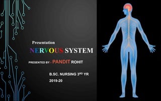
  • 1. NERVOUS SYSTEM PRESENTED BY:- PANDIT ROHIT A B.SC. NURSING 3RD YR 2019-20 Presentation
  • 2.
  • 3. INTRODUCTION • Nervous system is the chief controlling and coordinating system of the body. • It is the body’s most organised, complex, structural and functional system. • It is responsible for the judgement, intellegence and memory. • It detects and responds to changes occur inside and outside the body. • It consists :- brain, spinal cord and peripheral nerves.
  • 4. • With a mass of only 2kg, the nervous system is 3% of total body weight (almost). • The size of the nervous system ranges from a few hundred cells in the simplest worms, to around 300 billion cells in African elephants. • Neural signaling is capable of a much higher level. The fastest nerve signals travel at speeds that exceed 100 meters per second. • According to the U.S. National Library of Medicine there are more than 600 neurologic diseases.
  • 5. • Most neurons send signals via their axons, although some types are capable of dendrite-to-dendrite communication. (In fact, the types of neurons called amacrine cells have no axons, and communicate only via their dendrites). • Most amacrine cells are inhibitory neurons in the vertebrate retina, containing the common inhibitory neurotransmitters GABA. • The nervous system is made up of nervous tissues and nervous tissues made up of Neurons and Neuroglia(Neuroglial cells). • At the cellular level, the nervous system is defined by the presence of two special type of cells, called the NEURON and GLIAL cells
  • 6. • Neurology :- deals with normal functioning and disorders of the nervous system. • Neurologist :- physician who diagnose and treat disorders related to nervous system through various diagnostic tests like :- EEG, MRI, CAT Scan. • Neurosurgeon :- A neurosurgeon is a doctor having a degree of MS(master of surgery) and treat patients through surgery. • “In India, there is one neurosurgeon available per 10 lakh people. The ideal ratio should be at least one neurosurgeon for every one lakh population
  • 7.
  • 8. NERVE • A nerve is an enclosed, cable-like bundle of nerve fibers (called axons) in the peripheral nervous system. • A nerve transmits electrical impulses. • A nerve provides a common pathway for the electrochemical nerve impulses called action potentials that are transmitted along each of the axons. • Within a nerve, each axon is surrounded by a layer of connective tissue called the endoneurium.
  • 9.
  • 10. ENDONEURIUM • Also called Henle’s sheath. • It is a layer of delicate connective tissue around the myelin sheath of each myelinated nerve fiber in the PNS. • Its component cells are called endoneurial cells. • The endoneurium contains a liquid known as endoneurial fluid. • Peripheral nerve injuries commonly release increased amounts of endoneurial fluid into surrounding tissues; these can be detected by MRI.
  • 11. NERVE FASCICLE • A nerve fascicle, is a bundle of nerve fibers. • A nerve fascicle is enclosed by perineurium. • Fascicles enclose many thousands of axons.
  • 12. PERINEURIUM • The perineurium is a protective sheath that surrounds a nerve fascicle. • The perineurium is composed from fibroblasts.
  • 13. EPINEURIUM • The epineurium is the outermost layer of dense irregular connective tissue surrounding a peripheral nerve. • It usually surrounds multiple nerve fascicles as well as blood vessels which supply the nerve. • Epineurium is primarily made from collagen. • The epineurium is usually most abundant around joints, as its function is to protect the nerves from stretching.
  • 14.
  • 15. 1. NEURON • Neurons can be distinguished from other cells in a number of ways, but their most fundamental property is that they communicate with other cells via synapses. • Some nerve cells can be comparatively smaller by 0.1 millimeters or can be longer by 1 meter. The size of nerve cells is usually based on their functions i.e how long electrical impulse is transmitted within our body. • The length of a nerve cell can vary from nanometers to meters.
  • 16. • Each neuron is made up of 1. Cell body/ Cyton/ Soma/ Perikaryon 2. Dendrites 3. Axon • Neurotrophins: These are proteins which are required for the growth and survival of neurons. These are produced by astrocytes
  • 17.
  • 18.
  • 19. PROTEINS • Neurotrophins are a family of proteins that induce the survival, development, and function of neurons. • Glial cells(Astrocyte) that exist in the peripheral nervous system, which secrete NGF (Neurotrophins). • Neurotrophins are chemicals that help to stimulate and control neurogenesis.
  • 20. A)Cell body :- • It contains uni-nucleated cytoplasm. • Centriole is absent and thus cell division doesn’t take place. • Nissls’s granules are responsible for protein synthesis. • Collectively they form gray matter and are found at periphery of the brain and spinal cord. • The cell body contains genetic information, maintains the neuron's structure, and provides energy to drive activities.
  • 21. B) Dendrites :- Dendrites are many short richely branched and oftenly swallen branches of neuron, that recieve and carry incoming impulses towards cell body.They resemble a tree-like structure Conduct the electrochemical charge to the cell body
  • 22. C) Axon :- Axon, also called nerve fibre, portion of a nerve cell (neuron) that carries nerve impulses away from the cell body. Mostly(normally) a neuron has one axon that connects it with other neurons The longest axons of the human body are those that make up the sciatic nerve where the length can exceed one meter.
  • 23. MYELINOGENESIS 1. PNS • Axon is covered by a layer of phospholipids which is called myelin sheath • This myelin sheath is covered by thin membrane called neurilemma.
  • 24. • Where the sheath is discontious around axon, are called nodes of ranvier • This myelin sheath prevents leakage of ions. • The plasma membrane layer of Schwann cells is called the neurilemma.
  • 25. 2. CNS Neurilemma/Schwann cell are absent in CNS. Therefore, myelination occurs by Neuroglia (Oligodendrocytes). ✓Multiple sclerosis disease is caused by the demyelination of cranial nerves
  • 26. FUNCTIONS OF MYELIN SHEATH 1. Faster conduction (saltatory conduction) Myelin sheath is responsible for faster conduction of impulse through the nerve fibers. In myelinated nerve fibers, the impulses jump from one node to another node. Conduction of impulse is 50 times faster than unmyelinated neurons. 2. Insulating capacity Myelin sheath has a high insulating capacity. Because of this quality, myelin sheath restricts the nerve impulse within single nerve fiber and prevents the stimulation of
  • 27.
  • 28. Myelinated Neuron Unmyelinated Neuron 1. Large diameter of axon 1. Small 2. Nodes of Ranveir are present 2. Absent 3. Prevents loss of impulse 3. Can lose nerve impulse 4. Nerve fibres are white in colour 4. Gray in colour 5. Nerve impulse is faster 5. Slower 6. Salutatory conduction - present 6. Absent 7. Found in Spinal and cranial nerves 7. Found in ANS and SNS
  • 29. TYPES OF NEURONS A)Acc. to the number of their process 1.Multipolar neuron :- most neurons in humans are these. Commonly found in the cortex of the brain and the spinal cord. 2.Bipolar neuron :- Bipolar neurons are found in the retina of the eye, roof of the nasal cavity, and inner ear.
  • 30. 3. Pseudounipolar neuron :- Pseudounipolar neurons have one projection from the cell body, which splits into two axons: one that extends into the periphery and one that extends into the central nervous system. Found in dorsal nerve root ganglia and sensory ganglia of cranial nerve. 4. Unipolar neuron :- A unipolar neuron only has one axon that extends into dendrites. And are present in mesencephalic nucleus of trigeminal nerve.
  • 31.
  • 32. B) acc. to the length of axon(nerve fibre) 1.Golgi type 1 :- These neurons have long axons and short dendrites. These are seen in pyramidal cells of vertebral cortex, purkinje cells of cerebellum and anterior horn cells of spinal cord 2.Golgi type 2 :- neurons with short axons and establish synapse with neighbouring neurons. These are also seen in cerebral cortex. 3.Amacrine neurons :- These are one without axons(only dendrites are present). They are present in retina.
  • 33. CLASSIFICATION OF NERVE FIBRES 1. Depending upon structure 2. Depending upon diameter 3. Depending upon origin 4. Depending upon function 5. Depending upon secretion of neurotransmitter 6. Depending upon diameter and conduction of impulse
  • 34. 1. Depending upon structure a) myelinated , b) unmyelinated 2. Depending upon distribution a) somatic , b) visceral 3. Depending upon origin a) cranial , b) spinal 4. Depending upon function a) sensory , b) motor 5. Depending upon secretion of neurotransmitters a) adrenergic , b) cholinergic 4. Depending upon function a) sensory b)motor 5. Depending upon secretion of neurotransmitters a) adrenergic , b) cholinergic
  • 35. 6. Depending upon diameter and conduction of impulse Velocity of impulse through a nerve fiber is directly proportional to the thickness of the fiber.
  • 36. 2. NEUROGLIA • These are the cells that provide metabolic support and immune protection for neurons. • Ratio of neuron and neuroglia is 10:1 • Neuroglia don’t generate nerve impulses • Neuroglia can regenerate if required
  • 37.
  • 38. TYPES OF NEUROGLIA 1.Astrocyte :- • Provide energy and metabolic needs for neurons • Give structural support • When neurons are injured/destroyed, they are replaced with scar tissue made by astrocytes. This process is called gliosis. 2. Microglia :- Phagocytic cell ( similar to macrophages ). 3. Ependymal cells :- production of CSF.
  • 39. SYNAPSE ( NEURONAL JUNCTION ) • A synapse is the junction b/w two neurons. • Here a special activity occurs :- a weak signal may be allowed to die-out or amplified into a strong signal. Similarly, at synapse a signal can be both convergent as well as divergent. • The neurons are connected to one another by their processes forming long chains along which the impulses are conducted. • The site of contact b/w the nerve cells is known as synapse. • The impulse is transmitted across synapse through
  • 40. The synapse consists of :- 1.Presynaptic ending :- contains neurotransmitter 2.Postsynaptic ending :- contains receptors 3.Synaptic Cleft :- space b/w Presynaptic and Postsynaptic endings.
  • 41. • Name Synapse was given by Charles Sherrington. • Synapse = Presynaptic knob + synaptic cleft + Postsynaptic membrane • When action potential develops in Presynaptic membrane, it become permeable for calcium ions, that enter Presynaptic membrane and neurotransmitter comes out of synaptic vesicles.
  • 42.
  • 43. TYPES OF SYNAPSE A)Synapse with another neuron • Axodendritic :- most prominent kind of synapses that one neuron makes onto the dendrite of another neuron. • Axosomatic :- synapse b/w body and axon • Axoaxonic :- impulse carried by an axon onto axon of another neuron. It can induce inhibitory or excitatory effects in the postsynaptic neuron. B) Neuromuscular :- synapse of a motor neuron and a muscle C) Neuroglandular :- synapse of a neuron and a endo/exocrine gland.
  • 44. SYNAPTIC TRANSMISSION • It is the process where nerve cells communicate between them/muscle/glands TYPES OF TRANSMISSION 1.Electrical transmission :- In this, the two cells actually touch each other and they share proteins. Found only in heart and eye. 2. Chemical transmission :- This include the release of chemical neurotransmitters to propagate signals from one neuron (presynaptic) to another (postsynaptic). The two cells are separated by the synaptic cleft, a gap of approximately 40 nm between the presynaptic and
  • 45. CHEMICAL TRANSMISSION • A synaptic cleft is about 30-50nm( 1nm=10-9 ) which separates synaptic knob to the Postsynaptic cell. • There are three kinds of synaptic vesicles :- a) Small clear synaptic vesicles that contain neurotransmitters. b) Small vesicles with dense core that contain catecholamines. c) Large vesicles with dense core that contain neuropeptide. That are released after depolarization of the cell.
  • 46. • As the nerve reaches the axon terminal, it opens voltage gated calcium channels. This increased calcium concentration causes rupture of the synaptic vesicles. • In this, electrical activity in the presynaptic neuron is converted into the release of chemical called neurotransmitter, that bind to the receptors located in the plasma membrane of the postsynaptic cell. Reuptake of neurotransmitters :- In case of many neurotransmitters, most of the amount released into the synaptic cleft is actively transported back into the presynaptic nerve terminals and restored in the microvesicles.
  • 47.
  • 48.
  • 49. NEUROTRANSMITTERS • These are chemical messangers that transmits signals from a neuron to a target cell across a synapse. • Target cell may be a neuron/muscle cell/gland cells. Types Inhibitory Excitatory
  • 50. •Excitatory neurotransmitters have excitatory effects on the neuron. This means they increase the likelihood that the neuron will fire an action potential. •Inhibitory neurotransmitters have inhibitory effects on the neuron. This means they decrease the likelihood that the neuron will fire an action. *Some neurotransmitters, such as dopamine, depending on the receptors present, create both excitatory and inhibitory effects.
  • 51. TYPES OF NEUROTRANSMITTERS 1. INHIBITORY • GABA • Serotonin • Dopamine 2. EXCITATORY • Ach • Epinephrine • Norepinephrine • Histamine • Glutamate • Dopamine
  • 52. DISORDERS LINKED TO NEUROTRANSMITTERS 1. Alzheimer’s :- Lack of Ach and glutamte. 2. Schizophrenia :- Excessive Dopamine. 3. Parkinson’s disease :- lack of Dopamine. 4. Epilepsy :- lowered GABA. 5. Anxiety :- low serotonin.
  • 53. GENERATION AND CONDUCTION OF NERVE IMPULSE 1. RESTING POTENTIAL :- resting potential may be defined as the difference in voltage due to ion exchange between inside and outside of cell. ( In simple terms :- when a neron is at rest). Avg. Potential range ( -40mV to -90mV). 2. A resting neuron’s cell membrane is polarized(charged). 3. Outside of the membrane is more +vely charged. 4. Normal resting membrane has a potential
  • 54. • Depolarization occur when the Na+ channels open and they moved inside the cell. • The exit of K+ causes the membrane to once again become more +vely charged. • Action potential causes calcium to enter the synaptic knob. • Calcium entering the synaptic knob triggers the release of neurotransmitters. • Atomic mass of calcium is :- 22.98u and potassium is :- 39.09u • Depolarization : Na+ influx • Repolarization :- K+ efflux
  • 55. ACTION POTENTIAL • Action potential is basically the way of neurons to transport electrical signals from one cell to next. • The electrical signals are very rapid. Being measured in milliseconds and potential changes measured in miliVolts(mV). • Action potentials are generated by special types of voltage- gated ion channels • Voltage gated Sodium-ion channels are highly concentrated in the nodes of ranvier. RMP DEPOLARIZATION + REPOLARIZATION = ACTION POTENTIAL
  • 56. WHY THE IMPULSE JUMPS FROM NODE TO NODE ( SALTATORY CONDUCTION ) • The myelin sheath is wrapped around an axon in such a fashion, that there are a few gaps(node of ranvier). • The nodes of Ranvier have 2,000 to 12,000 voltage- gated channels per square micrometer (μm²) of membrane. • Now, there are alot of ion channels on the cell membrane (neurilemma) of the nerve cells. These ion channels selectively allow some ions to pass through them, and prevent some of the ions.
  • 57. • If the membrane is preventing certain positively charged ions to come inside the cell, there will be more positive charge outside the cell. • Now when a signal reaches the cell body through its dendrites, this potential is disturbed. • This disturbance goes in a linear fashion in an unmyelinated neuron, so should pass the entire neuron.
  • 58. • In a myelinated neuron, the disturbance does not have to pass along the entire length. • The signal jumps from one node to another. • Hence myelinated neurons are much much faster than unmyelinated neurons in terms of conduction.
  • 59. REFRACTORY PERIOD • Refractory period is the period at which the nerve does not give any response to a stimulus. Types of Refractory period 1. Absolute Refractory Period 2. Relative Refractory Period
  • 60. • Absolute Refractory Period is the period during which the nerve does not show any response at all, whatever may be the strength of stimulus come. • Relative Refractory Period is the period during which the nerve fiber shows response, if/when strength of stimulus is increased. • If a stimulus is applied, of any strength, the muscle responds to the max. or it doesn’t respond at all. This is the law. ALL OR NONE LAW
  • 61. SUMMATION • When one subliminal stimulus is applied ( when we are aware ), it doesn’t produce any response in the nerve fiber. • However, if two or more stimuli are applied within a short period (~0.5 msec.) the response is produced. • This is because the subliminal stimuli are summed up together. This is called summation.
  • 62.
  • 63.
  • 64.
  • 65.
  • 66. DALE’S PRINCIPLE Henry Hallett Dale • Dale’s principle states that one type of neuron contains and releases only one type of neurotransmitter but this does not mean that every neuron exerts same effects on all of its targets, because the effect of a synapse depends not on the neurotransmitter but on the receptors that it activates. • Dale’s principle ensures that biological neurons are either exclusively excitatory or inhibitory.
  • 67. • Because different targets can use different types of receptors. Example:- motor neuron contains Ach sensory neuron contains glutamate • Some modern writers have understood the principle to state that neurons release one and only one transmitter at all of their synapses, which is false. • In later publications, have taken it to mean that neurons release the same set of transmitters at all of their synapses.
  • 68. CENTRAL NERVOUS SYSTEM • It includes brain(Cranial cavity) and spinal cord. • These structures are protected by rigid bony encasement. • Spinal cord is connected to the brain through ‘Foramen Magnum’ and is encircled by the bones of vertebral column. • Brain is covered by meninges :- Dura mater, Arachnoid mater and Pia mater.
  • 69. • The blood supply to the brain originates from the first major arterial branches from the heart, ensuring that over 20% of the entire supply of oxygenated blood flows directly into the brain. • Thee structures of brain and spinal cord are arranged in two layers (gray matter and white matter). • Gray matter is formed by cell bodies. • White matter is formed by nerve fibres. • In Brain, the white matter is centrally placed and gray matter is in the outer part. But in spinal cord it is vice-
  • 70. • Brain and Spinal cord are surrounded by three layers of meninges(Dura+Arachnoid+Pia mater). • The space b/w Dura mater and Arachnoid Mater is called ‘Subdural Space’, between Arachnoid mater and Pia mater is called Sub-Arachnoid space. • The Sub-arachnoid space is filled with CSF(Cerebrospinal fluid).
  • 71.
  • 72. 1. BRAIN • Brain is the one of the largest and most complex part of the nervous system. • Brain weighs around 1300-1400g in an adult and lies within the cranial cavity. • Circulus Arteriosus and contributing arteries play vital role in maintaining supply of blood to brain. • Venous blood from brain drain into the Dural venous sinuses, and then downwards into the jugular veins. • The brain itself contains more than 100 billion neurons. ✓ Isotropic Fractionator: A Simple, Rapid Method for the Quantification of Total Cell and Neuron Numbers in the Brain.
  • 73.
  • 74.
  • 75.
  • 76.
  • 77. FOREBRAIN(PROSENCEPHALON) • The forebrain plays a central role in the processing activities:- sensory and associative functions, and voluntary motor activities. 1. TELENCEPHALON A)Cerebral hemispheres make up the uppermost portion of the brain and are involved in sensory integration, control of voluntary movement, and higher intellectual functions, such as speech and abstract thought. B)Basal Ganglia are a group of structures near the center of the brain that allow different areas of the brain to work together. The basal ganglia manage the signals the brain
  • 78. BASAL GANGLIA • Ganglia is a group of neurons, so we can call it as Basal Nuclei. • The basal ganglia are situated at the base of the forebrain and top of the midbrain. • The main components of the basal ganglia :- striatum, globus pallidus, ventral pallidum, substantia nigra, and the sub-thalamic nucleus.
  • 79. C) LIMBIC SYSTEM • Is a set of brain structures located on both sides of the thalamus. • The structures and interacting areas of the limbic system are involved in motivation, emotion, learning, and memory. • It is highly interconnected with the nucleus accumbens, which plays a role in sexual arousal. • It has two major parts :- a) Hippocampus b) Amygdala
  • 80. Hippocampus is a complex brain structure embedded deep into temporal lobe. It has a major role in learning and memory. Amygdala, region of the brain primarily associated with emotional processes. The name amygdala is derived from the Greek word amygdale, meaning “almond, leads to almond like shape.
  • 81. FUNCTIONS OF LIMBIC SYSTEM 1. Olfaction 2. Regulation of endocrine glands 3. Regulation of autonomic functions :- heart rate, bp, body temp. 4. Regulation of food intake. 5. Control of circadian rhythm. 6. Regulation of sexual function. 7. Role in memory and motivation. 8. Role in emotional state.
  • 82. 2. DIENCIPHALON A)THALAMUS • Is a large mass of gray matter located in the dorsal part of the diencephalon. • It has several functions, such as the relaying of sensory signals, including motor signals to the cerebral cortex and the regulation of consciousness, sleep, and alertness. • Anatomically, it is a paramedian symmetrical structure of two halves (left and right),situated between the cerebral cortex and the midbrain. • Damage to the thalamus can lead to permanent coma.
  • 83.
  • 84. Blood Supply To The Thalamus • Mainly basilar artery supplies to the thalamus. • The thalamus derives its blood supply from a number of arteries. a) The polar(posterior communicating) artery b) Paramedian thalamic-subthalamic arteries. c) Inferolateral(thalamogeniculate) arteries d) Posterior choroidal arteries
  • 85. FUNCTIONS OF THALAMUS • Act as a relay station • Plays an important role in regulating states of sleep and wakefulness. • This is the case for many of the sensory systems :- auditory, somatic, visceral, gustatory and visual system
  • 86. THALAMOTOMY • Is a surgical procedure in which an opening is made into the thalamus. • It is primarily effective for patients with severe Parkinson’s disease, where a selected portion of the thalamus is surgically destroyed.
  • 87.
  • 88. B) HYPOTHALAMUS • One of the most important functions is to link the nervous system to the endocrine system via the pituitary gland. • The hypothalamus is located below the thalamus and is part of the limbic system. • In humans, it is the size of an almond. • It synthesizes and secretes certain neurohormones, called releasing hormone.
  • 89. • The hypothalamus is divided into 3 regionsevents.praoptic, tuberal and mammillary. A) Supraoptic Hypothalamus Produce and secrete the peptide hormone vasopressin, also known as antidiuretic hormone (ADH) and oxytocin. B) Tuberal Hypothalamus Is mainly involved in feeding behavior and is said to be a “satiety center”. C) Mammillary Hypothalamus Important for episodic memory. It is the memory of everyday events.
  • 90. MID-BRAIN(MESENCEPHALON) • It is 2cm long and connects b/w forebrain and hindbrai. • It is divided into two parts :- a) The tectum, b) Cerebral Peduncle A)The Tectum is the posterior part to aqueduct. *Aqueduct = allows CSF to flow between the 3rd and 4th ventricle. B) Cerebral Peduncle are the two structures located each half of the mid-brain anterior to the aqueduct. a) Crus cerbri(crura), b) Substantia nigra, c) Tegmentum
  • 91. a)Crus Cerebri :- The crura transmit axons of the primary motor pathway descending from the cerebral cortex to the spinal cord. b)Substantia Nigra :- The substantia nigra is a critical brain region for the production of dopamine. The substantia nigra consists of two parts, the pars reticulata and the pars compacta. Cells of the pars compacta contain the dark pigment melanin; these cells synthesize dopamine. c) Tegmentum :- It is a motor center that relays inhibitory signals to the thalamus and prevent unwanted body movement.
  • 92. HIND-BRAIN(RHOMBENCEPHALON) 1. METENCEPHALON a) Pons b) Cerebellum A)Pons :- A structure that links brain to the spinal cord. It handles unconscious processes and jobs, such as your sleep-wake cycle and breathing. B)Cerebellum :- Composed of gray and white matter. Maintaimg balance, co-ordimating movements, Vision, Motor learning, Ect.
  • 93. 2. Myelencephalon( Medulla Oblongata) The medulla oblongata is divided into two parts: •Superior section :- connects to brain’s 4th ventricle •Inferior section :- connects to the spinal cord. ✓Also helps control vital processes like heartbeat, breathing and blood pressure. ✓ Out of 12 cranial nerves, 9 to 12 Cranial nerves starts from Medulla Oblongata.
  • 94. BLOOD SUPPLY TO THE BRAIN • The entire blood supply to the brain, depends on two branches of Aorta. a) Internal Carotid Arteries b) Vertebral Arteries • The brain recieves about 15-20% blood supply of total body and receives 20-25% oxygen of the total inhaled oxygen. • 10 seconds of brain ischemia leads to unconciousness. • 20 seconds brain ischemia leads to cessation of electrical activity of the brain. • 3-4min of brain ischemia leads to permanent brain damage.
  • 95. 1. ARTERIAL BLOOD SUPPLY TO THE BRAIN • The arterial blood reaches to the brain through the :- a) Internal Carotid Arteries (80% to Forebrain). b) Vertebral Arteries(20% to brainstem and cerebellum).
  • 96.
  • 97. 2. VENOUS DRAINAGE OF THE BRAIN • Runs in Sub-arachnoid space. • It includes dural sinus and cerebral sinus(Superficial and Deep). A) Cerebral Sinus :- • Superficial/External veins :- Drains the cortex of the brain. • Deep :- This system drains the thalamus, hypothalamus, etc. B) Dural Sinus :- subsequently drain to the internal jugular vein.
  • 98.
  • 99. BRAINSTEM •Brainstem is the part of brain formed by Medulla Oblongata, Pons, And mid-brain. •It is the pathway between brain and spinal cord. •It is the location of many important centers for the regulation of vital functions in the body.
  • 100. MEDULLA OBLONGATA • Medulla Oblongata or Medulla is the lowermost part of the brain. • It is situated below pons. • It contains centres for heartbeat, breathing, and blood pressure ✓ Controls vital functions 1. Respiratory center🫁 2. Vasomotor center 3. Deglutition center 4. Vomiting center 🫁 5. Salivation center🫁
  • 101. PONS • Pons means “bridge”, it is the bridge between Medulla and Mid-brain. • The pons can be broadly divided into two parts : ventral and dorsal • The pons in humans measures about 2.5 centimetres (0.98 in) in length. • Four cranial nerves are present here. ( 5th-8th ) • It handles unconscious processes and jobs, such as our sleep-wake cycle and breathing.
  • 102. 2. SPINAL CORD ✓ INTRODUCTION • The spinal cord is the long cylindrical lower part of central nervous system. • It is the main pathway for information between brain and PNS. • Spinal cord is 17-18inch long ( in an adult normally ). • Though spinal cord is surrounded by three meninges. But below the level of conus( L1 starts), only Pia mater is continued. • The spinal cord is enclosed by Dura mater and the space between dura mater and vertebral canal is called
  • 103.
  • 104.
  • 105. • Spinal cord is made up of 31 segments : cervical = 8 thoracic = 12 lumbar = 5 sacral = 5 coccyx = 1 • It gives rise to 31 pairs of spinal nerves. • Golgi type 1(in Anterior horns) and Golgi 2(in Posterior horns) are found in Gray matter of spinal cord. Both are multipolar neurons. • Length of spinal cord :- 45cm • Weight of spinal cord :- 35cm
  • 106. DEVELOPMENT OF SPINAL CORD IN EMBRYO • During the initial third month of embryonic development, the spinal cord extends the entire length of the vertebral canal and both grow at about the same rate. • As development continues, the body and the vertebral column continue to grow at a much greater rate than the spinal cord. This results in displacement of the lower parts of the spinal cord with relation to the vertebrae column.
  • 107. FUNCTIONS OF SPINAL CORD • Protection of spinal cord, nerves and internal organs. • Flexibility of motion • Pass information from and to brain
  • 108. ASCENDING TRACTS OF SPINAL CORD • Carries the impulses to the brain. • The pathway is formed by three types of neurons :- a) first order neurons b) second order neurons c) third order neurons
  • 109. • 1st order neurons receive sensory impulses from the receptors and send them to the sensory neurons(2nd order) present in the posterior gray horn of spinal cord. • 2nd order neurons present in posterior gray horn carry impulses to the subcortical area (thalamus),where 3rd order neurons are present. • 3rd order neurons carry impulses from subcortical area(thalamus) to the cerebral cortex.
  • 110. Diastematomyelia • Diastematomyelia (occasionally diastomyelia) is a congenital disorder in which a part of the spinal cord is split • Usually at the level of the upper lumbar vertebra in the longitudinal (sagittal) direction. • Diastematomyelia, a type of spina bifida, resulting in a split cord malformation. • The spinal cord is longitudinally divided into two ‘hemicords,’ each surrounded by its own dural tube and separated by a midline bony spur or cartilaginous or
  • 111. CEREBRO-SPINAL FLUID(CSF) • The CSF is a colorless fluid. • Amount :- 100-200ml. • Lies within the Sub-arachnoid space. • Rate of formation :- 0.3-0.4 ml/min. • Slightly alkaline in nature. • Composition :- water (99.13%), solids (0.87%) • CSF also contains lymphocytes. • CSF is formed by Epithelium and Ependymal cells in Coroid Plexus.
  • 112. FORMATION OF CSF • Inside the Ependymal cell sodium efflux from the cell by using ATP due to sodium-potassium pump. • With the efflux of sodium, chlorine also efflux from the cell. • This creates the osmotic gradient, which drives the secretion of H2O(water). • Glucose channel is also present and glucose is secreted from the cells of choroid capillaries.
  • 113. CIRCULATION OF CSF Lateral ventricles Third ventricle Fourth ventricle Sub-Arachnoid space Cerebral hemisphere Spinal cord
  • 114.
  • 115. ✓ FUNCTIONS OF CSF • Protects brain and spinal cord from shock. • Prevents the movements of brain against the bones of skull. • Medium of exchange through which substances • Excretion of waste products. • It is the procedure of removing CSF through a needle to examine CSF in a lab. • Needle is inserted into Sub-arachnoid space. • Spinal cord ends at L1-L2, so the ideal site for sample collection is L2-L4 • Lateral recumbent position ✓ LUMBAR PUNCTURE
  • 116. MENINGES OF BRAIN AND SPINAL CORD 1. Dura mater ( Pachymeninx ) 2. Arachnoid mater 3. Pia mater
  • 117. 1. Dura mater • It is made up of two layers :- an outer endosteal layer and an inner meningeal layer • The periosteal or endosteal layer of the dura mater is simply a layer of periosteum that covers the inner surface of the skull. • A deep layer called the meningeal layer. When it covers the spinal cord it is known as the dural sac or thecal sac. • Unlike cranial dura mater, spinal dura mater only has one layer, known as the meningeal layer. The potential space between these two layers is known as the epidural space. • The dura mater is the outermost, thickest and toughest membrane covering the brain.
  • 118. DURAL FOLDS • The function of the dural folds is to limit the rotational displacement of the brain. • The dura separates into four layers at dural reflections (also known as dural folds). 1. Tentorium cerebri 2. Falx cerebri 3. Cerebellar Falx 4. Diaphragm Sellar
  • 119.
  • 121. HEMMORHAGE IN DURA MATER • Epidural hematoma is when bleeding occurs between the tough outer membrane covering the brain (dura mater) and the skull. • Often there is loss of consciousness following a head injury, a brief regaining of consciousness, and then loss of consciousness again. ✓ Symptoms :- headache, confusion, vomiting, and an inability to move parts of body, seizures. Causes :- Head injury, bleeding disorder, blood vessel malformation
  • 122. •Diagnosis :- CT-Scan, MRI •Treatment : A) It may not require surgery when :- • If glasgow coma score is above 8. • If the volume of the epidural hematoma is less than 30 ml. • The clot diameter less than 15 mm. B) Epidural hematoma is a surgical emergency. Delayed surgery can result in permanent brain damage or death. The hematoma is evacuated through a burr hole or
  • 123. Epidural hematoma as seen on a CT scan with overlying skull fracture. Note the biconvex shaped collection of blood.
  • 124. LUCID INTERVAL • Lucid interval is the period between initial unconsciousness, which occurs immediately after accident and after some time of consciousness then again unconciousness at later stage. Unconciousness Consciousness Unconsciousness
  • 125. 2. Arachnoid mater • The Arachnoid mater is a thin transparent membrane that loosely surrounds the brain without dipping into sulci. • The arachnoid mater makes arachnoid villi, small protrusions through the dura mater into the venous system of the brain, which allow CSF to exit the subarachnoid space and enter the blood stream. • They may, however, play an important role in the absorption of CSF’s waste products of neuronal metabolism (mainly CSF proteins) under normal physiological conditions.
  • 126. 3. Pia Mater • Thin vascular membrane which closely invests the brain, dipping into various sulci and other irregularities of it’s surface. • Pia mater is the delicate innermost layer of the meninges, the membranes surrounding the brain and spinal cord. • The pia mater is a thin fibrous tissue that is permeable to water and small solutes. • A network of blood vessels travels to the brain and spinal cord by interlacing through the pia membrane. These capillaries are responsible for nourishing the brain.
  • 127. • At the point where the pia mater reaches the conus medullaris or medullary cone at the end of the spinal cord, the membrane extends as a thin filament called the filum terminale or terminal filum(Figure on next slide). • The pia mater attaches to the dura mater through 21 pairs of denticulate ligaments that pass through the arachnoid mater and dura mater of the spinal cord.
  • 128.
  • 129.
  • 130. BLOOD BRAIN BARRIER • As the name suggests, this is a barrier between the brain and blood vessels • To maintain normal brain functions, the neural environment must be in homeostasis. • This requires a tight regulation and transportation of cells, molecules and ions between the blood and the brain. • There are three barrier layers contribute to the separation of blood and neural tissues.
  • 131. • BBB is the highly selective semipermeable membrane barrier that separates the circulating blood from the brain and extracellular fluid • A few water soluble substances such as glucose croas BBB by active transport. • The purpose of the blood–brain barrier is to protect against circulating toxins or pathogens that could cause brain infections, while at the same time allowing vital nutrients to reach the brain.
  • 132. • When a nerve fiber is injured, various changes occur in the nerve fibers and nerve cell body(Soma). • All these changes are together called the degenerative changes. • The injury occurs due to the obstruction of blood flow, local injection of toxins, crushing/transection of nerve fiber. DEGENERATION AND REGENERATION OF NERVE FIBRES
  • 133. DEGREES OF INJURY 1. First degree • Most common type of injury • Caused by applying pressure over nerves for a short period of time • Leads to hypoxia • But the axon is not destroyed • The normal functioning returns within few hours. • Also called Seddon’s Neuropraxia.
  • 134. 2. Second degree • Due to prolonged severe pressure • Repair and restoration take about 18 months. • Also called Axonotmesis. 3. Third degree • After degeneration, the recovery is slow, poor and incomplete.
  • 135. 4. Fourth degree • Injury is so severe. • Regeneration is poor/incomplete. 5. Fifth degree • Involves complete transaction of the nerve with loss of continuity. • Regeneration is not possible unless the cutends are rearranged by surgery.
  • 136. DEGENERATION • Degeneration refers to impairment or pathological changes of an injured tissue. • The degeneraive changes are classified into three types. 1. Wallarian degeneration 2. Retrograde degeneration 3. Transneural degeneration
  • 137. 1.Wallarian/Orthograde Degeneration :- • Wallarian degeneration is the pathological change that occurs in the distal cut end of nerve fiber(axon). • Wallarian degenration starts within 24hrs. of injury. • Axon cylinder swells and breaks up into small pieces and then appear as debris into the space. • The myelin sheath is slowly disintegrated into fat droplets. These changes occur from 8th to 35th day. • The macrophages invade from outside and remove the debris of axon. • Later the void(empty space) is filled by the cytoplasm of Schwann cell.
  • 138. 2. Retrograde degeneration :- • Retrograde degenration is the pathological changes which occur in the nerve cell body and axon. • Retrograde degeneration starts within 48hrs. • First, the Nissl’s granules disintegrate into fragments. • Then the Golgi apparatus is disintegrated. • The cell body(Soma) swells due to accumulation of fluid and becomes round. • *Sometimes, the nucleus is extruded out of the cell resulting permanent death of neuron and thus regeneration is impossible.
  • 139. 3. Transneuronal degenration :- • If an Afferent nerve fiber is cut, the degenrative changes occur in the Neuron with which the Afferent nerve fiber synapses. • Some researchers believe that transneuronal degeneration leads to degeneration and atrophy of retinal cells and photoreceptors, resulting in damage to vision loss.
  • 140. REGENERATION • Regeneration means regrowth of lost or destroyed part of tissue. • The injured and degenrated nerve fiber can regenerate. • It starts as earliest as 4th day after injury but becomes more effective only after 30 days and is completed in about 80 days.
  • 141. CRITERIA FOR REGENERATION Regeneration can only be possible if :- • Gap between cut-ends shouln’t exceed 3mm. • Neurilemma should be present (As neurilemma is absent in CNS, the regeneration of nerve doest not occur in CNS). • Nucleus must be intact(unbroken). • Two ends should remain in the same line.
  • 142. STAGES OF REGENERATION • Schwann cell synthesize nerve growth factors and myelin sheath It takes about a year. • The newly formed internodes are shorter than original ones. • In the nerve cell body, first the Nissl’s granules appear and then Golgi body. • The cell loose excess fluid and nucleus occupies the central portion.
  • 143.
  • 144. PERIPHERAL NERVOUS SYSTEM • The PNS consists of nerves and ganglia, which lie outside the brain and the spinal cord. • The main function of the PNS is to connect the CNS to the limbs and organs • Unlike the CNS, the PNS is not protected by the vertebral column and skull • The peripheral nervous system is divided into the somatic nervous system and the autonomic nervous system
  • 145. AUTONOMIC (SPLENCHNIC) NERVOUS SYSTEM • ANS is concerned with the regulation of visceral functions of the body. • Involuntary in control. • Connected with organs that have smooth muscle, such as the heart, bladder, and other cardiac, exocrine, and endocrine related organs • Divisions of ANS :- a) Sympathetic division b) Parasympathetic division • There are 31 pair of spinal nerves :- 8 cervical, 12 thoracic, 5 lumbar, 5 sacral, 1 coccygeal
  • 146.
  • 147. 1. SYMPATHETIC NERVOUS SYSTEM • When the body requires sympathy to help the body to face and overcome critical situations, it is called SNS of body. • The sympathetic system is activated during a “fight or flight” situation in which mental stress or physical danger is encountered.[ • There are two kinds of neurons involved in the transmission of any signal through the sympathetic system: pre-ganglionic and post- ganglionic.
  • 148. 2. PARASYMPATHETIC NERVOUS SYSTEM • The parasympathetic system allows the body to function in a “rest and digest” state. • When the parasympathetic system dominates the body, there are increases in salivation and activities in digestion, while heart rate and other sympathetic response decrease.
  • 149. SYMPATHETIC NERVOUS SYSTEM PARA-SYMPATHETIC NERVOUS SYSTEM 1. Pupil dilates 1. Constricts 2. Heart rate increase 2. Decrease 3. Inhibits salivary secretion 3. Stimulation 4. Dilated lungs 4. Constrict lungs 5. Peristaltic movt. inhibited 5. Stimulation of peristaltic movt. 6. Sweat secretion 6. Inhibits sweat secretion 7. Inhibits micturition 7. Micturition 8. Closes anus by contracting anal sphincters 8. Relaxes anal sphincters and opens the anus. 9. Stimulation of adrenal hormones 9. Inhibits adrenal secretion 10. Increased activity of digestive system 10. Decreased activity
  • 150. SOMATIC NERVOUS SYSTEM • The somatic nervous system (SNS), or voluntary nervous system is the part of the peripheral nervous system associated with the voluntary control of body movements. • The SNS has further two divisions :- a) motor division b) sensory division Sound, smell, Vision, Taste and Touch
  • 151. 1.Motor division • Motor neurins are those who recieve signals from CNS and send them to tissues, glands, or organs. • The dendrites and cell body of the motor neuron originate in the spinal cord, once the signal is picked up by the dendrites it sends it to the axon and axon to axon terminal upto the site. 2. Sensory division • Receptors pick up stimulus from the environment and that stimulus is transformed into an electrical signal by sensory neurons. • These neurons carry signals upto the spinal cord and to
  • 152. REFLEX ACTIVITY • Reflex activity is the response to a peripheral nervous stimulation that occurs without our consciousness. • It is a type of protective mechanism that protects us from damages. ✓ Reflex Arc • Reflex Arc is the anatomical nervous pathway for a reflex action • A simple reflex Arc includes five components Receptors, Afferent Nerve, Center(Spinal cord), Efferent
  • 153. CLASSIFICATION OF REFLEXES • Reflexes are classified by five different methods depending upon various factors :- 1. Whether inborn or acquired 2. Situation of the center 3. Purpose 4. Number of synapse 5. Clinical basis
  • 154. 1. Depending upon whether inborn or acquired A)Unconditional Reflex/Inborn reflex B)Conditioned Reflex/Acquired Reflex 2. Depending upon situation of the center A)Cerebellar Reflexes(Cerebellum) B)Cortical Reflexes(Cerebral cortex) C)Mid-brain Reflexes D)Bulbar Reflexes(Center in Medulla Oblongata) E)Spinal reflexes
  • 155. 3. Depending upon purpose A)Protective Reflexes( to protect body from harmful stimuli ) B)Antigravity Reflexes( protect the body against gravity, muscles contraction ) 4. Depending upon the no. of synapse A)Monosynaptic( Reflexes having one synapse in reflex Arc ) B)Polysynaptic( Reflexes having more than one synapse in reflex arc )
  • 156. 5. Depending upon clinical basis • Superficial Reflexes ; Eye blinking • Deep Reflexes ; Closure of the mouth • Visceral Reflexes ; Brady/Tachy-cardia, Dilation of pupil • Pathological Reflexes ; Babinski’s sign
  • 157.
  • 158. GLASGOW COMA SCALE • The Glasgow Coma Scale(GCS) is a clinical scale used to reliably measure a person’s level of consciousness after a brain injury. • A patient is accessed against the criteria of the scale, and the resulting points give a patient score between 3(min) to 15(max). • GSC check three components 1. Eye response (1-4) 2. Verbal response (1-5) 3. Motor response (1-6)
  • 159. EYE RESPONSE (1-4) 1. No eye opening 2. Eyes opening in response to pain 3. Eye opening in response to a speech 4. Eyes opening spontaneously
  • 160. VERBAL RESPONSE (1-5) 1. No verbal response 2. Incomprehensible sound(moaning only) 3. Speaks but inappropriate and random words 4. The patient responds to questions but the patient is little bit confused 5. Patient responds correctly and appropriately
  • 161. MOTOR RESPONSE (1-6) 1. No motor response 2. Decerebrate posture ( Abnormal extension ) 3. Decorticate posture ( Abnormal flexion ) 4. Withdrwal from painful stimuli 5. Purposeful movement towards painful stimuli 6. Obeys commands (the patient does the things simply as asked to do)
  • 162. ✓ Generally brain injury is classified as :- • Severe ; GCS ≤ 8 • Moderate ; GCS (9-12) • Mild ; GCS ≥ 13
  • 163. BIBLIOGRAPHY • Brunner Siddarth’s textbook of medical surgical nursing 14th edition • Gerard J. Tortora, Principles of Anatomy And Physiology • Bd Chaurasia, Human Anatomy Vol.3 • Textbook of medical physiology, GK Pal • Dr. Najeeb Lectures on Youtube
  • 164. • Ross and Wilson, Ross & Wilson Anatomy and Physiology in Health and Illness • Google Wikipedia • SlideShare • Essentials Of Medical Physiology Fifth Edition, K sembulingam and Prema Sembulingam • Guyton and Hall textbook of medical physiology
  • 165. Thank you || जय श्री राम ||