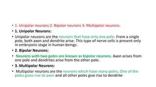
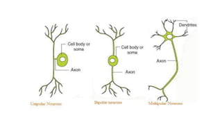
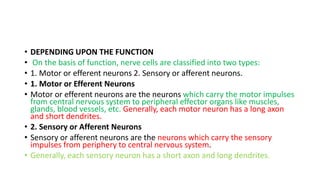
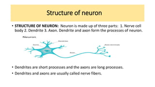
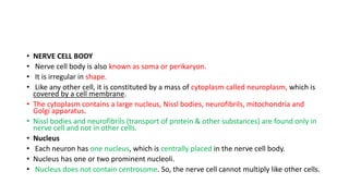
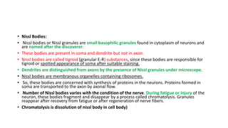
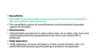
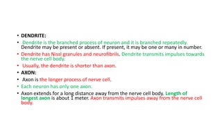
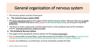
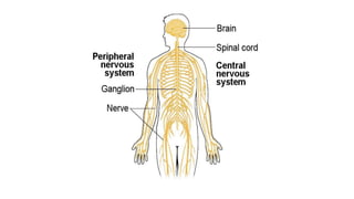
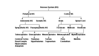
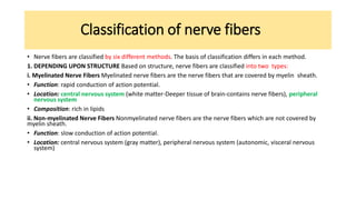
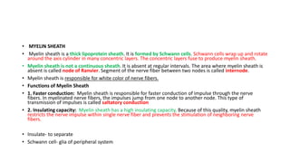
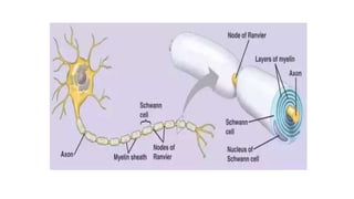
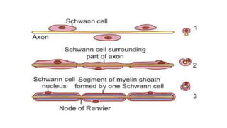
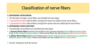
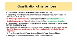
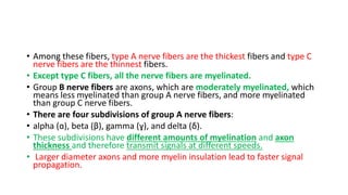
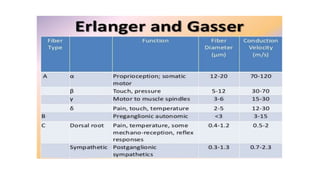
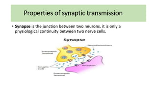
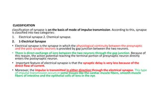
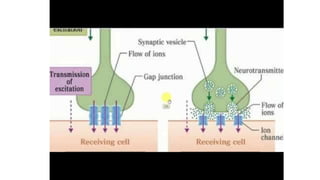
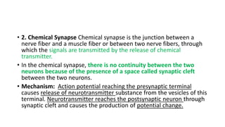
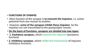
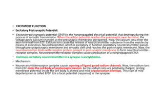
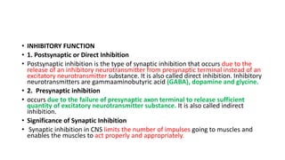
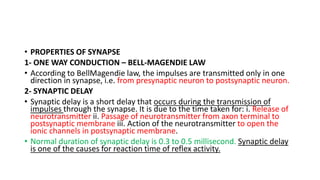
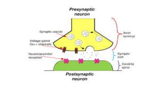
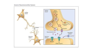
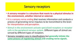
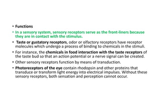
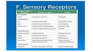
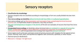
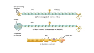
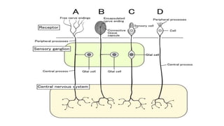
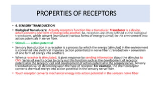
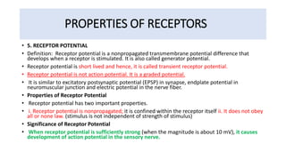
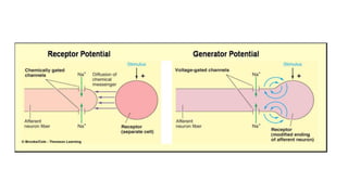
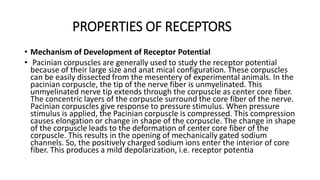
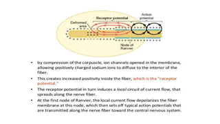
SlideShare a Scribd company logo
1 of 74
Physiology-3
Department of physiotherapy
Central Nervous System
BY
Dr.Laraib jameel Rph
Follow me on slideshare.net
https://www.slideshare.net/
Nervous system
• The nervous system is a complex network of nerves and cells that carry
messages to and from the brain and spinal cord to various parts of the
body.
• The nervous system includes both the Central nervous system and
Peripheral nervous system
• Difference between nerve & neuron:
• Nerve: bundle of neurons with fascia and blood supply. it is whitish fiber of
neuron cell which carry impulses to CNS & from CNS to effector organ.
• (fiber+vessel+ lymphatic) (in CNS+PNS)(act as conducting zone)
• Neuron: it is single nerve cell. it is specialized cell involved in transmitting
nerve impulses (chemical & electrical signal generated)
Nervous System
• Neuron or nerve cell :
• Neuron or nerve cell is defined as the structural and functional unit of nervous system. Neuron is similar to
any other cell in the body, having nucleus and all the organelles in cytoplasm.
• However, it is different from other cells by two ways:
• 1. Neuron has branches or processes called axon and dendrites
• 2. Neuron does not have centrosome. (help to replicate the chromosome into daughters) So, it cannot
undergo division.
• „CLASSIFICATION OF NEURON
• Neurons are classified by three different methods.
• A. Depending upon the number of poles
• B. Depending upon the function
• C. Depending upon the length of axon.
• „DEPENDING UPON THE NUMBER OF POLES
• Based on the number of poles from which the nerve fibers arise, neurons are divided into three types:
• 1. Unipolar neurons 2. Bipolar neurons 3. Multipolar neurons.
• 1. Unipolar Neurons:
• Unipolar neurons are the neurons that have only one pole. From a single
pole, both axon and dendrite arise. This type of nerve cells is present only
in embryonic stage in human beings.
• 2. Bipolar Neurons:
• Neurons with two poles are known as bipolar neurons. Axon arises from
one pole and dendrites arise from the other pole.
• 3. Multipolar Neurons:
• Multipolar neurons are the neurons which have many poles. One of the
poles gives rise to axon and all other poles give rise to dendrite
• DEPENDING UPON THE FUNCTION
• On the basis of function, nerve cells are classified into two types:
• 1. Motor or efferent neurons 2. Sensory or afferent neurons.
• 1. Motor or Efferent Neurons
• Motor or efferent neurons are the neurons which carry the motor impulses
from central nervous system to peripheral effector organs like muscles,
glands, blood vessels, etc. Generally, each motor neuron has a long axon
and short dendrites.
• 2. Sensory or Afferent Neurons
• Sensory or afferent neurons are the neurons which carry the sensory
impulses from periphery to central nervous system.
• Generally, each sensory neuron has a short axon and long dendrites.
• DEPENDING UPON THE LENGTH OF AXON
• Depending upon the length of axon, neurons are divided into two types:
• 1. Golgi type I neurons 2. Golgi type II neurons.
• 1. Golgi Type I Neurons: Golgi type I neurons have long axons. Cell body of
these neurons is in different parts of central nervous system (begins from
grey matter of CNS and extend from there) and their axons reach the
peripheral organs.
• 2. Golgi Type II Neurons: Neurons of this type have short axons. These
neurons are present in cerebral cortex and spinal cord. It has star like
appearance.
• These neuron distinction was introduced by Camillo Golgi he used golgi
stain under microscope that he had invented
Structure of neuron
• STRUCTURE OF NEURON: Neuron is made up of three parts: 1. Nerve cell
body 2. Dendrite 3. Axon. Dendrite and axon form the processes of neuron.
• Dendrites are short processes and the axons are long processes.
• Dendrites and axons are usually called nerve fibers.
• NERVE CELL BODY
• Nerve cell body is also known as soma or perikaryon.
• It is irregular in shape.
• Like any other cell, it is constituted by a mass of cytoplasm called neuroplasm, which is
covered by a cell membrane.
• The cytoplasm contains a large nucleus, Nissl bodies, neurofibrils, mitochondria and
Golgi apparatus.
• Nissl bodies and neurofibrils (transport of protein & other substances) are found only in
nerve cell and not in other cells.
• Nucleus
• Each neuron has one nucleus, which is centrally placed in the nerve cell body.
• Nucleus has one or two prominent nucleoli.
• Nucleus does not contain centrosome. So, the nerve cell cannot multiply like other cells.
• Nissl Bodies:
• Nissl bodies or Nissl granules are small basophilic granules found in cytoplasm of neurons and
are named after the discoverer.
• These bodies are present in soma and dendrite but not in axon.
• Nissl bodies are called tigroid (granular E-R) substances, since these bodies are responsible for
tigroid or spotted appearance of soma after suitable staining.
• Dendrites are distinguished from axons by the presence of Nissl granules under microscope.
• Nissl bodies are membranous organelles containing ribosomes.
• So, these bodies are concerned with synthesis of proteins in the neurons. Proteins formed in
soma are transported to the axon by axonal flow.
• Number of Nissl bodies varies with the condition of the nerve. During fatigue or injury of the
neuron, these bodies fragment and disappear by a process called chromatolysis. Granules
reappear after recovery from fatigue or after regeneration of nerve fibers.
• Chromatolysis is dissolution of nissl body in cell body)
• Neurofibrils:
• Neurofibrils are thread-like structures present in the form of network in
the soma and the nerve processes.
• The neurofibrils consist of microfilaments and microtubules (provides
support & strength).
• Mitochondria:
• Mitochondria are present in soma and in axon. As in other cells, here also
mitochondria form the powerhouse of the nerve cell, where ATP is
produced.
• Golgi Apparatus:
• Golgi apparatus of nerve cell body is similar to that of other cells. It is
concerned with processing and packing of proteins into granules.
• DENDRITE:
• Dendrite is the branched process of neuron and it is branched repeatedly.
Dendrite may be present or absent. If present, it may be one or many in number.
• Dendrite has Nissl granules and neurofibrils. Dendrite transmits impulses towards
the nerve cell body.
• Usually, the dendrite is shorter than axon.
• AXON:
• Axon is the longer process of nerve cell.
• Each neuron has only one axon.
• Axon extends for a long distance away from the nerve cell body. Length of
longest axon is about 1 meter. Axon transmits impulses away from the nerve cell
body.
General organization of nervous system
• The nervous system consists of two parts
1. The central nervous system (CNS)
• The brain and spinal cord are the organs of the central nervous system. Because they are so vitally
important, the brain and spinal cord, located in the dorsal body cavity, are encased in bone for
protection.
• The brain is in the cranial vault, and the spinal cord is in the vertebral canal of the vertebral
column. (provide protection, shape to trunk)
2. The Peripheral Nervous System
• The organs of the peripheral nervous system are the nerves and ganglia.
• Nerves are bundles of nerve fibers, much like muscles are bundles of muscle fibers. Cranial nerves
and spinal nerves extend from the CNS to peripheral organs such as muscles and glands.
• Ganglia are collections, or small knots, of nerve cell bodies outside the CNS.
• Cranial- relating to cranium & skull
General organization of nervous system
• The autonomic nervous system has two divisions:
• The sympathetic nervous system: (FIGHT & FLIGHT) is involved in the stimulation of activities
that prepare the body for action, such as increasing the heart rate, increasing the release of sugar
from the liver into the blood, and other activities generally considered as fight‐or‐flight responses
(responses that serve to fight off or retreat from danger).
• The parasympathetic nervous system (PEACE) activates tranquil functions, such as stimulating
the secretion of saliva or digestive enzymes into the stomach and small intestine.
• Generally, both sympathetic and parasympathetic systems target the same organs, but often
work antagonistically. For example, the sympathetic system accelerates the heartbeat, while the
parasympathetic system slows the heartbeat. Each system is stimulated as is appropriate to
maintain homeostasis.
• The somatic nervous system, also called the somatomotor or somatic efferent nervous system,
supplies motor impulses to the skeletal muscles. Because these nerves permit conscious control
of the skeletal muscles, it is sometimes called the voluntary nervous system.
Classification of nerve fibers
• Nerve fibers are classified by six different methods. The basis of classification differs in each method.
1. DEPENDING UPON STRUCTURE Based on structure, nerve fibers are classified into two types:
i. Myelinated Nerve Fibers Myelinated nerve fibers are the nerve fibers that are covered by myelin sheath.
• Function: rapid conduction of action potential.
• Location: central nervous system (white matter-Deeper tissue of brain-contains nerve fibers), peripheral
nervous system
• Composition: rich in lipids
ii. Non-myelinated Nerve Fibers Nonmyelinated nerve fibers are the nerve fibers which are not covered by
myelin sheath.
• Function: slow conduction of action potential.
• Location: central nervous system (gray matter), peripheral nervous system (autonomic, visceral nervous
system)
• MYELIN SHEATH
• Myelin sheath is a thick lipoprotein sheath. It is formed by Schwann cells. Schwann cells wrap up and rotate
around the axis cylinder in many concentric layers. The concentric layers fuse to produce myelin sheath.
• Myelin sheath is not a continuous sheath. It is absent at regular intervals. The area where myelin sheath is
absent is called node of Ranvier. Segment of the nerve fiber between two nodes is called internode.
• Myelin sheath is responsible for white color of nerve fibers.
• Functions of Myelin Sheath
• 1. Faster conduction: Myelin sheath is responsible for faster conduction of impulse through the nerve
fibers. In myelinated nerve fibers, the impulses jump from one node to another node. This type of
transmission of impulses is called saltatory conduction
• 2. Insulating capacity: Myelin sheath has a high insulating capacity. Because of this quality, myelin sheath
restricts the nerve impulse within single nerve fiber and prevents the stimulation of neighboring nerve
fibers.
• Insulate- to separate
• Schwann cell- glia of peripheral system
Classification of nerve fibers
2. DEPENDING UPON DISTRIBUTION
Nerve fibers are classified into two types
i. Somatic Nerve Fibers
• Somatic nerve fibers supply the skeletal muscles of the body.
• They are commonly referred to as motor neurons due to their termination
in skeletal muscle.
• ii. Visceral or Autonomic Nerve Fibers
• The visceral motor fibers (those supplying smooth muscle, cardiac muscle,
& glands) make up the Autonomic Nervous System.
• Viscera-internal organs of body(located in chest and abdomen)
Classification of nerve fibers
3. DEPENDING UPON ORIGIN
• On the basis of origin, nerve fibers are divided into two types:
• i. Cranial Nerve Fibers Nerve fibers arising from brain are called cranial nerve fibers.
• ii. Spinal Nerve Fibers Nerve fibers arising from spinal cord are called spinal nerve fibers.
4. DEPENDING UPON FUNCTION
• Functionally, nerve fibers are classified into two types
• i. Sensory Nerve Fibers Sensory nerve fibers carry sensory impulses from different parts of the
body to the central nervous system. These nerve fibers are also known as afferent nerve fibers.
• ii. Motor Nerve Fibers Motor nerve fibers carry motor impulses from central nervous system to
different parts of the body. These nerve fibers are also called efferent nerve fibers.
• Cranial- relating to skull & cranium
Classification of nerve fibers
5. DEPENDING UPON SECRETION OF NEUROTRANSMITTER
• Depending upon the neurotransmitter substance secreted, nerve fibers are
divided into two types:
• i. Adrenergic Nerve Fibers Adrenergic nerve fibers secrete noradrenaline.
• ii. Cholinergic Nerve Fibers Cholinergic nerve fibers secrete acetylcholine.
6. DEPENDING UPON DIAMETER AND CONDUCTION OF IMPULSE (ERLANGER-
GASSER CLASSIFICATION)
• Erlanger and Gasser classified the nerve fibers into three major types, on the
basis of diameter (thickness) of the fibers and velocity of conduction of
impulses:
• i. Type A nerve fibers ii. Type B nerve fibers iii. Type C nerve fibers.
• These groups include both sensory fibers and motor fibers
• Among these fibers, type A nerve fibers are the thickest fibers and type C
nerve fibers are the thinnest fibers.
• Except type C fibers, all the nerve fibers are myelinated.
• Group B nerve fibers are axons, which are moderately myelinated, which
means less myelinated than group A nerve fibers, and more myelinated
than group C nerve fibers.
• There are four subdivisions of group A nerve fibers:
• alpha (ɑ), beta (β), gamma (ɣ), and delta (δ).
• These subdivisions have different amounts of myelination and axon
thickness and therefore transmit signals at different speeds.
• Larger diameter axons and more myelin insulation lead to faster signal
propagation.
Properties of synaptic transmission
• Synapse is the junction between two neurons. it is only a
physiological continuity between two nerve cells.
CLASSIFICATION
classification of synapse is on the basis of mode of impulse transmission. According to this, synapse
is classified into two categories:
1. Electrical synapse 2. Chemical synapse.
2. 1-Electrical Synapse
• Electrical synapse is the synapse in which the physiological continuity between the presynaptic
and the post synaptic neurons is provided by gap junction between the two neurons.
• There is direct exchange of ions between the two neurons through the gap junction. Because of
this reason, the action potential reaching the terminal portion of presynaptic neuron directly
enters the postsynaptic neuron.
• Important feature of electrical synapse is that the synaptic delay is very less because of the
direct flow of current.
• Moreover, the impulse is transmitted in either direction through the electrical synapse. This type
of impulse transmission occurs in some tissues like the cardiac muscle fibers, smooth muscle
fibers of intestine and the epithelial cells of lens in the eye.
• 2. Chemical Synapse Chemical synapse is the junction between a
nerve fiber and a muscle fiber or between two nerve fibers, through
which the signals are transmitted by the release of chemical
transmitter.
• In the chemical synapse, there is no continuity between the two
neurons because of the presence of a space called synaptic cleft
between the two neurons.
• Mechanism: Action potential reaching the presynaptic terminal
causes release of neurotransmitter substance from the vesicles of this
terminal. Neurotransmitter reaches the postsynaptic neuron through
synaptic cleft and causes the production of potential change.
• FUNCTIONS OF SYNAPSE
• Main function of the synapse is to transmit the impulses, i.e. action
potential from one neuron to another.
• However, some of the synapses inhibit these impulses. So the
impulses are not transmitted to the postsynaptic neuron.
• On the basis of functions, synapses are divided into two types:
• 1. Excitatory synapses, which transmit the impulses (excitatory
function)
• 2. Inhibitory synapses, which inhibit the transmission of impulses
(inhibitory function).
• EXCITATORY FUNCTION
• Excitatory Postsynaptic Potential:
• Excitatory postsynaptic potential (EPSP) is the nonpropagated electrical potential that develops during the
process of synaptic transmission. When the action potential reaches the presynaptic axon terminal, the
voltage gated calcium channels at the presynaptic membrane are opened. Now, the calcium ions enter the
axon terminal from ECF. Calcium ions cause the release of neurotransmitter substance from the vesicles by
means of exocytosis. Neurotransmitter, which is excitatory in function (excitatory neurotransmitter) passes
through presynapticnaptic membrane and synaptic cleft and reaches the postsynaptic membrane. Now, the
neurotransmitter binds with receptor protein present in postsynaptic membrane to form neurotransmitter-
receptor complex. Neurotransmitterreceptor complex causes production of a nonpropagated EPSP.
• Common excitatory neurotransmitter in a synapse is acetylcholine.
• Mechanism:
• Neurotransmitterreceptor complex causes opening of ligand gated sodium channels. Now, the sodium ions
from ECF enter the cell body of postsynaptic neuron. As the sodium ions are positively charged, resting
membrane potential inside the cell body is altered and mild depolarization develops. This type of mild
depolarization is called EPSP. It is a local potential (response) in the synapse.
• INHIBITORY FUNCTION
• 1. Postsynaptic or Direct Inhibition
• Postsynaptic inhibition is the type of synaptic inhibition that occurs due to the
release of an inhibitory neurotransmitter from presynaptic terminal instead of an
excitatory neurotransmitter substance. It is also called direct inhibition. Inhibitory
neurotransmitters are gammaaminobutyric acid (GABA), dopamine and glycine.
• 2. Presynaptic inhibition
• occurs due to the failure of presynaptic axon terminal to release sufficient
quantity of excitatory neurotransmitter substance. It is also called indirect
inhibition.
• Significance of Synaptic Inhibition
• Synaptic inhibition in CNS limits the number of impulses going to muscles and
enables the muscles to act properly and appropriately.
• PROPERTIES OF SYNAPSE
1- ONE WAY CONDUCTION – BELL-MAGENDIE LAW
• According to BellMagendie law, the impulses are transmitted only in one
direction in synapse, i.e. from presynaptic neuron to postsynaptic neuron.
2- SYNAPTIC DELAY
• Synaptic delay is a short delay that occurs during the transmission of
impulses through the synapse. It is due to the time taken for: i. Release of
neurotransmitter ii. Passage of neurotransmitter from axon terminal to
postsynaptic membrane iii. Action of the neurotransmitter to open the
ionic channels in postsynaptic membrane.
• Normal duration of synaptic delay is 0.3 to 0.5 millisecond. Synaptic delay
is one of the causes for reaction time of reflex activity.
• 3- FATIGUE
• During continuous muscular activity, synapse becomes the seat of fatigue along with Betz
cells present in motor area of frontal lobe of cerebral cortex.
• Fatigue at synapse is due to the depletion of neurotransmitter substance, acetylcholine.
• Depletion of acetylcholine occurs because of two factors: i. Soon after the action,
acetylcholine is destroyed by acetylcholinesterase ii. Due to continuous action, new
acetylcholine is not synthesized.
• 4- SUMMATION
• Summation is the fusion of effects or progressive increase in the excitatory postsynaptic
potential in post synaptic neuron when many presynaptic excitatory terminals are
stimulated simultaneously or when single presynaptic terminal is stimulated repeatedly.
Increased EPSP triggers the axon potential in the initial segment of axon of postsynaptic
neuron. Summation is of two types:
• i. Spatial Summation Spatial summation occurs when many
presynaptic terminals are stimulated simultaneously.
• Temporal Summation Temporal summation occurs when one
presynaptic terminal is stimulated repeatedly.
• Thus, both spatial summation and temporal summation play an
important role in facilitation of response
• 5. ELECTRICAL PROPERTY
• Electrical properties of the synapse are the EPSP and IPSP
Function of neurotransmitters
• Neurotransmitter is a chemical substance that acts as a mediator for the transmission of
nerve impulse from one neuron to another neuron through a synapse
• DISCOVERY: Existence of neurotransmitter was first discovered by an Austrian scientist
named Otto Loewi in 1921. He dreamt of an experiment, which he did practically and
came out with the discovery.
• CRITERIA FOR NEUROTRANSMITTER
• Nowadays, many substances are categorized as neuro - transmitters. To consider a
substance as a neurotransmitter, it should fulfill certain criteria as given below:
• 1. It must be found in a neuron
• 2. It must be produced by a neuron
• 3. It must be released by a neuron
• 4. After release, it must act on a target area and produce some biological effect
• 5. After the action, it must be inactivated.
Neurotransmitter
• CLASSIFICATION OF NEUROTRANSMITTERS
• DEPENDING UPON CHEMICAL NATURE
• Many substances of different chemical nature are identified as
neurotransmitters. Depending upon their chemical nature, neurotransmitters
are classified into three groups.
• 1. Amino Acids: Neurotransmitters of this group are involved in fast synaptic
transmission and are inhibitory and excitatory in action. GABA, glycine, glutamate
(glutamic acid) and aspartate (aspartic acid) belong to this group.
• 2. Amines: Amines are the modified amino acids. These neurotransmitters
involve in slow synaptic transmission. These neurotransmitters are also inhibitory
and excitatory in action. Noradrenaline, adrenaline, dopamine, serotonin and
histamine belong to this group.
• 3. Other: One such substance is acetylcholine. It is formed from the choline and
acetyl coenzyme A in the presence of the enzyme called choline acetyltransferase
• DEPENDING UPON FUNCTION
• Some of the neurotransmitters cause excitation of postsynaptic
neuron while others cause inhibition.
• Thus, neurotransmitters are classified into two types:
• 1. Excitatory neurotransmitters (Depolarization)
• Ex- anxiety
• 2. Inhibitory neurotransmitters (Repolarization)
• Ex- anesthesia, depression
Neurotransmitter
• TRANSPORT AND RELEASE OF NEUROTRANSMITTER
• Neurotransmitter is produced in the cell body of the neuron and is transported through axon. At
the axon terminal, the neurotransmitter is stored in small packets called vesicles. Under the
influence of a stimulus, these vesicles open and release the neurotransmitter into synaptic cleft. It
binds to specific receptors on the surface of the postsynaptic cell.
• Receptors are G proteins, protein kinase or ligand-gated receptors.
• INACTIVATION OF NEUROTRANSMITTER
• After the execution of the action, neurotransmitter is inactivated by four different mechanisms:
• 1. It diffuses out of synaptic cleft to the area where it has no action
• 2. It is destroyed or disintegrated by specific enzymes
• 3. It is engulfed and removed by astrocytes (macrophages)
• 4. It is removed by means of reuptake into the axon terminal.
• Ex- epinephrine, dopamine, serotonin (involved in memory function)
Neuropeptides
• Neuropeptides are small proteins produced by neurons that act on G protein-coupled
receptors and are responsible for slow-onset, long-lasting modulation of synaptic
transmission.
• Neuropeptides often coexist with each other or with other neurotransmitters in single
neurons.
• According to their chemical nature, coexisting messengers are localized to different cell
compartments:
• neuropeptides are packaged in large granular vesicles (LGVs),
• whereas low-molecular weight neurotransmitters are stored in small synaptic vesicles.
• This compartmentalization allows selective release in response to stimuli.
• Individual LGVs may store several different neuropeptides, at times
with monoamines or neurotrophic factors.
• LGVs release their neuropeptide cargo by full-fusion exocytosis
Synthesis storage and release of
neuropeptides
• Neuropeptides are synthesized from larger inactive precursor proteins in
the lumen of the endoplasmic reticulum
• After a series of modification steps common to many proteins in the
endoplasmic reticulum, inactive pro-peptides are packaged at the trans-
Golgi network into immature secretory granules along with peptide-
specific biosynthetic processing enzymes.
• As the granules mature into large dense-core vesicles, processing to
smaller, further modified, active neuropeptides occurs.
• Neuropeptides are then stored, transported to distal parts of the neuron,
and eventually released in response to a stimulus.
• Different peptides can be sorted into separate granules, and granules are
trafficked to both axons and dendrites.
Sensory receptors
• A sensory receptor is a structure that reacts to a physical stimulus in
the environment, whether internal or external.
• It is a sensory nerve ending that receives information and conducts a
process of generating nerve impulses to be transmitted to the brain
for interpretation and perception.
• Stimuli in the environment activate specialized receptors or receptor
cells in the peripheral nervous system. Different types of stimuli are
sensed by different types of receptors.
• Sensory receptors vary in classifications but generally initiate the
same process of registering stimuli and creating nerve signals.
• Functions
• In a sensory system, sensory receptors serve as the front-liners because
they are in contact with the stimulus.
• Taste or gustatory receptors, odor or olfactory receptors have receptor
molecules which undergo a process of binding to chemicals in the stimuli.
• For instance, the chemicals in food interaction with the taste receptors of
the taste bud so that an action potential or a nerve signal can be created.
• Other sensory receptors function by means of transduction.
• Photoreceptors of the eye contain rhodopsin and other proteins that
transduce or transform light energy into electrical impulses. Without these
sensory receptors, both sensation and perception cannot occur.
Sensory receptors
• Classification by morphology:
• Sensory receptors that are classified according to morpohology or form are usually divided into two main
groups
• Free nerve endings are dendrites whose terminal ends have little or no physical specialization.
• Free nerve endings such as thermoreceptors and nociceptors have unmyelinated terminal neuronal
branches (i.e. no myelinated sheath or protection, thus they are bare).
• The pain and temperature receptors in the dermis of the skin are examples of neurons that have free nerve
endings.
• Encapsulated nerve endings are dendrites whose terminal ends are enclosed in a capsule of connective
tissue. Encapsulated receptors such as Meissner's and Pacinian corpuscles are protected by layered
connective neurons with encapsulated nerve endings that respond to pressure and touch
• Sense organs (such as the eyes and ears) consist of sensory neurons with receptors for the special senses
(vision, hearing, smell, taste, and equilibrium) together with connective, epithelial, or other tissues.
• Pacinian+pressure &vibration)
• Meissner's = George = for light touch
• Classification by location:
Exteroceptors occur at or near the surface of the skin and are sensitive to
stimuli occurring outside or on the surface of the body. These receptors
include those for tactile sensations, such as touch, pain, and temperature,
as well as those for vision, hearing, smell, and taste.
• Interoceptors (visceroceptors) respond to stimuli occurring in the body
from visceral organs and blood vessels. These receptors are the sensory
neurons associated with the autonomic nervous system.
• Proprioceptors respond to stimuli occurring in skeletal muscles, tendons,
ligaments, and joints. These receptors collect information concerning body
position and the physical conditions of these locations.
• By Location
• The types of sensory receptors according to location include cutaneous
receptors and mechanoreceptors.
• Sensory receptors located in the dermis or epidermis of the skin are called
cutaneous receptors. These include nociceptors and thermoreceptors.
• Mechanoreceptors, on the other hand, are located in muscle spindles,
enabling them to detect muscle stretch. Other receptors are located inside
the body, such as the baroceptors in the blood vessels.
• Telereceptors:
• Telereceptors are the receptors that give response to stimuli arising away
from the body. These receptors are also called the distance receptors
• Classification by type of stimulus detected:
• Mechanoreceptors respond to physical force such as pressure (touch
or blood pressure) and stretch.
• Photoreceptors respond to light.
• Thermoreceptors respond to temperature changes.
• Chemoreceptors respond to dissolved chemicals during sensations of
taste and smell and to changes in internal body chemistry such as
variations of O 2, CO 2, or H + in the blood.
• Nociceptors respond to a variety of stimuli associated with tissue
damage. The brain interprets the pain.
• Pathway
• Graded potentials in free and encapsulated nerve endings are called generator
potentials.
• When strong enough to reach threshold they can directly trigger an action
potential along the axon of the sensory neuron.
• Action potentials triggered by receptor cells, however, are indirect.
• Graded potentials in receptor cells are called receptor potentials. These graded
potentials cause neurotransmitter to be released onto a sensory neuron causing a
graded post-synaptic potential. If this graded post-synaptic potential is strong
enough to reach threshold it will trigger an action potential along the axon of the
sensory neuron.
• (graded potential- generate potential--------- reach the threshold level-------trigger
action potential by receptor cells------ neurotransmitter release by sensory
neuron------ graded post synaptic potential------ reach on threshold level)
PROPERTIES OF RECEPTORS
• PROPERTIES OF RECEPTORS
• 1. SPECIFICITY OF RESPONSE – MÜLLER LAW Specificity of response
or Müller law refers to the response given by a particular type of
receptor to a specific sensation. For example:
• pain receptors give response only to pain sensation.
• Similarly, temperature receptors give response only to temperature
sensation. In addition, each type of sensation depends upon the part
of the brain in which its fibers terminate. Specificity of response is
also called Müller’s doctrine of specific nerve energies.
PROPERTIES OF RECEPTORS
• 2. ADAPTATION – SENSORY ADAPTATION
• Adaptation is the decline in discharge of sensory impulses when a receptor is stimulated
continuously with constant strength. It is also called sensory adaptation or desensitization.
• Depending upon adaptation time, receptors are divided into two types:
• i. Phasic receptors – rapidly adapt, useful in situations where it is important to signal a change in
stimulus – tactile (touch) receptors. Touch and pressure receptors are the phasic receptors
• ii. Tonic receptors, which adapt slowly. Muscle spindle, pain receptors and cold receptors are the
tonic receptors. Tonic receptors do not adapt at all or very slowly, important when maintaining
information about a stimulus is valuable – stretch, pain receptors
• 3. RESPONSE TO INCREASE IN STRENGTH OF STIMULUS – WEBERFECHNER LAW
• During the stimulation of a receptor, if the response given by the receptor is to be doubled, the
strength of stimulus must be increased 100 times. This phenomenon is called WeberFechner law,
which states that intensity of response (sensation) of a receptor is directly proportional to
logarithmic increase in the intensity of stimulus.
PROPERTIES OF RECEPTORS
• 4. SENSORY TRANSDUCTION
• Biological Transducers: Actually receptors function like a transducer. Transducer is a device,
which converts one form of energy into another. So, receptors are often defined as the biological
transducers, which convert (transducer) various forms of energy (stimuli) in the environment into
action potentials in nerve fiber.
• Stimuli----- action potential
• Sensory transduction in a receptor is a process by which the energy (stimulus) in the environment
is converted into electrical impulses (action potentials) in nerve fiber (transduction = conversion
of one form of energy into another).
• When a receptor is stimulated, it gives response by sending information about the stimulus to
CNS. Series of events occur to carry out this function such as the development of receptor
potential in the receptor cell and development of action potential in the sensory nerve. Sensory
transduction varies depending upon the type of receptor. For example, the chemoreceptor
converts chemical energy into action potential in the sensory nerve fiber.
• Touch receptor converts mechanical energy into action potential in the sensory nerve fiber
PROPERTIES OF RECEPTORS
• 5. RECEPTOR POTENTIAL
• Definition: Receptor potential is a nonpropagated transmembrane potential difference that
develops when a receptor is stimulated. It is also called generator potential.
• Receptor potential is short lived and hence, it is called transient receptor potential.
• Receptor potential is not action potential. It is a graded potential.
• It is similar to excitatory postsynaptic potential (EPSP) in synapse, endplate potential in
neuromuscular junction and electric potential in the nerve fiber.
• Properties of Receptor Potential
• Receptor potential has two important properties.
• i. Receptor potential is nonpropagated; it is confined within the receptor itself ii. It does not obey
all or none law. (stimulus is not independent of strength of stimulus)
• Significance of Receptor Potential
• When receptor potential is sufficiently strong (when the magnitude is about 10 mV), it causes
development of action potential in the sensory nerve.
PROPERTIES OF RECEPTORS
• Mechanism of Development of Receptor Potential
• Pacinian corpuscles are generally used to study the receptor potential
because of their large size and anat mical configuration. These corpuscles
can be easily dissected from the mesentery of experimental animals. In the
pacinian corpuscle, the tip of the nerve fiber is unmyelinated. This
unmyelinated nerve tip extends through the corpuscle as center core fiber.
The concentric layers of the corpuscle surround the core fiber of the nerve.
Pacinian corpuscles give response to pressure stimulus. When pressure
stimulus is applied, the Pacinian corpuscle is compressed. This compression
causes elongation or change in shape of the corpuscle. The change in shape
of the corpuscle leads to the deformation of center core fiber of the
corpuscle. This results in the opening of mechanically gated sodium
channels. So, the positively charged sodium ions enter the interior of core
fiber. This produces a mild depolarization, i.e. receptor potentia
PROPERTIES OF RECEPTORS
• 6. LAW OF PROJECTION
• When a sensory pathway from receptor to cerebral cortex is
stimulated on any particular site along its course, the sensation
caused by stimulus is always felt at the location of receptor,
irrespective of site stimulated. This phenomenon is known as law of
projection.
• Examples of Law of Projection
• If sometic area in right cerebral cortex, which receives sensation from
left hand is stimulated, sensations are felt in left hand and not in
head.
Graded potentials Action potentials
Depending on the stimulus, graded potentials can
be depolarizing or hyperpolarizing.
Action potentials always lead to depolarization of
membrane and reversal of the membrane
potential.
Amplitude is proportional to the strength of the
stimulus.
Amplitude is all-or-none; strength of the stimulus is
coded in the frequency of all-or-none action
potentials generated.
Amplitude is generally small (a few mV to tens of
mV).
Large amplitude of ~100 mV.
Duration of graded potentials may be a few
milliseconds to seconds.
Action potential duration is relatively short; 3-5 ms.
Ion channels responsible for graded potentials
may be ligand-gated (extracellular ligands such as
neurotransmitters), mechanosensitive, or
temperature sensitive channels, or may be channels
that are gated by cytoplasmic signaling molecules.
Voltage-gated Na+
and voltage-gated K+
channels
are responsible for the neuronal action potential.
The ions involved are usually Na+
, K+
, or Cl−
.
The ions involved are Na+
and K+
(for neuronal
action potentials).

More Related Content

What's hot

Nervous System
Nervous System Nervous System
Nervous System EduFlash
 
Ch11 Ppt Lect
Ch11 Ppt LectCh11 Ppt Lect
Ch11 Ppt Lectbholmes
 
1. Central Nervous System - Functional Organization (Physiology)
1. Central Nervous System - Functional Organization  (Physiology)1. Central Nervous System - Functional Organization  (Physiology)
1. Central Nervous System - Functional Organization (Physiology)DrANITA4
 
Nervous System-Review
Nervous System-ReviewNervous System-Review
Nervous System-ReviewTosca Torres
 
The central nervous system
The central nervous systemThe central nervous system
The central nervous systemraj kumar
 
Anatomy and physiology of nervous system
Anatomy and physiology of nervous systemAnatomy and physiology of nervous system
Anatomy and physiology of nervous systemShweta Sharma
 
Nervous system anatomy and physiology
Nervous system anatomy and physiologyNervous system anatomy and physiology
Nervous system anatomy and physiologyManthan Kapadiya
 
Nervous system: part I
Nervous system: part INervous system: part I
Nervous system: part IFawaz A.M.
 
The autonomic nervous system
The autonomic nervous systemThe autonomic nervous system
The autonomic nervous systemSahar Hafeez
 
C Ns Discuss Ist
C Ns Discuss IstC Ns Discuss Ist
C Ns Discuss Istmycomic
 
What Is A Neuron
What Is A NeuronWhat Is A Neuron
What Is A Neuronneurosciust
 

What's hot (20)

Nervous system PART 1
Nervous  system PART 1Nervous  system PART 1
Nervous system PART 1
 
Nervous System
Nervous System Nervous System
Nervous System
 
Ch11 Ppt Lect
Ch11 Ppt LectCh11 Ppt Lect
Ch11 Ppt Lect
 
1. Central Nervous System - Functional Organization (Physiology)
1. Central Nervous System - Functional Organization  (Physiology)1. Central Nervous System - Functional Organization  (Physiology)
1. Central Nervous System - Functional Organization (Physiology)
 
Nervous System-Review
Nervous System-ReviewNervous System-Review
Nervous System-Review
 
Central nervous system
Central nervous systemCentral nervous system
Central nervous system
 
Chapter 12 the nervous system
Chapter 12 the nervous systemChapter 12 the nervous system
Chapter 12 the nervous system
 
NERVOUS SYSTEM
NERVOUS SYSTEMNERVOUS SYSTEM
NERVOUS SYSTEM
 
Nervous system ppt
Nervous system pptNervous system ppt
Nervous system ppt
 
The central nervous system
The central nervous systemThe central nervous system
The central nervous system
 
Anatomy and physiology of nervous system
Anatomy and physiology of nervous systemAnatomy and physiology of nervous system
Anatomy and physiology of nervous system
 
Nervous system anatomy and physiology
Nervous system anatomy and physiologyNervous system anatomy and physiology
Nervous system anatomy and physiology
 
Nervous system: part I
Nervous system: part INervous system: part I
Nervous system: part I
 
Central nervous system
Central nervous systemCentral nervous system
Central nervous system
 
The autonomic nervous system
The autonomic nervous systemThe autonomic nervous system
The autonomic nervous system
 
Nervous system
Nervous systemNervous system
Nervous system
 
Nervous system
Nervous systemNervous system
Nervous system
 
C Ns Discuss Ist
C Ns Discuss IstC Ns Discuss Ist
C Ns Discuss Ist
 
What Is A Neuron
What Is A NeuronWhat Is A Neuron
What Is A Neuron
 
Nervous system
Nervous systemNervous system
Nervous system
 

Similar to central nervous system (20)

NERVOUS SYSTEM
NERVOUS SYSTEMNERVOUS SYSTEM
NERVOUS SYSTEM
 
CNS.pptx
CNS.pptxCNS.pptx
CNS.pptx
 
NERVOUS SYSTEM 2022
NERVOUS SYSTEM 2022NERVOUS SYSTEM 2022
NERVOUS SYSTEM 2022
 
Anatomy and physiology of brain ppt, its organization
Anatomy and physiology of brain ppt,  its organizationAnatomy and physiology of brain ppt,  its organization
Anatomy and physiology of brain ppt, its organization
 
Nervous System-ppt.pptx
Nervous System-ppt.pptxNervous System-ppt.pptx
Nervous System-ppt.pptx
 
Lect 1 introduction to nerve
Lect 1 introduction to nerveLect 1 introduction to nerve
Lect 1 introduction to nerve
 
Nervous system PPT for grade 10 (basic concepts regarding human nervous system)
Nervous system PPT for grade 10 (basic concepts regarding human nervous system)Nervous system PPT for grade 10 (basic concepts regarding human nervous system)
Nervous system PPT for grade 10 (basic concepts regarding human nervous system)
 
Nerve and muscle
Nerve and muscleNerve and muscle
Nerve and muscle
 
Nervous System .pdf
Nervous System .pdfNervous System .pdf
Nervous System .pdf
 
Nervous system.pdf
Nervous system.pdfNervous system.pdf
Nervous system.pdf
 
U4-Neurone System.pdf
U4-Neurone System.pdfU4-Neurone System.pdf
U4-Neurone System.pdf
 
central nervous system
central nervous systemcentral nervous system
central nervous system
 
The nervous system
The nervous systemThe nervous system
The nervous system
 
CNS.pdf
CNS.pdfCNS.pdf
CNS.pdf
 
Nervous system unit iii stds
Nervous system unit iii stdsNervous system unit iii stds
Nervous system unit iii stds
 
NERVE CELLS FINAL( NEURON AND GLIAL CELLS.pptx FOR NURSING STUDENTS
NERVE CELLS FINAL( NEURON AND GLIAL CELLS.pptx FOR NURSING STUDENTSNERVE CELLS FINAL( NEURON AND GLIAL CELLS.pptx FOR NURSING STUDENTS
NERVE CELLS FINAL( NEURON AND GLIAL CELLS.pptx FOR NURSING STUDENTS
 
Nervous system
Nervous systemNervous system
Nervous system
 
Ns3
Ns3Ns3
Ns3
 
structure of neuron
structure of neuronstructure of neuron
structure of neuron
 
Nervous System 1 (63).pptx
Nervous System 1 (63).pptxNervous System 1 (63).pptx
Nervous System 1 (63).pptx
 

More from laraib jameel

More from laraib jameel (20)

Cell & its organelles
Cell & its organellesCell & its organelles
Cell & its organelles
 
Spinal cord
Spinal cord Spinal cord
Spinal cord
 
Radiopharmaceutical
Radiopharmaceutical Radiopharmaceutical
Radiopharmaceutical
 
validation process
  validation process  validation process
validation process
 
Principal of genetic engineering & its applications
Principal of genetic engineering & its applications Principal of genetic engineering & its applications
Principal of genetic engineering & its applications
 
Blood disorders
Blood disordersBlood disorders
Blood disorders
 
Diseases of conjunctiva
Diseases of conjunctivaDiseases of conjunctiva
Diseases of conjunctiva
 
Aqueous Humour
Aqueous HumourAqueous Humour
Aqueous Humour
 
Eye Injuries
Eye InjuriesEye Injuries
Eye Injuries
 
Pustular lesions
Pustular lesionsPustular lesions
Pustular lesions
 
Ophthalmology
OphthalmologyOphthalmology
Ophthalmology
 
Ophthalmology
OphthalmologyOphthalmology
Ophthalmology
 
Ophthalmology
OphthalmologyOphthalmology
Ophthalmology
 
Tumor
TumorTumor
Tumor
 
Diseases of intestine
Diseases of intestineDiseases of intestine
Diseases of intestine
 
Inflammatory diseases
Inflammatory diseasesInflammatory diseases
Inflammatory diseases
 
Intestinal diseases
Intestinal diseasesIntestinal diseases
Intestinal diseases
 
diseases of intestine
diseases of intestinediseases of intestine
diseases of intestine
 
Skin Diseases
Skin DiseasesSkin Diseases
Skin Diseases
 
Radiopharmaceutical presentation
Radiopharmaceutical presentationRadiopharmaceutical presentation
Radiopharmaceutical presentation
 

Recently uploaded

Hubble Asteroid Hunter III. Physical properties of newly found asteroids
Hubble Asteroid Hunter III. Physical properties of newly found asteroidsHubble Asteroid Hunter III. Physical properties of newly found asteroids
Hubble Asteroid Hunter III. Physical properties of newly found asteroidsSérgio Sacani
 
Unlocking the Potential: Deep dive into ocean of Ceramic Magnets.pptx
Unlocking  the Potential: Deep dive into ocean of Ceramic Magnets.pptxUnlocking  the Potential: Deep dive into ocean of Ceramic Magnets.pptx
Unlocking the Potential: Deep dive into ocean of Ceramic Magnets.pptxanandsmhk
 
Biopesticide (2).pptx .This slides helps to know the different types of biop...
Biopesticide (2).pptx  .This slides helps to know the different types of biop...Biopesticide (2).pptx  .This slides helps to know the different types of biop...
Biopesticide (2).pptx .This slides helps to know the different types of biop...RohitNehra6
 
Presentation Vikram Lander by Vedansh Gupta.pptx
Presentation Vikram Lander by Vedansh Gupta.pptxPresentation Vikram Lander by Vedansh Gupta.pptx
Presentation Vikram Lander by Vedansh Gupta.pptxgindu3009
 
Call Girls in Munirka Delhi 💯Call Us 🔝9953322196🔝 💯Escort.
Call Girls in Munirka Delhi 💯Call Us 🔝9953322196🔝 💯Escort.Call Girls in Munirka Delhi 💯Call Us 🔝9953322196🔝 💯Escort.
Call Girls in Munirka Delhi 💯Call Us 🔝9953322196🔝 💯Escort.aasikanpl
 
Formation of low mass protostars and their circumstellar disks
Formation of low mass protostars and their circumstellar disksFormation of low mass protostars and their circumstellar disks
Formation of low mass protostars and their circumstellar disksSérgio Sacani
 
All-domain Anomaly Resolution Office U.S. Department of Defense (U) Case: “Eg...
All-domain Anomaly Resolution Office U.S. Department of Defense (U) Case: “Eg...All-domain Anomaly Resolution Office U.S. Department of Defense (U) Case: “Eg...
All-domain Anomaly Resolution Office U.S. Department of Defense (U) Case: “Eg...Sérgio Sacani
 
Recombinant DNA technology (Immunological screening)
Recombinant DNA technology (Immunological screening)Recombinant DNA technology (Immunological screening)
Recombinant DNA technology (Immunological screening)PraveenaKalaiselvan1
 
Isotopic evidence of long-lived volcanism on Io
Isotopic evidence of long-lived volcanism on IoIsotopic evidence of long-lived volcanism on Io
Isotopic evidence of long-lived volcanism on IoSérgio Sacani
 
Nightside clouds and disequilibrium chemistry on the hot Jupiter WASP-43b
Nightside clouds and disequilibrium chemistry on the hot Jupiter WASP-43bNightside clouds and disequilibrium chemistry on the hot Jupiter WASP-43b
Nightside clouds and disequilibrium chemistry on the hot Jupiter WASP-43bSérgio Sacani
 
Disentangling the origin of chemical differences using GHOST
Disentangling the origin of chemical differences using GHOSTDisentangling the origin of chemical differences using GHOST
Disentangling the origin of chemical differences using GHOSTSérgio Sacani
 
PossibleEoarcheanRecordsoftheGeomagneticFieldPreservedintheIsuaSupracrustalBe...
PossibleEoarcheanRecordsoftheGeomagneticFieldPreservedintheIsuaSupracrustalBe...PossibleEoarcheanRecordsoftheGeomagneticFieldPreservedintheIsuaSupracrustalBe...
PossibleEoarcheanRecordsoftheGeomagneticFieldPreservedintheIsuaSupracrustalBe...Sérgio Sacani
 
Bentham & Hooker's Classification. along with the merits and demerits of the ...
Bentham & Hooker's Classification. along with the merits and demerits of the ...Bentham & Hooker's Classification. along with the merits and demerits of the ...
Bentham & Hooker's Classification. along with the merits and demerits of the ...Nistarini College, Purulia (W.B) India
 
G9 Science Q4- Week 1-2 Projectile Motion.ppt
G9 Science Q4- Week 1-2 Projectile Motion.pptG9 Science Q4- Week 1-2 Projectile Motion.ppt
G9 Science Q4- Week 1-2 Projectile Motion.pptMAESTRELLAMesa2
 
Call Girls in Mayapuri Delhi 💯Call Us 🔝9953322196🔝 💯Escort.
Call Girls in Mayapuri Delhi 💯Call Us 🔝9953322196🔝 💯Escort.Call Girls in Mayapuri Delhi 💯Call Us 🔝9953322196🔝 💯Escort.
Call Girls in Mayapuri Delhi 💯Call Us 🔝9953322196🔝 💯Escort.aasikanpl
 
Traditional Agroforestry System in India- Shifting Cultivation, Taungya, Home...
Traditional Agroforestry System in India- Shifting Cultivation, Taungya, Home...Traditional Agroforestry System in India- Shifting Cultivation, Taungya, Home...
Traditional Agroforestry System in India- Shifting Cultivation, Taungya, Home...jana861314
 

Recently uploaded (20)

Hubble Asteroid Hunter III. Physical properties of newly found asteroids
Hubble Asteroid Hunter III. Physical properties of newly found asteroidsHubble Asteroid Hunter III. Physical properties of newly found asteroids
Hubble Asteroid Hunter III. Physical properties of newly found asteroids
 
Unlocking the Potential: Deep dive into ocean of Ceramic Magnets.pptx
Unlocking  the Potential: Deep dive into ocean of Ceramic Magnets.pptxUnlocking  the Potential: Deep dive into ocean of Ceramic Magnets.pptx
Unlocking the Potential: Deep dive into ocean of Ceramic Magnets.pptx
 
Biopesticide (2).pptx .This slides helps to know the different types of biop...
Biopesticide (2).pptx  .This slides helps to know the different types of biop...Biopesticide (2).pptx  .This slides helps to know the different types of biop...
Biopesticide (2).pptx .This slides helps to know the different types of biop...
 
The Philosophy of Science
The Philosophy of ScienceThe Philosophy of Science
The Philosophy of Science
 
Presentation Vikram Lander by Vedansh Gupta.pptx
Presentation Vikram Lander by Vedansh Gupta.pptxPresentation Vikram Lander by Vedansh Gupta.pptx
Presentation Vikram Lander by Vedansh Gupta.pptx
 
Call Girls in Munirka Delhi 💯Call Us 🔝9953322196🔝 💯Escort.
Call Girls in Munirka Delhi 💯Call Us 🔝9953322196🔝 💯Escort.Call Girls in Munirka Delhi 💯Call Us 🔝9953322196🔝 💯Escort.
Call Girls in Munirka Delhi 💯Call Us 🔝9953322196🔝 💯Escort.
 
Formation of low mass protostars and their circumstellar disks
Formation of low mass protostars and their circumstellar disksFormation of low mass protostars and their circumstellar disks
Formation of low mass protostars and their circumstellar disks
 
All-domain Anomaly Resolution Office U.S. Department of Defense (U) Case: “Eg...
All-domain Anomaly Resolution Office U.S. Department of Defense (U) Case: “Eg...All-domain Anomaly Resolution Office U.S. Department of Defense (U) Case: “Eg...
All-domain Anomaly Resolution Office U.S. Department of Defense (U) Case: “Eg...
 
Recombinant DNA technology (Immunological screening)
Recombinant DNA technology (Immunological screening)Recombinant DNA technology (Immunological screening)
Recombinant DNA technology (Immunological screening)
 
Isotopic evidence of long-lived volcanism on Io
Isotopic evidence of long-lived volcanism on IoIsotopic evidence of long-lived volcanism on Io
Isotopic evidence of long-lived volcanism on Io
 
Engler and Prantl system of classification in plant taxonomy
Engler and Prantl system of classification in plant taxonomyEngler and Prantl system of classification in plant taxonomy
Engler and Prantl system of classification in plant taxonomy
 
9953056974 Young Call Girls In Mahavir enclave Indian Quality Escort service
9953056974 Young Call Girls In Mahavir enclave Indian Quality Escort service9953056974 Young Call Girls In Mahavir enclave Indian Quality Escort service
9953056974 Young Call Girls In Mahavir enclave Indian Quality Escort service
 
CELL -Structural and Functional unit of life.pdf
CELL -Structural and Functional unit of life.pdfCELL -Structural and Functional unit of life.pdf
CELL -Structural and Functional unit of life.pdf
 
Nightside clouds and disequilibrium chemistry on the hot Jupiter WASP-43b
Nightside clouds and disequilibrium chemistry on the hot Jupiter WASP-43bNightside clouds and disequilibrium chemistry on the hot Jupiter WASP-43b
Nightside clouds and disequilibrium chemistry on the hot Jupiter WASP-43b
 
Disentangling the origin of chemical differences using GHOST
Disentangling the origin of chemical differences using GHOSTDisentangling the origin of chemical differences using GHOST
Disentangling the origin of chemical differences using GHOST
 
PossibleEoarcheanRecordsoftheGeomagneticFieldPreservedintheIsuaSupracrustalBe...
PossibleEoarcheanRecordsoftheGeomagneticFieldPreservedintheIsuaSupracrustalBe...PossibleEoarcheanRecordsoftheGeomagneticFieldPreservedintheIsuaSupracrustalBe...
PossibleEoarcheanRecordsoftheGeomagneticFieldPreservedintheIsuaSupracrustalBe...
 
Bentham & Hooker's Classification. along with the merits and demerits of the ...
Bentham & Hooker's Classification. along with the merits and demerits of the ...Bentham & Hooker's Classification. along with the merits and demerits of the ...
Bentham & Hooker's Classification. along with the merits and demerits of the ...
 
G9 Science Q4- Week 1-2 Projectile Motion.ppt
G9 Science Q4- Week 1-2 Projectile Motion.pptG9 Science Q4- Week 1-2 Projectile Motion.ppt
G9 Science Q4- Week 1-2 Projectile Motion.ppt
 
Call Girls in Mayapuri Delhi 💯Call Us 🔝9953322196🔝 💯Escort.
Call Girls in Mayapuri Delhi 💯Call Us 🔝9953322196🔝 💯Escort.Call Girls in Mayapuri Delhi 💯Call Us 🔝9953322196🔝 💯Escort.
Call Girls in Mayapuri Delhi 💯Call Us 🔝9953322196🔝 💯Escort.
 
Traditional Agroforestry System in India- Shifting Cultivation, Taungya, Home...
Traditional Agroforestry System in India- Shifting Cultivation, Taungya, Home...Traditional Agroforestry System in India- Shifting Cultivation, Taungya, Home...
Traditional Agroforestry System in India- Shifting Cultivation, Taungya, Home...
 

central nervous system

  • 1. Physiology-3 Department of physiotherapy Central Nervous System BY Dr.Laraib jameel Rph Follow me on slideshare.net https://www.slideshare.net/
  • 2. Nervous system • The nervous system is a complex network of nerves and cells that carry messages to and from the brain and spinal cord to various parts of the body. • The nervous system includes both the Central nervous system and Peripheral nervous system • Difference between nerve & neuron: • Nerve: bundle of neurons with fascia and blood supply. it is whitish fiber of neuron cell which carry impulses to CNS & from CNS to effector organ. • (fiber+vessel+ lymphatic) (in CNS+PNS)(act as conducting zone) • Neuron: it is single nerve cell. it is specialized cell involved in transmitting nerve impulses (chemical & electrical signal generated)
  • 3. Nervous System • Neuron or nerve cell : • Neuron or nerve cell is defined as the structural and functional unit of nervous system. Neuron is similar to any other cell in the body, having nucleus and all the organelles in cytoplasm. • However, it is different from other cells by two ways: • 1. Neuron has branches or processes called axon and dendrites • 2. Neuron does not have centrosome. (help to replicate the chromosome into daughters) So, it cannot undergo division. • „CLASSIFICATION OF NEURON • Neurons are classified by three different methods. • A. Depending upon the number of poles • B. Depending upon the function • C. Depending upon the length of axon. • „DEPENDING UPON THE NUMBER OF POLES • Based on the number of poles from which the nerve fibers arise, neurons are divided into three types:
  • 4. • 1. Unipolar neurons 2. Bipolar neurons 3. Multipolar neurons. • 1. Unipolar Neurons: • Unipolar neurons are the neurons that have only one pole. From a single pole, both axon and dendrite arise. This type of nerve cells is present only in embryonic stage in human beings. • 2. Bipolar Neurons: • Neurons with two poles are known as bipolar neurons. Axon arises from one pole and dendrites arise from the other pole. • 3. Multipolar Neurons: • Multipolar neurons are the neurons which have many poles. One of the poles gives rise to axon and all other poles give rise to dendrite
  • 5.
  • 6. • DEPENDING UPON THE FUNCTION • On the basis of function, nerve cells are classified into two types: • 1. Motor or efferent neurons 2. Sensory or afferent neurons. • 1. Motor or Efferent Neurons • Motor or efferent neurons are the neurons which carry the motor impulses from central nervous system to peripheral effector organs like muscles, glands, blood vessels, etc. Generally, each motor neuron has a long axon and short dendrites. • 2. Sensory or Afferent Neurons • Sensory or afferent neurons are the neurons which carry the sensory impulses from periphery to central nervous system. • Generally, each sensory neuron has a short axon and long dendrites.
  • 7. • DEPENDING UPON THE LENGTH OF AXON • Depending upon the length of axon, neurons are divided into two types: • 1. Golgi type I neurons 2. Golgi type II neurons. • 1. Golgi Type I Neurons: Golgi type I neurons have long axons. Cell body of these neurons is in different parts of central nervous system (begins from grey matter of CNS and extend from there) and their axons reach the peripheral organs. • 2. Golgi Type II Neurons: Neurons of this type have short axons. These neurons are present in cerebral cortex and spinal cord. It has star like appearance. • These neuron distinction was introduced by Camillo Golgi he used golgi stain under microscope that he had invented
  • 8.
  • 9. Structure of neuron • STRUCTURE OF NEURON: Neuron is made up of three parts: 1. Nerve cell body 2. Dendrite 3. Axon. Dendrite and axon form the processes of neuron. • Dendrites are short processes and the axons are long processes. • Dendrites and axons are usually called nerve fibers.
  • 10. • NERVE CELL BODY • Nerve cell body is also known as soma or perikaryon. • It is irregular in shape. • Like any other cell, it is constituted by a mass of cytoplasm called neuroplasm, which is covered by a cell membrane. • The cytoplasm contains a large nucleus, Nissl bodies, neurofibrils, mitochondria and Golgi apparatus. • Nissl bodies and neurofibrils (transport of protein & other substances) are found only in nerve cell and not in other cells. • Nucleus • Each neuron has one nucleus, which is centrally placed in the nerve cell body. • Nucleus has one or two prominent nucleoli. • Nucleus does not contain centrosome. So, the nerve cell cannot multiply like other cells.
  • 11. • Nissl Bodies: • Nissl bodies or Nissl granules are small basophilic granules found in cytoplasm of neurons and are named after the discoverer. • These bodies are present in soma and dendrite but not in axon. • Nissl bodies are called tigroid (granular E-R) substances, since these bodies are responsible for tigroid or spotted appearance of soma after suitable staining. • Dendrites are distinguished from axons by the presence of Nissl granules under microscope. • Nissl bodies are membranous organelles containing ribosomes. • So, these bodies are concerned with synthesis of proteins in the neurons. Proteins formed in soma are transported to the axon by axonal flow. • Number of Nissl bodies varies with the condition of the nerve. During fatigue or injury of the neuron, these bodies fragment and disappear by a process called chromatolysis. Granules reappear after recovery from fatigue or after regeneration of nerve fibers. • Chromatolysis is dissolution of nissl body in cell body)
  • 12. • Neurofibrils: • Neurofibrils are thread-like structures present in the form of network in the soma and the nerve processes. • The neurofibrils consist of microfilaments and microtubules (provides support & strength). • Mitochondria: • Mitochondria are present in soma and in axon. As in other cells, here also mitochondria form the powerhouse of the nerve cell, where ATP is produced. • Golgi Apparatus: • Golgi apparatus of nerve cell body is similar to that of other cells. It is concerned with processing and packing of proteins into granules.
  • 13. • DENDRITE: • Dendrite is the branched process of neuron and it is branched repeatedly. Dendrite may be present or absent. If present, it may be one or many in number. • Dendrite has Nissl granules and neurofibrils. Dendrite transmits impulses towards the nerve cell body. • Usually, the dendrite is shorter than axon. • AXON: • Axon is the longer process of nerve cell. • Each neuron has only one axon. • Axon extends for a long distance away from the nerve cell body. Length of longest axon is about 1 meter. Axon transmits impulses away from the nerve cell body.
  • 14.
  • 15. General organization of nervous system • The nervous system consists of two parts 1. The central nervous system (CNS) • The brain and spinal cord are the organs of the central nervous system. Because they are so vitally important, the brain and spinal cord, located in the dorsal body cavity, are encased in bone for protection. • The brain is in the cranial vault, and the spinal cord is in the vertebral canal of the vertebral column. (provide protection, shape to trunk) 2. The Peripheral Nervous System • The organs of the peripheral nervous system are the nerves and ganglia. • Nerves are bundles of nerve fibers, much like muscles are bundles of muscle fibers. Cranial nerves and spinal nerves extend from the CNS to peripheral organs such as muscles and glands. • Ganglia are collections, or small knots, of nerve cell bodies outside the CNS. • Cranial- relating to cranium & skull
  • 16.
  • 17.
  • 18. General organization of nervous system • The autonomic nervous system has two divisions: • The sympathetic nervous system: (FIGHT & FLIGHT) is involved in the stimulation of activities that prepare the body for action, such as increasing the heart rate, increasing the release of sugar from the liver into the blood, and other activities generally considered as fight‐or‐flight responses (responses that serve to fight off or retreat from danger). • The parasympathetic nervous system (PEACE) activates tranquil functions, such as stimulating the secretion of saliva or digestive enzymes into the stomach and small intestine. • Generally, both sympathetic and parasympathetic systems target the same organs, but often work antagonistically. For example, the sympathetic system accelerates the heartbeat, while the parasympathetic system slows the heartbeat. Each system is stimulated as is appropriate to maintain homeostasis. • The somatic nervous system, also called the somatomotor or somatic efferent nervous system, supplies motor impulses to the skeletal muscles. Because these nerves permit conscious control of the skeletal muscles, it is sometimes called the voluntary nervous system.
  • 19.
  • 20.
  • 21. Classification of nerve fibers • Nerve fibers are classified by six different methods. The basis of classification differs in each method. 1. DEPENDING UPON STRUCTURE Based on structure, nerve fibers are classified into two types: i. Myelinated Nerve Fibers Myelinated nerve fibers are the nerve fibers that are covered by myelin sheath. • Function: rapid conduction of action potential. • Location: central nervous system (white matter-Deeper tissue of brain-contains nerve fibers), peripheral nervous system • Composition: rich in lipids ii. Non-myelinated Nerve Fibers Nonmyelinated nerve fibers are the nerve fibers which are not covered by myelin sheath. • Function: slow conduction of action potential. • Location: central nervous system (gray matter), peripheral nervous system (autonomic, visceral nervous system)
  • 22. • MYELIN SHEATH • Myelin sheath is a thick lipoprotein sheath. It is formed by Schwann cells. Schwann cells wrap up and rotate around the axis cylinder in many concentric layers. The concentric layers fuse to produce myelin sheath. • Myelin sheath is not a continuous sheath. It is absent at regular intervals. The area where myelin sheath is absent is called node of Ranvier. Segment of the nerve fiber between two nodes is called internode. • Myelin sheath is responsible for white color of nerve fibers. • Functions of Myelin Sheath • 1. Faster conduction: Myelin sheath is responsible for faster conduction of impulse through the nerve fibers. In myelinated nerve fibers, the impulses jump from one node to another node. This type of transmission of impulses is called saltatory conduction • 2. Insulating capacity: Myelin sheath has a high insulating capacity. Because of this quality, myelin sheath restricts the nerve impulse within single nerve fiber and prevents the stimulation of neighboring nerve fibers. • Insulate- to separate • Schwann cell- glia of peripheral system
  • 23.
  • 24.
  • 25. Classification of nerve fibers 2. DEPENDING UPON DISTRIBUTION Nerve fibers are classified into two types i. Somatic Nerve Fibers • Somatic nerve fibers supply the skeletal muscles of the body. • They are commonly referred to as motor neurons due to their termination in skeletal muscle. • ii. Visceral or Autonomic Nerve Fibers • The visceral motor fibers (those supplying smooth muscle, cardiac muscle, & glands) make up the Autonomic Nervous System. • Viscera-internal organs of body(located in chest and abdomen)
  • 26. Classification of nerve fibers 3. DEPENDING UPON ORIGIN • On the basis of origin, nerve fibers are divided into two types: • i. Cranial Nerve Fibers Nerve fibers arising from brain are called cranial nerve fibers. • ii. Spinal Nerve Fibers Nerve fibers arising from spinal cord are called spinal nerve fibers. 4. DEPENDING UPON FUNCTION • Functionally, nerve fibers are classified into two types • i. Sensory Nerve Fibers Sensory nerve fibers carry sensory impulses from different parts of the body to the central nervous system. These nerve fibers are also known as afferent nerve fibers. • ii. Motor Nerve Fibers Motor nerve fibers carry motor impulses from central nervous system to different parts of the body. These nerve fibers are also called efferent nerve fibers. • Cranial- relating to skull & cranium
  • 27. Classification of nerve fibers 5. DEPENDING UPON SECRETION OF NEUROTRANSMITTER • Depending upon the neurotransmitter substance secreted, nerve fibers are divided into two types: • i. Adrenergic Nerve Fibers Adrenergic nerve fibers secrete noradrenaline. • ii. Cholinergic Nerve Fibers Cholinergic nerve fibers secrete acetylcholine. 6. DEPENDING UPON DIAMETER AND CONDUCTION OF IMPULSE (ERLANGER- GASSER CLASSIFICATION) • Erlanger and Gasser classified the nerve fibers into three major types, on the basis of diameter (thickness) of the fibers and velocity of conduction of impulses: • i. Type A nerve fibers ii. Type B nerve fibers iii. Type C nerve fibers. • These groups include both sensory fibers and motor fibers
  • 28. • Among these fibers, type A nerve fibers are the thickest fibers and type C nerve fibers are the thinnest fibers. • Except type C fibers, all the nerve fibers are myelinated. • Group B nerve fibers are axons, which are moderately myelinated, which means less myelinated than group A nerve fibers, and more myelinated than group C nerve fibers. • There are four subdivisions of group A nerve fibers: • alpha (ɑ), beta (β), gamma (ɣ), and delta (δ). • These subdivisions have different amounts of myelination and axon thickness and therefore transmit signals at different speeds. • Larger diameter axons and more myelin insulation lead to faster signal propagation.
  • 29.
  • 30. Properties of synaptic transmission • Synapse is the junction between two neurons. it is only a physiological continuity between two nerve cells.
  • 31. CLASSIFICATION classification of synapse is on the basis of mode of impulse transmission. According to this, synapse is classified into two categories: 1. Electrical synapse 2. Chemical synapse. 2. 1-Electrical Synapse • Electrical synapse is the synapse in which the physiological continuity between the presynaptic and the post synaptic neurons is provided by gap junction between the two neurons. • There is direct exchange of ions between the two neurons through the gap junction. Because of this reason, the action potential reaching the terminal portion of presynaptic neuron directly enters the postsynaptic neuron. • Important feature of electrical synapse is that the synaptic delay is very less because of the direct flow of current. • Moreover, the impulse is transmitted in either direction through the electrical synapse. This type of impulse transmission occurs in some tissues like the cardiac muscle fibers, smooth muscle fibers of intestine and the epithelial cells of lens in the eye.
  • 32.
  • 33. • 2. Chemical Synapse Chemical synapse is the junction between a nerve fiber and a muscle fiber or between two nerve fibers, through which the signals are transmitted by the release of chemical transmitter. • In the chemical synapse, there is no continuity between the two neurons because of the presence of a space called synaptic cleft between the two neurons. • Mechanism: Action potential reaching the presynaptic terminal causes release of neurotransmitter substance from the vesicles of this terminal. Neurotransmitter reaches the postsynaptic neuron through synaptic cleft and causes the production of potential change.
  • 34. • FUNCTIONS OF SYNAPSE • Main function of the synapse is to transmit the impulses, i.e. action potential from one neuron to another. • However, some of the synapses inhibit these impulses. So the impulses are not transmitted to the postsynaptic neuron. • On the basis of functions, synapses are divided into two types: • 1. Excitatory synapses, which transmit the impulses (excitatory function) • 2. Inhibitory synapses, which inhibit the transmission of impulses (inhibitory function).
  • 35. • EXCITATORY FUNCTION • Excitatory Postsynaptic Potential: • Excitatory postsynaptic potential (EPSP) is the nonpropagated electrical potential that develops during the process of synaptic transmission. When the action potential reaches the presynaptic axon terminal, the voltage gated calcium channels at the presynaptic membrane are opened. Now, the calcium ions enter the axon terminal from ECF. Calcium ions cause the release of neurotransmitter substance from the vesicles by means of exocytosis. Neurotransmitter, which is excitatory in function (excitatory neurotransmitter) passes through presynapticnaptic membrane and synaptic cleft and reaches the postsynaptic membrane. Now, the neurotransmitter binds with receptor protein present in postsynaptic membrane to form neurotransmitter- receptor complex. Neurotransmitterreceptor complex causes production of a nonpropagated EPSP. • Common excitatory neurotransmitter in a synapse is acetylcholine. • Mechanism: • Neurotransmitterreceptor complex causes opening of ligand gated sodium channels. Now, the sodium ions from ECF enter the cell body of postsynaptic neuron. As the sodium ions are positively charged, resting membrane potential inside the cell body is altered and mild depolarization develops. This type of mild depolarization is called EPSP. It is a local potential (response) in the synapse.
  • 36. • INHIBITORY FUNCTION • 1. Postsynaptic or Direct Inhibition • Postsynaptic inhibition is the type of synaptic inhibition that occurs due to the release of an inhibitory neurotransmitter from presynaptic terminal instead of an excitatory neurotransmitter substance. It is also called direct inhibition. Inhibitory neurotransmitters are gammaaminobutyric acid (GABA), dopamine and glycine. • 2. Presynaptic inhibition • occurs due to the failure of presynaptic axon terminal to release sufficient quantity of excitatory neurotransmitter substance. It is also called indirect inhibition. • Significance of Synaptic Inhibition • Synaptic inhibition in CNS limits the number of impulses going to muscles and enables the muscles to act properly and appropriately.
  • 37. • PROPERTIES OF SYNAPSE 1- ONE WAY CONDUCTION – BELL-MAGENDIE LAW • According to BellMagendie law, the impulses are transmitted only in one direction in synapse, i.e. from presynaptic neuron to postsynaptic neuron. 2- SYNAPTIC DELAY • Synaptic delay is a short delay that occurs during the transmission of impulses through the synapse. It is due to the time taken for: i. Release of neurotransmitter ii. Passage of neurotransmitter from axon terminal to postsynaptic membrane iii. Action of the neurotransmitter to open the ionic channels in postsynaptic membrane. • Normal duration of synaptic delay is 0.3 to 0.5 millisecond. Synaptic delay is one of the causes for reaction time of reflex activity.
  • 38. • 3- FATIGUE • During continuous muscular activity, synapse becomes the seat of fatigue along with Betz cells present in motor area of frontal lobe of cerebral cortex. • Fatigue at synapse is due to the depletion of neurotransmitter substance, acetylcholine. • Depletion of acetylcholine occurs because of two factors: i. Soon after the action, acetylcholine is destroyed by acetylcholinesterase ii. Due to continuous action, new acetylcholine is not synthesized. • 4- SUMMATION • Summation is the fusion of effects or progressive increase in the excitatory postsynaptic potential in post synaptic neuron when many presynaptic excitatory terminals are stimulated simultaneously or when single presynaptic terminal is stimulated repeatedly. Increased EPSP triggers the axon potential in the initial segment of axon of postsynaptic neuron. Summation is of two types:
  • 39. • i. Spatial Summation Spatial summation occurs when many presynaptic terminals are stimulated simultaneously. • Temporal Summation Temporal summation occurs when one presynaptic terminal is stimulated repeatedly. • Thus, both spatial summation and temporal summation play an important role in facilitation of response • 5. ELECTRICAL PROPERTY • Electrical properties of the synapse are the EPSP and IPSP
  • 40.
  • 41. Function of neurotransmitters • Neurotransmitter is a chemical substance that acts as a mediator for the transmission of nerve impulse from one neuron to another neuron through a synapse • DISCOVERY: Existence of neurotransmitter was first discovered by an Austrian scientist named Otto Loewi in 1921. He dreamt of an experiment, which he did practically and came out with the discovery. • CRITERIA FOR NEUROTRANSMITTER • Nowadays, many substances are categorized as neuro - transmitters. To consider a substance as a neurotransmitter, it should fulfill certain criteria as given below: • 1. It must be found in a neuron • 2. It must be produced by a neuron • 3. It must be released by a neuron • 4. After release, it must act on a target area and produce some biological effect • 5. After the action, it must be inactivated.
  • 42. Neurotransmitter • CLASSIFICATION OF NEUROTRANSMITTERS • DEPENDING UPON CHEMICAL NATURE • Many substances of different chemical nature are identified as neurotransmitters. Depending upon their chemical nature, neurotransmitters are classified into three groups. • 1. Amino Acids: Neurotransmitters of this group are involved in fast synaptic transmission and are inhibitory and excitatory in action. GABA, glycine, glutamate (glutamic acid) and aspartate (aspartic acid) belong to this group. • 2. Amines: Amines are the modified amino acids. These neurotransmitters involve in slow synaptic transmission. These neurotransmitters are also inhibitory and excitatory in action. Noradrenaline, adrenaline, dopamine, serotonin and histamine belong to this group. • 3. Other: One such substance is acetylcholine. It is formed from the choline and acetyl coenzyme A in the presence of the enzyme called choline acetyltransferase
  • 43. • DEPENDING UPON FUNCTION • Some of the neurotransmitters cause excitation of postsynaptic neuron while others cause inhibition. • Thus, neurotransmitters are classified into two types: • 1. Excitatory neurotransmitters (Depolarization) • Ex- anxiety • 2. Inhibitory neurotransmitters (Repolarization) • Ex- anesthesia, depression
  • 44.
  • 45. Neurotransmitter • TRANSPORT AND RELEASE OF NEUROTRANSMITTER • Neurotransmitter is produced in the cell body of the neuron and is transported through axon. At the axon terminal, the neurotransmitter is stored in small packets called vesicles. Under the influence of a stimulus, these vesicles open and release the neurotransmitter into synaptic cleft. It binds to specific receptors on the surface of the postsynaptic cell. • Receptors are G proteins, protein kinase or ligand-gated receptors. • INACTIVATION OF NEUROTRANSMITTER • After the execution of the action, neurotransmitter is inactivated by four different mechanisms: • 1. It diffuses out of synaptic cleft to the area where it has no action • 2. It is destroyed or disintegrated by specific enzymes • 3. It is engulfed and removed by astrocytes (macrophages) • 4. It is removed by means of reuptake into the axon terminal. • Ex- epinephrine, dopamine, serotonin (involved in memory function)
  • 46.
  • 47.
  • 48.
  • 49.
  • 50. Neuropeptides • Neuropeptides are small proteins produced by neurons that act on G protein-coupled receptors and are responsible for slow-onset, long-lasting modulation of synaptic transmission. • Neuropeptides often coexist with each other or with other neurotransmitters in single neurons. • According to their chemical nature, coexisting messengers are localized to different cell compartments: • neuropeptides are packaged in large granular vesicles (LGVs), • whereas low-molecular weight neurotransmitters are stored in small synaptic vesicles. • This compartmentalization allows selective release in response to stimuli. • Individual LGVs may store several different neuropeptides, at times with monoamines or neurotrophic factors. • LGVs release their neuropeptide cargo by full-fusion exocytosis
  • 51.
  • 52. Synthesis storage and release of neuropeptides • Neuropeptides are synthesized from larger inactive precursor proteins in the lumen of the endoplasmic reticulum • After a series of modification steps common to many proteins in the endoplasmic reticulum, inactive pro-peptides are packaged at the trans- Golgi network into immature secretory granules along with peptide- specific biosynthetic processing enzymes. • As the granules mature into large dense-core vesicles, processing to smaller, further modified, active neuropeptides occurs. • Neuropeptides are then stored, transported to distal parts of the neuron, and eventually released in response to a stimulus. • Different peptides can be sorted into separate granules, and granules are trafficked to both axons and dendrites.
  • 53.
  • 54.
  • 55. Sensory receptors • A sensory receptor is a structure that reacts to a physical stimulus in the environment, whether internal or external. • It is a sensory nerve ending that receives information and conducts a process of generating nerve impulses to be transmitted to the brain for interpretation and perception. • Stimuli in the environment activate specialized receptors or receptor cells in the peripheral nervous system. Different types of stimuli are sensed by different types of receptors. • Sensory receptors vary in classifications but generally initiate the same process of registering stimuli and creating nerve signals.
  • 56. • Functions • In a sensory system, sensory receptors serve as the front-liners because they are in contact with the stimulus. • Taste or gustatory receptors, odor or olfactory receptors have receptor molecules which undergo a process of binding to chemicals in the stimuli. • For instance, the chemicals in food interaction with the taste receptors of the taste bud so that an action potential or a nerve signal can be created. • Other sensory receptors function by means of transduction. • Photoreceptors of the eye contain rhodopsin and other proteins that transduce or transform light energy into electrical impulses. Without these sensory receptors, both sensation and perception cannot occur.
  • 57.
  • 58. Sensory receptors • Classification by morphology: • Sensory receptors that are classified according to morpohology or form are usually divided into two main groups • Free nerve endings are dendrites whose terminal ends have little or no physical specialization. • Free nerve endings such as thermoreceptors and nociceptors have unmyelinated terminal neuronal branches (i.e. no myelinated sheath or protection, thus they are bare). • The pain and temperature receptors in the dermis of the skin are examples of neurons that have free nerve endings. • Encapsulated nerve endings are dendrites whose terminal ends are enclosed in a capsule of connective tissue. Encapsulated receptors such as Meissner's and Pacinian corpuscles are protected by layered connective neurons with encapsulated nerve endings that respond to pressure and touch • Sense organs (such as the eyes and ears) consist of sensory neurons with receptors for the special senses (vision, hearing, smell, taste, and equilibrium) together with connective, epithelial, or other tissues. • Pacinian+pressure &vibration) • Meissner's = George = for light touch
  • 59.
  • 60.
  • 61. • Classification by location: Exteroceptors occur at or near the surface of the skin and are sensitive to stimuli occurring outside or on the surface of the body. These receptors include those for tactile sensations, such as touch, pain, and temperature, as well as those for vision, hearing, smell, and taste. • Interoceptors (visceroceptors) respond to stimuli occurring in the body from visceral organs and blood vessels. These receptors are the sensory neurons associated with the autonomic nervous system. • Proprioceptors respond to stimuli occurring in skeletal muscles, tendons, ligaments, and joints. These receptors collect information concerning body position and the physical conditions of these locations.
  • 62. • By Location • The types of sensory receptors according to location include cutaneous receptors and mechanoreceptors. • Sensory receptors located in the dermis or epidermis of the skin are called cutaneous receptors. These include nociceptors and thermoreceptors. • Mechanoreceptors, on the other hand, are located in muscle spindles, enabling them to detect muscle stretch. Other receptors are located inside the body, such as the baroceptors in the blood vessels. • Telereceptors: • Telereceptors are the receptors that give response to stimuli arising away from the body. These receptors are also called the distance receptors
  • 63. • Classification by type of stimulus detected: • Mechanoreceptors respond to physical force such as pressure (touch or blood pressure) and stretch. • Photoreceptors respond to light. • Thermoreceptors respond to temperature changes. • Chemoreceptors respond to dissolved chemicals during sensations of taste and smell and to changes in internal body chemistry such as variations of O 2, CO 2, or H + in the blood. • Nociceptors respond to a variety of stimuli associated with tissue damage. The brain interprets the pain.
  • 64.
  • 65. • Pathway • Graded potentials in free and encapsulated nerve endings are called generator potentials. • When strong enough to reach threshold they can directly trigger an action potential along the axon of the sensory neuron. • Action potentials triggered by receptor cells, however, are indirect. • Graded potentials in receptor cells are called receptor potentials. These graded potentials cause neurotransmitter to be released onto a sensory neuron causing a graded post-synaptic potential. If this graded post-synaptic potential is strong enough to reach threshold it will trigger an action potential along the axon of the sensory neuron. • (graded potential- generate potential--------- reach the threshold level-------trigger action potential by receptor cells------ neurotransmitter release by sensory neuron------ graded post synaptic potential------ reach on threshold level)
  • 66. PROPERTIES OF RECEPTORS • PROPERTIES OF RECEPTORS • 1. SPECIFICITY OF RESPONSE – MÜLLER LAW Specificity of response or Müller law refers to the response given by a particular type of receptor to a specific sensation. For example: • pain receptors give response only to pain sensation. • Similarly, temperature receptors give response only to temperature sensation. In addition, each type of sensation depends upon the part of the brain in which its fibers terminate. Specificity of response is also called Müller’s doctrine of specific nerve energies.
  • 67. PROPERTIES OF RECEPTORS • 2. ADAPTATION – SENSORY ADAPTATION • Adaptation is the decline in discharge of sensory impulses when a receptor is stimulated continuously with constant strength. It is also called sensory adaptation or desensitization. • Depending upon adaptation time, receptors are divided into two types: • i. Phasic receptors – rapidly adapt, useful in situations where it is important to signal a change in stimulus – tactile (touch) receptors. Touch and pressure receptors are the phasic receptors • ii. Tonic receptors, which adapt slowly. Muscle spindle, pain receptors and cold receptors are the tonic receptors. Tonic receptors do not adapt at all or very slowly, important when maintaining information about a stimulus is valuable – stretch, pain receptors • 3. RESPONSE TO INCREASE IN STRENGTH OF STIMULUS – WEBERFECHNER LAW • During the stimulation of a receptor, if the response given by the receptor is to be doubled, the strength of stimulus must be increased 100 times. This phenomenon is called WeberFechner law, which states that intensity of response (sensation) of a receptor is directly proportional to logarithmic increase in the intensity of stimulus.
  • 68. PROPERTIES OF RECEPTORS • 4. SENSORY TRANSDUCTION • Biological Transducers: Actually receptors function like a transducer. Transducer is a device, which converts one form of energy into another. So, receptors are often defined as the biological transducers, which convert (transducer) various forms of energy (stimuli) in the environment into action potentials in nerve fiber. • Stimuli----- action potential • Sensory transduction in a receptor is a process by which the energy (stimulus) in the environment is converted into electrical impulses (action potentials) in nerve fiber (transduction = conversion of one form of energy into another). • When a receptor is stimulated, it gives response by sending information about the stimulus to CNS. Series of events occur to carry out this function such as the development of receptor potential in the receptor cell and development of action potential in the sensory nerve. Sensory transduction varies depending upon the type of receptor. For example, the chemoreceptor converts chemical energy into action potential in the sensory nerve fiber. • Touch receptor converts mechanical energy into action potential in the sensory nerve fiber
  • 69. PROPERTIES OF RECEPTORS • 5. RECEPTOR POTENTIAL • Definition: Receptor potential is a nonpropagated transmembrane potential difference that develops when a receptor is stimulated. It is also called generator potential. • Receptor potential is short lived and hence, it is called transient receptor potential. • Receptor potential is not action potential. It is a graded potential. • It is similar to excitatory postsynaptic potential (EPSP) in synapse, endplate potential in neuromuscular junction and electric potential in the nerve fiber. • Properties of Receptor Potential • Receptor potential has two important properties. • i. Receptor potential is nonpropagated; it is confined within the receptor itself ii. It does not obey all or none law. (stimulus is not independent of strength of stimulus) • Significance of Receptor Potential • When receptor potential is sufficiently strong (when the magnitude is about 10 mV), it causes development of action potential in the sensory nerve.
  • 70.
  • 71. PROPERTIES OF RECEPTORS • Mechanism of Development of Receptor Potential • Pacinian corpuscles are generally used to study the receptor potential because of their large size and anat mical configuration. These corpuscles can be easily dissected from the mesentery of experimental animals. In the pacinian corpuscle, the tip of the nerve fiber is unmyelinated. This unmyelinated nerve tip extends through the corpuscle as center core fiber. The concentric layers of the corpuscle surround the core fiber of the nerve. Pacinian corpuscles give response to pressure stimulus. When pressure stimulus is applied, the Pacinian corpuscle is compressed. This compression causes elongation or change in shape of the corpuscle. The change in shape of the corpuscle leads to the deformation of center core fiber of the corpuscle. This results in the opening of mechanically gated sodium channels. So, the positively charged sodium ions enter the interior of core fiber. This produces a mild depolarization, i.e. receptor potentia
  • 72.
  • 73. PROPERTIES OF RECEPTORS • 6. LAW OF PROJECTION • When a sensory pathway from receptor to cerebral cortex is stimulated on any particular site along its course, the sensation caused by stimulus is always felt at the location of receptor, irrespective of site stimulated. This phenomenon is known as law of projection. • Examples of Law of Projection • If sometic area in right cerebral cortex, which receives sensation from left hand is stimulated, sensations are felt in left hand and not in head.
  • 74. Graded potentials Action potentials Depending on the stimulus, graded potentials can be depolarizing or hyperpolarizing. Action potentials always lead to depolarization of membrane and reversal of the membrane potential. Amplitude is proportional to the strength of the stimulus. Amplitude is all-or-none; strength of the stimulus is coded in the frequency of all-or-none action potentials generated. Amplitude is generally small (a few mV to tens of mV). Large amplitude of ~100 mV. Duration of graded potentials may be a few milliseconds to seconds. Action potential duration is relatively short; 3-5 ms. Ion channels responsible for graded potentials may be ligand-gated (extracellular ligands such as neurotransmitters), mechanosensitive, or temperature sensitive channels, or may be channels that are gated by cytoplasmic signaling molecules. Voltage-gated Na+ and voltage-gated K+ channels are responsible for the neuronal action potential. The ions involved are usually Na+ , K+ , or Cl− . The ions involved are Na+ and K+ (for neuronal action potentials).