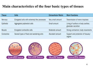
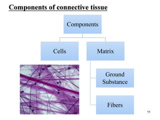
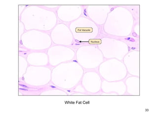
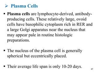
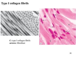
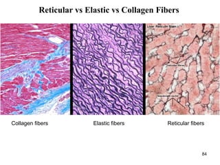
This document provides an overview of connective tissue. It discusses the main cells found in connective tissue, including fibroblasts, adipocytes, macrophages, mast cells, plasma cells, and leukocytes. It also describes the main fibers - collagen, reticular, and elastic fibers - and ground substance. Finally, it outlines the main types of connective tissue, including connective tissue proper (loose and dense connective tissue), supporting connective tissue (cartilage and bone), and fluid connective tissue (blood). Loose connective tissue, also called areolar tissue, is characterized as having cells, fibers and ground substance in roughly equal parts and serving to bind organs and fill spaces between tissues.
2. ØIntroduction
ØFinctions of connective tissue
ØCells of connective tissue
ØFibers of connective tissue
ØGround substance
ØTypes of connective tissue
ØLab quiz
2
3. § Connective tissue is one of the four tissues found
in the human body.
§ Connective tissue provides a matrix that supports
and physically connects other tissues and cells
together to form the organs of the body.
§ Connective tissue is found in between other
tissues everywhere in the body, including the
nervous system.
3
6. 6
Organ Components:
§ Most organs can be divided into:
ØThe parenchyma, which is composed of the
cells responsible for the organ’s specialized
functions.
ØThe stroma, the cells of which have a
supporting role in the organ. Except in the
brain and spinal cord, the stroma is always
connective tissue.
8. § Connective tissue originates from the
mesenchyme.
§ A tissue developing mainly from the middle layer of the
embryo, the mesoderm.
§ Mesenchymal cells are undifferentiated and have large nuclei,
with prominent nucleoli and fine chromatin. They are
“spindle-shaped,” with their scant cytoplasm extended as two
or more thin cytoplasmic processes.
§ Embryonic mesenchyme producing all types of connective
tissue proper and the specialized connective tissues bone and
cartilage, they also include stem cells for other tissues such as
blood, the vascular endothelium, and muscle.
8
12. § Connective tissue is composed of cells and an
extracellular matrix that binds the cells and organs,
integrating all parts of the body.
§ Connective tissue is the most widespread tissue of
the body and can be found in every organ.
§ A variety of cell types are found in connective
tissues.
12
15. Connective tissue vs Epithelium:
§ Unlike the other tissue types (epithelium,
muscle, and nerve), which consist mainly of
cells, the major constituent of connective tissue
is the extracellular matrix (ECM).
15
17. § Connective tissue that connects, supports, binds, or
separates other tissues or organs, typically having
relatively few cells embedded in an amorphous
matrix, often with or other fibres, and
including cartilaginous, fatty, and elastic tissues.
§ The variety of connective tissue types in the body
reflects differences in composition and amount of
the cells, fibers, and ground substance which
together are responsible for the remarkable
structural, functional, and pathologic diversity of
connective tissue.
17
23. § Fibroblasts are the key cells in connective tissue
proper. Fibroblasts originate locally from
mesenchymal cells and are permanent residents of
connective tissue.
§ Other cells found here, such as macrophages,
plasma cells, and mast cells, originate from
hematopoietic stem cells in bone marrow, circulate
in the blood, and then move into connective tissue
where they function.
23
24. § The most common cells in connective tissue
proper.
§ Produce and maintain most of the tissue’s
extracellular components.
§ Fibroblasts synthesize and secrete collagen (the
most abundant protein of the body) and elastin,
which both form large fibers, as well as the
GAGs, proteoglycans, and multiadhesive
glycoproteins that comprise the ground substance.
24
31. § Adipocytes, or fat cells, are found in the
connective tissue of many organs.
§ These large, mesenchymally derived cells are
specialized for cytoplasmic storage of lipid as
neutral fats, or less commonly for the production
of heat.
§ Tissue with a large population of adipocytes,
called adipose connective tissue, serves to
cushion and insulate the skin and other organs.
31
35. Ø Macrophages & the Mononuclear
Phagocyte System
§ Macrophages a large phagocytic cell found in
stationary form in the tissues or as a mobile white
blood cell, especially at sites of infection.
§ Size and shape vary considerably, corresponding
to their state of functional activity.
§ A typical macrophage measures between 10 and
30 μm in diameter and has an eccentrically located,
oval or kidney-shaped nucleus. 35
38. § Macrophages arise from precursor cells called
monocytes. Monocytes originate in the bone
marrow from where they are released into the
blood stream. They are actively mobile and leave
the blood stream to enter connective tissues, where
they differentiate into macrophages.
38
41. Ø Mast Cells
§ Mast cells are oval or irregularly shaped cells of
connective tissue, between 7 and 20 μm in
diameter.
§ Mast cells filled with basophilic secretory
granules that often obscure the central nucleus.
§ Mast cells are components of loose connective
tissues, often located near small blood vessels.
41
45. Function of Mast cells
§ Localized release of many bioactive substances
important in the local inflammatory response,
innate immunity, and tissue repair.
§ A partial list of molecules released from these
cells’ secretory granules includes the following:
ØHeparin
ØHistamine
ØHistamine
ØEosinophil and neutrophil chemotactic factors
ØCytokines
ØPhospholipid 45
46. Like macrophages, mast cells originate from
progenitor cells in the bone marrow, which circulate
in the blood, cross the wall of small vessels called
venules, and enter connective tissues, where they
differentiate.
46
47. Ø Plasma Cells
§ Plasma cells are lymphocyte-derived, antibody-
producing cells. These relatively large, ovoid
cells have basophilic cytoplasm rich in RER and
a large Golgi apparatus near the nucleus that
may appear pale in routine histologic
preparations.
§ The nucleus of the plasma cell is generally
spherical but eccentrically placed.
§ Their average life span is only 10-20 days. 47
51. Ø Leukocytes
§ leukocyte is a colourless cell which circulates in
the blood and body fluids and is involved in
counteracting foreign substances and disease; a
white (blood) cell. There are several types, all
amoeboid cells with a nucleus, including
lymphocytes, granulocytes, and monocytes.
§ leukocytes, besides macrophages and plasma
cells normally comprise a population of
wandering cells in connective tissue.
51
55. § The fibrous components of connective tissue
are elongated structures formed from proteins
that polymerize after secretion from fibroblasts.
§ The three main types of fibers are:
§ Collagen
§ Reticular fibers
§ Elastic fibers
55
56. 56
• is a type of fiber found in
connective tissue that provides
strength
Collagen
fiber
• is a type of fiber found in
connective tissue that has a netlike
formation and provides support
Reticular
fiber
• is a type of fiber found in
connective tissue that can stretch
and recoil
Elastic
fiber
57. § Collagen and reticular fibers are both formed by
proteins of the collagen family, and elastic fibers
are composed mainly of the protein elastin.
§ These fibers are distributed unequally among the
different types of connective tissue, with the
predominant fiber type conferring most specific
tissue properties.
57
61. Ø Collagen
§ The collagens constitute a family of proteins
selected during evolution for their ability to
form various extracellular fibers, sheets, and
networks, all of which extremely strong and
resistant to normal shearing and tearing forces.
§ Collagen is a key element of all connective
tissues, as well as epithelial basement
membranes and the external laminae of muscle
and nerve cells.
61
62. § Collagen is the most abundant protein in the
human body, representing 30% of its dry weight.
§ A family of 28 collagens exists in vertebrates,
numbered in the order they were identified, and
the most important are listed in table in the next
slide. They can be categorized according to the
structures formed by their interacting α-chains
subunits:
62
64. § Type I is the most common - 90% of all
collagens are type I. This type is found in bone,
skin, tendon, ligaments, cornea etc.
§ Types I, II, III, V and IX are fibrillar, which
means that the protein chains assemble into
fibrils, 10-300nm in diameter.
§ Types V & VI form networks in the basal
lamina.
§ Type VI helps to anchor basal lamina of skin to
underlying connective tissue. 64
65. 65
C: type I collagen fibrils
arrows: fibroblast
Type I collagen fibrils
66. Collagen synthesis:
§ Collagen synthesis occurs in many cell types but is
a specialty of fibroblasts. The initial procollagen α
chains are polypeptides made in the RER.
66
The Collagen subunit
71. Ø Reticular fibers
§ Reticular fibers found in delicate connective
tissue of many organs, notably in the immune
system.
§ Reticular fibers consist mainly of collagen type
III, which forms an extensive network
(reticulum) of thin (diameter 0.5-2 μm) fibers
for the support of many different cells.
71
72. § Reticular fibers are seldom visible in hematoxylin
and eosin (H&E) preparations but are
characteristically stained black after impregnation
with silver salts and are thus termed argyrophilic.
72
73. § Reticular fibers produced by fibroblasts.
§ Reticular fibers occur in the reticular lamina of
basement membranes and typically also surround
adipocytes, smooth muscle and nerve fibers, and
small blood vessels.
§ Abundant reticular fibers also characterize the
stroma of hemopoietic tissue (bone marrow), the
spleen, and lymph nodes where they support
rapidly changing populations of proliferating cells
and phagocytic cells.
73
77. Ø Elastic fibers
§ Elastic fibers (or yellow fibers) are bundles of
proteins (elastin) found in extracellular matrix of
connective tissue.
§ Elastic fibers produced by fibroblasts and smooth
muscle cells in arteries.
77
Elastin An elastic fiber
78. § Elastic fibers are thinner than the type I collagen
fibers and form sparse networks interspersed with
collagen bundles in many organs, particularly
those subject to regular stretching or bending.
§ Elastic fibers can stretch up to 1.5 times their
length, and snap back to their original length when
relaxed (e.g. the stroma of the lungs) 78
86. § Ground substance is an amorphous gel-like
substance in the extracellular space.
§ The ground substance of the ECM is a highly
hydrated (with much bound water), transparent,
complex mixture of three major kinds of
macromolecules:
§ Glycosaminoglycans (GAGs)
§ Proteoglycans
§ Multiadhesive glycoproteins 86
87. Ground substance functions:
§ Ground substance filling the space between cells
and fibers in connective tissue.
§ Ground substance allows diffusion of small
molecules and, because it is viscous, acts as both a
lubricant and a barrier to the penetration of
invaders.
87
89. Ø Glycosaminoglycans (GAGs)
§ GAGs (also called mucopolysaccharides) are
long polymers of repeating disaccharide units,
usually a hexosamine and uronic acid.
§ The hexosamine can be glucosamine or
galactosamine, and the uronic acid can be
glucuronate or iduronate.
§ The largest and most ubiquitous GAG is
hyaluronan (also called hyaluronate or hyaluronic
acid). 89
90. § Hyaluronan forms a viscous, pericellular network
that binds a considerable amount of water, giving
it an important role in allowing molecular
diffusion through connective tissue and in
lubricating various organs and joints.
90
93. 93
§ Different combinations and densities of the cells,
fibers, and other ECM components produce
graded variations in histological structure within
connective tissue.
§ Descriptive names or classifications used for the
various types of connective tissue typically denote
either a structural characteristic or a major
component.
98. § Loose connective tissue
§ Also called areolar tissue, is common, a layer
beneath the epithelial lining of many organs and
filling the spaces between fibers of muscle and
nerve.
§ The loose connective tissue typically contains
cells, fibers, and ground substance in roughly
equal parts.
98
99. Charastaristics of Loose connective tissue:
§ Delicate, flexible, not very resistant to stress.
§ Well vascularized.
§ All types of connective tissue cells present.
(Majority are fibroblasts and macropahges)
§ Collagen, elastic, and reticular fibers present.
§ In certain organs (intestine) and in certain disease
conditions, numerous lymphocytes may be present.
99
104. § Reticular tissue: a specialized loose connective
tissue with reticular cells that form a fine matrix
of reticular fibers. Provides a structural
framework for hematopoietic organs such as
bone marrow and spleen.
§ Reticular tissue is characterized by abundant
fibers of type III collagen forming a delicate
network that supports various types of cells. This
collagen is also known as reticulin and is
produced by modified fibroblasts often called
reticular cells that remain associated with and
partially cover the fibers.
§ Reticular tissues have a high cellular content. 104
107. § Dense connective tissue
§ Dense connective tissue has similar components
as loose connective tissue, but with fewer cells,
mostly fibroblasts, and a clear predominance of
bundled type I collagen fibers over ground
substance.
§ Less flexible and more resistant to stress
§ When collagen bundles are present without
apparent orientation, called dense irregular
connective tissue.
§ When oriented in parallel arrays, called dense
regular connective tissue. 107
112. § The best examples of dense regular connective
tissue are the very strong and flexible tendons,
cords connecting muscles to bones; aponeuroses,
which are sheetlike tendons; and ligaments,
bands or sheets that hold together components of
the skeletal system.
§ On their outer surface tendons and ligaments
have a layer of dense irregular connective tissue.
112
113. § Elastic connective tissue: is a modified dense
connective tissue that contains numerous elastic
fibers in addition to collagen fibers, which allows
the tissue to return to its original length after
stretching.
113
115. § Loose Connective Tissue: The major function of
the loose connective tissue is to serve as a
supporting matrix for the blood vessels,
lymphatic vessels, nerves, muscle fibers, organs,
and the skin.
§ Dense Connective Tissue: Dense connective
tissue produces tendon and ligaments by forming
strong, rope-like structures.
115
116. Ø Mucous tissue
§ Mucoid (or mucous) connective tissue is the
principal component of the fetal umbilical cord,
where it is referred to as Wharton’s jelly. With
abundant ground substance composed chiefly of
hyaluronan, mucoid tissue is gelatinous, with
sparse collagen fibers and scattered fibroblasts.
116Wharton's jelly is a gelatinous substance within the umbilical cord.