Downloaded 262 times
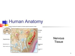


The document describes the structure and function of the nervous system. It discusses how the nervous system can be divided structurally into the central nervous system (CNS) and peripheral nervous system (PNS), and functionally into the sensory and motor divisions. Neurons are the basic functional units that initiate and transmit nerve impulses, while glial cells provide support and protection. The document provides details on the anatomy and classifications of neurons, glial cells, nerves, and synapses.


