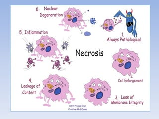
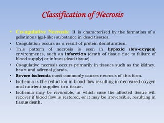
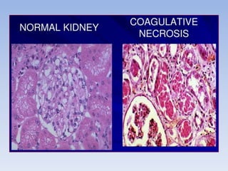
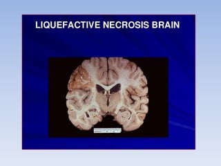
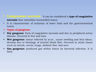
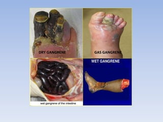
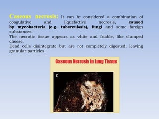
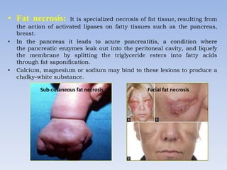
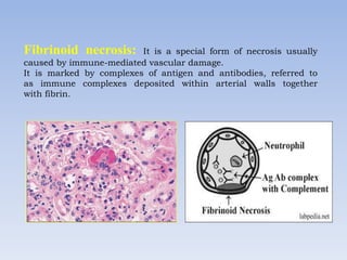
NECROSIS
Mrs. Praveen Garg
VITS College, Satna
Introduction
• Necrosis is the death of body tissue through disease or injury.
• It occurs when too little blood flows to the tissue.
• Necrosis is the death of cells in living tissue caused by external factors
such as infection, trauma, or toxins.
• Necrosis is the pattern of cell death that occurs in response to injuries
such as hypoxia, extremes of temperature, toxins, physical trauma, and
infection with lytic viruses. The injury to a cell is said to be irreversible if
it kills the cell.
• Necrosis is a form of cell injury which results in the premature death
of cells in living tissue by autolysis.
• Necrosis cannot be reversed. When large areas of tissue die due to a lack
of blood supply, the condition is called gangrene.
• Pain
• Redness of the skin
• Swelling
• Blisters (is a pocket of fluid between the upper layers of skin)
• Fluid collection
• Skin discolouration
• Sensation
• Numbness (a loss of sensation or feeling in a part of your body)
Symptoms of Necrosis
Types of necrosis
• Coagulative necrosis – eg. Myocardial infarction, renal infarction.
• Liquefactive necrosis – eg. Infarct brain , Abscess.
• Caseous necrosis – eg. Tuberculosis.
• Fat necrosis – eg. Acute pancreatitis, traumatic fat necrosis of
breast.
• Fibrinoid necrosis – eg. Immune complex deposited in arterial wall.
• Gangrenous necrosis- Ischemia due to lack of blood supply
Classification of Necrosis
• Co-agulative Necrosis: It is characterized by the formation of a
gelatinous (gel-like) substance in dead tissues.
• Coagulation occurs as a result of protein denaturation.
• This pattern of necrosis is seen in hypoxic (low-oxygen)
environments, such as infarction (death of tissue due to failure of
blood supply) or infract (dead tissue).
• Coagulative necrosis occurs primarily in tissues such as the kidney,
heart and adrenal glands.
• Severe ischemia most commonly causes necrosis of this form.
• Ischemia is the reduction in blood flow resulting in decreased oxygen
and nutrient supplies to a tissue.
• Ischemia may be reversible, in which case the affected tissue will
recover if blood flow is restored, or it may be irreversible, resulting in
tissue death.
• Liquefactive necrosis: It is characterized by the digestion of
dead cells to form a viscous liquid mass.
• This is occur due to bacterial, or fungal, infections because of their
ability to stimulate an inflammatory response.
• The necrotic liquid mass is frequently creamy yellow due to the
presence of dead leukocytes and is commonly known as pus.
• Hypoxic infarcts in the brain presents as this type of necrosis,
because the brain contains little connective tissue but high amounts
of digestive enzymes and lipids, and cells therefore can be readily
digested by their own enzymes.
• Gangrenous necrosis: It can be considered a type of coagulative
necrosis that resembles mummified tissue.
• It is characteristic of ischemia of lower limb and the gastrointestinal
tracts.
 Types of gangrene:
 Dry gangrene: form of coagulative necrosis and due to peripheral artery
disease. Occured in foot and toes.
 Wet gangrene: tissue infected by m.or., cause swelling and foul odour,
develop due to blockage of arterial blood flow. Occured in moist tissue
such as mouth, cervix, lungs, diabetic foot, bed sore.
 Gas gangrene: produced gas within tissue by bacterial infection. It is
fatal.
GAS GANGRENE
DRY GANGRENE
Caseous necrosis: It can be considered a combination of
coagulative and liquefactive necrosis, caused
by mycobacteria (e.g. tuberculosis), fungi and some foreign
substances.
The necrotic tissue appears as white and friable, like clumped
cheese.
Dead cells disintegrate but are not completely digested, leaving
granular particles.
• Fat necrosis: It is specialized necrosis of fat tissue, resulting from
the action of activated lipases on fatty tissues such as the pancreas,
breast.
• In the pancreas it leads to acute pancreatitis, a condition where
the pancreatic enzymes leak out into the peritoneal cavity, and liquefy
the membrane by splitting the triglyceride esters into fatty acids
through fat saponification.
• Calcium, magnesium or sodium may bind to these lesions to produce a
chalky-white substance.
Sub-cutaneous fat necrosis Facial fat necrosis
Fibrinoid necrosis: It is a special form of necrosis usually
caused by immune-mediated vascular damage.
It is marked by complexes of antigen and antibodies, referred to
as immune complexes deposited within arterial walls together
with fibrin.
• There are also very specific forms of necrosis:
 Gangrene (type of tissue death, suffered severe hypoxia)
 Treatment of gangrene is the removal of dead tissue by surgery
called Debridement, Amputation (Removal of limb by trauma and
surgery).
 Gummatous necrosis (due to spirochaetal infections)
 Hemorrhagic necrosis (due to the blockage of venous drainage of
an organ or tissue).
• Necrosis may occur due to external or internal factors.
External factors
• External factors may involve mechanical trauma (physical damage to
the body which causes cellular breakdown), damage to blood vessels
(which may disrupt blood supply to associated tissue),
and ischemia. Thermal effects can result in necrosis due to the
disruption of cells.
Internal factors
• Internal factors causing necrosis include: trophoneurotic disorders
(diseases that occur due to defective nerve action in a part of an organ
which results in failure of nutrition); injury and paralysis of nerve
cells. Pancreatic enzymes (lipases) are the major cause of fat necrosis.
Causes
Treatment
• Treatment may include surgery to restore blood flow or to remove
the damaged tissues, antibiotics to prevent or treat infection.
• Debridement, refers to the removal of dead tissue by surgical or
non-surgical means, is the standard therapy for necrosis.
• Depending on the severity of the necrosis, included removal of small
patches of skin to complete amputation of affected limbs or organs.
Thank You

Necrosis

  • 1.
  • 2.
    Introduction • Necrosis isthe death of body tissue through disease or injury. • It occurs when too little blood flows to the tissue. • Necrosis is the death of cells in living tissue caused by external factors such as infection, trauma, or toxins. • Necrosis is the pattern of cell death that occurs in response to injuries such as hypoxia, extremes of temperature, toxins, physical trauma, and infection with lytic viruses. The injury to a cell is said to be irreversible if it kills the cell. • Necrosis is a form of cell injury which results in the premature death of cells in living tissue by autolysis. • Necrosis cannot be reversed. When large areas of tissue die due to a lack of blood supply, the condition is called gangrene.
  • 4.
    • Pain • Rednessof the skin • Swelling • Blisters (is a pocket of fluid between the upper layers of skin) • Fluid collection • Skin discolouration • Sensation • Numbness (a loss of sensation or feeling in a part of your body) Symptoms of Necrosis
  • 5.
    Types of necrosis •Coagulative necrosis – eg. Myocardial infarction, renal infarction. • Liquefactive necrosis – eg. Infarct brain , Abscess. • Caseous necrosis – eg. Tuberculosis. • Fat necrosis – eg. Acute pancreatitis, traumatic fat necrosis of breast. • Fibrinoid necrosis – eg. Immune complex deposited in arterial wall. • Gangrenous necrosis- Ischemia due to lack of blood supply
  • 7.
    Classification of Necrosis •Co-agulative Necrosis: It is characterized by the formation of a gelatinous (gel-like) substance in dead tissues. • Coagulation occurs as a result of protein denaturation. • This pattern of necrosis is seen in hypoxic (low-oxygen) environments, such as infarction (death of tissue due to failure of blood supply) or infract (dead tissue). • Coagulative necrosis occurs primarily in tissues such as the kidney, heart and adrenal glands. • Severe ischemia most commonly causes necrosis of this form. • Ischemia is the reduction in blood flow resulting in decreased oxygen and nutrient supplies to a tissue. • Ischemia may be reversible, in which case the affected tissue will recover if blood flow is restored, or it may be irreversible, resulting in tissue death.
  • 9.
    • Liquefactive necrosis:It is characterized by the digestion of dead cells to form a viscous liquid mass. • This is occur due to bacterial, or fungal, infections because of their ability to stimulate an inflammatory response. • The necrotic liquid mass is frequently creamy yellow due to the presence of dead leukocytes and is commonly known as pus. • Hypoxic infarcts in the brain presents as this type of necrosis, because the brain contains little connective tissue but high amounts of digestive enzymes and lipids, and cells therefore can be readily digested by their own enzymes.
  • 11.
    • Gangrenous necrosis:It can be considered a type of coagulative necrosis that resembles mummified tissue. • It is characteristic of ischemia of lower limb and the gastrointestinal tracts.  Types of gangrene:  Dry gangrene: form of coagulative necrosis and due to peripheral artery disease. Occured in foot and toes.  Wet gangrene: tissue infected by m.or., cause swelling and foul odour, develop due to blockage of arterial blood flow. Occured in moist tissue such as mouth, cervix, lungs, diabetic foot, bed sore.  Gas gangrene: produced gas within tissue by bacterial infection. It is fatal.
  • 12.
  • 13.
    Caseous necrosis: Itcan be considered a combination of coagulative and liquefactive necrosis, caused by mycobacteria (e.g. tuberculosis), fungi and some foreign substances. The necrotic tissue appears as white and friable, like clumped cheese. Dead cells disintegrate but are not completely digested, leaving granular particles.
  • 14.
    • Fat necrosis:It is specialized necrosis of fat tissue, resulting from the action of activated lipases on fatty tissues such as the pancreas, breast. • In the pancreas it leads to acute pancreatitis, a condition where the pancreatic enzymes leak out into the peritoneal cavity, and liquefy the membrane by splitting the triglyceride esters into fatty acids through fat saponification. • Calcium, magnesium or sodium may bind to these lesions to produce a chalky-white substance. Sub-cutaneous fat necrosis Facial fat necrosis
  • 15.
    Fibrinoid necrosis: Itis a special form of necrosis usually caused by immune-mediated vascular damage. It is marked by complexes of antigen and antibodies, referred to as immune complexes deposited within arterial walls together with fibrin.
  • 16.
    • There arealso very specific forms of necrosis:  Gangrene (type of tissue death, suffered severe hypoxia)  Treatment of gangrene is the removal of dead tissue by surgery called Debridement, Amputation (Removal of limb by trauma and surgery).  Gummatous necrosis (due to spirochaetal infections)  Hemorrhagic necrosis (due to the blockage of venous drainage of an organ or tissue).
  • 17.
    • Necrosis mayoccur due to external or internal factors. External factors • External factors may involve mechanical trauma (physical damage to the body which causes cellular breakdown), damage to blood vessels (which may disrupt blood supply to associated tissue), and ischemia. Thermal effects can result in necrosis due to the disruption of cells. Internal factors • Internal factors causing necrosis include: trophoneurotic disorders (diseases that occur due to defective nerve action in a part of an organ which results in failure of nutrition); injury and paralysis of nerve cells. Pancreatic enzymes (lipases) are the major cause of fat necrosis. Causes
  • 18.
    Treatment • Treatment mayinclude surgery to restore blood flow or to remove the damaged tissues, antibiotics to prevent or treat infection. • Debridement, refers to the removal of dead tissue by surgical or non-surgical means, is the standard therapy for necrosis. • Depending on the severity of the necrosis, included removal of small patches of skin to complete amputation of affected limbs or organs.
  • 19.