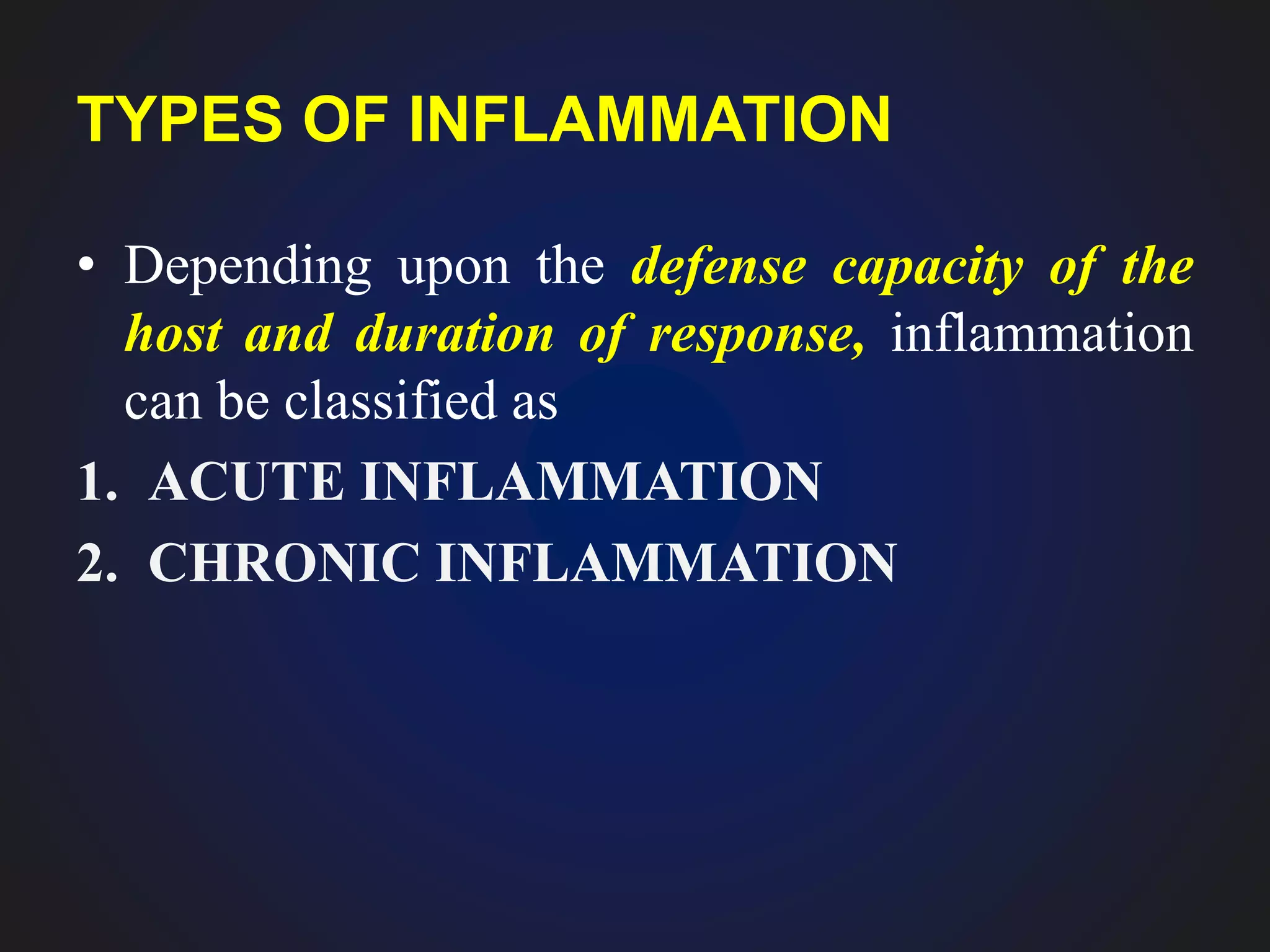
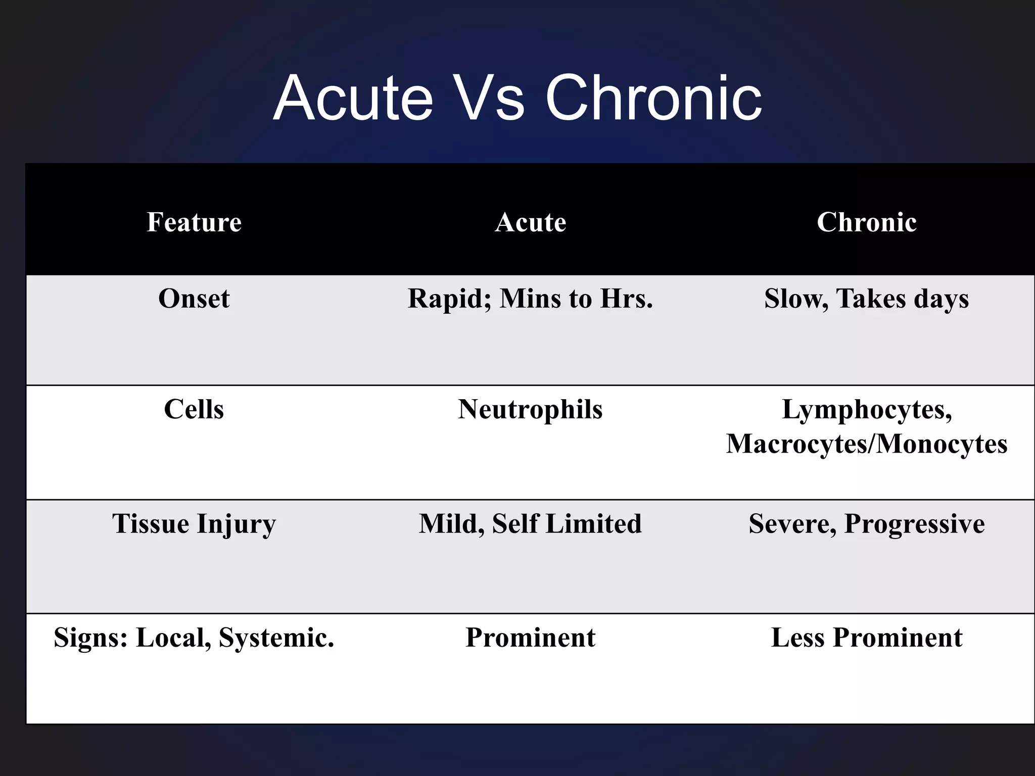
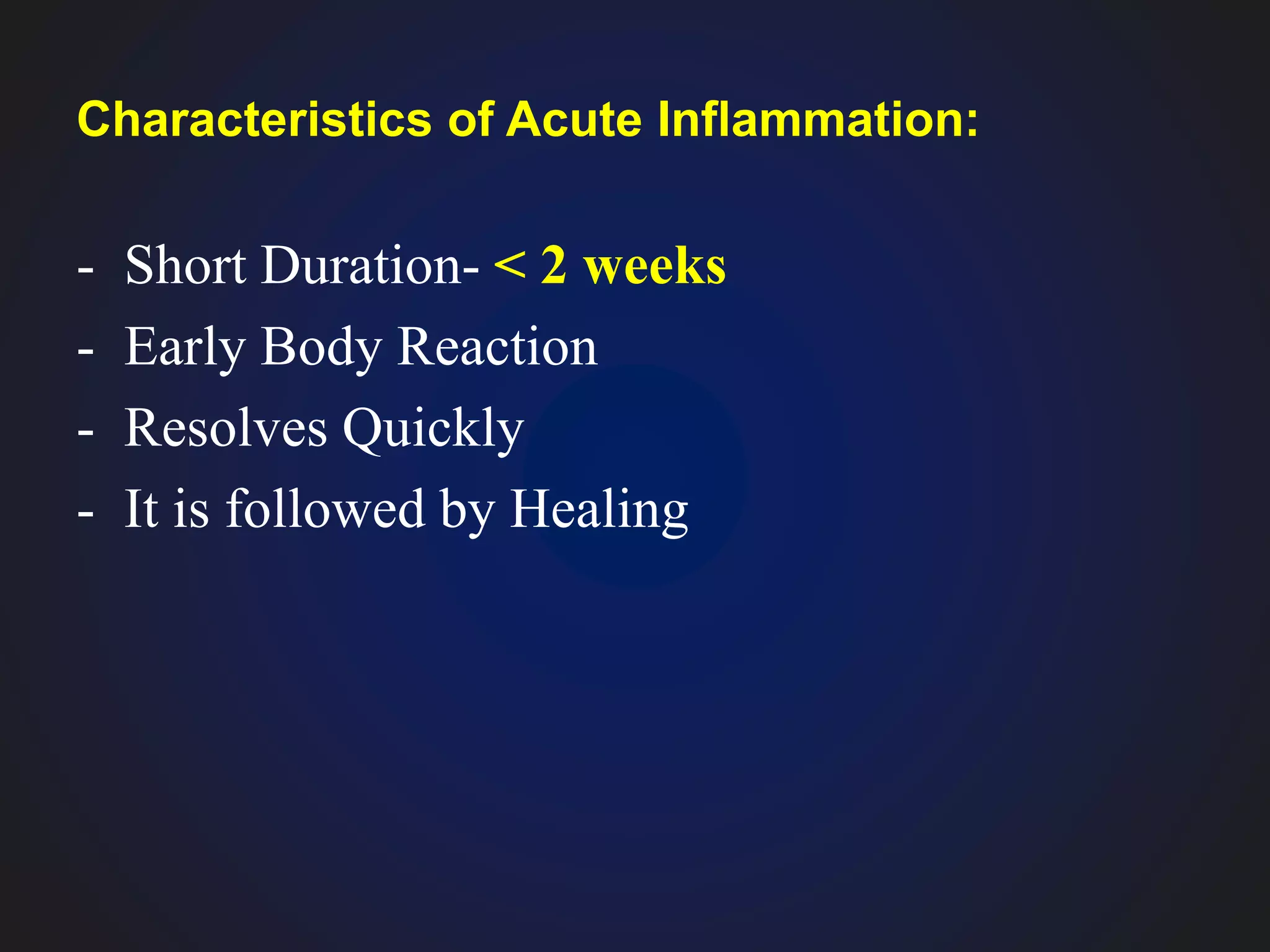
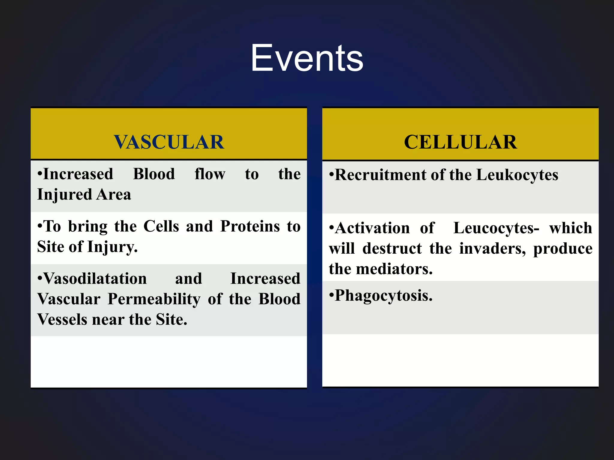
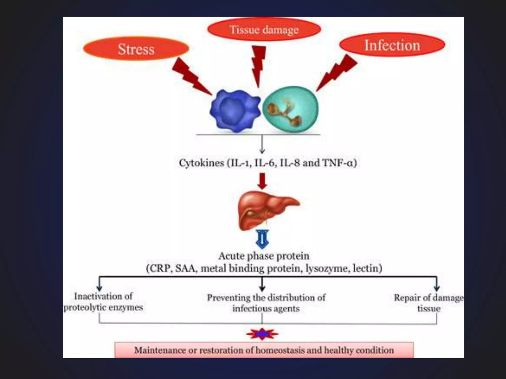
Inflammation
Dr. Dussa Vamshikrishna MD (H)
Learning Objectives
• What is Inflammation?
• Its Types.
• Etiology/Causes of Inflammation.
• Steps of Inflammatory Response.
• Acute Inflammation-
1. Vascular Events
2. Cellular Events
Inflammation: Definition.
• Inflammation is defined as the local response
of living mammalian tissues to injury from
any agent.
Etiological Agent
Defense System
(Leukocytes,
Antibodies,
Complement etc..)
WAR
• It is a body defense reaction.
– Eliminates injurious agent,
– Limits the spread of Injurious Agent.
– Followed by removal of the necrosed cells and
tissues.
INJURIOUS AGENTS CAUSING INFLAMMATION:
Infective agents : Bacteria, Viruses And Their Toxins, Fungi,
Parasites.
Immunological agents like cell-mediated and antigen-antibody
reactions.
Physical agents like Heat, Cold, Radiation, Mechanical Trauma
Chemical agents like organic and inorganic poisons
Inert materials such as foreign bodies.
SIGNS OF INFLAMMATION
• The Roman writer Celsus in 1st century A.D. named
the famous 4 cardinal signs of inflammation as:
I) Rubor (Redness)
II) Tumor (Swelling)
III) Calor (Heat)
IV) Dolor (Pain).
• To these, fifth sign Functio Laesa (loss of function)
was later added by Virchow.
Steps of Inflammatory Response.- 5 R's
1. Recognition of Injurious Agent.
2. Recruitment of Inflammatory Cells.
3. Removal of Injurious Agent.
4. Regulation of the Inflammatory Response.
5. Resolution/Repair.
TYPES OF INFLAMMATION
• Depending upon the defense capacity of the
host and duration of response, inflammation
can be classified as
1. ACUTE INFLAMMATION
2. CHRONIC INFLAMMATION
Acute Vs Chronic
Feature Acute Chronic
Onset Rapid; Mins to Hrs. Slow, Takes days
Cells Neutrophils Lymphocytes,
Macrocytes/Monocytes
Tissue Injury Mild, Self Limited Severe, Progressive
Signs: Local, Systemic. Prominent Less Prominent
ACUTE INFLAMMATION
Characteristics of Acute Inflammation:
- Short Duration- < 2 weeks
- Early Body Reaction
- Resolves Quickly
- It is followed by Healing
• It can be divided into following two events:
I. Vascular events
II. Cellular events
Events
VASCULAR
•Increased Blood flow to the
Injured Area
•To bring the Cells and Proteins to
Site of Injury.
•Vasodilatation and Increased
Vascular Permeability of the Blood
Vessels near the Site.
CELLULAR
•Recruitment of the Leukocytes
•Activation of Leucocytes- which
will destruct the invaders, produce
the mediators.
•Phagocytosis.
I. VASCULAR EVENTS
• Alteration in the microvasculature (arterioles,
capillaries and venules) is the earliest response
to tissue injury.
• These alterations include:
A. Haemodynamic changes.
B. Changes in Vascular Permeability.
A. Haemodynamic Changes:
• Sequence of changes are under:
1. Transient vasoconstriction of arterioles.
• Immediate vascular response.
3-5 SECONDS FOR MILD INJURY
5 MINS FOR SEVERE INJURY
2. Persistent Progressive Vasodilatation:
• Mainly Arterioles, less extent Venules and
Capillaries.
• Occurs after 30 mins/ Half an Hour of Injury.
• Causes increased blood volume in the injured area
• Responsible for the signs: RUBOR AND CALOR
3. Progressive Vasodilatation- Increased Local
Hydrostatic Pressure:
• Results in transudation of fluid into Extracellular
Space.
• This leads to local swelling- TUMOR
4. Stasis of Microcirculation:
• Increased concentration of RED CELLS
• Leading to Increased Viscosity of the Blood at
the site of Inflammation.
5. Leucocytic Margination:
• Peripheral orientation of leucocytes (mainly
neutrophils) along the vascular endothelium.
• The leucocytes stick to the vascular
endothelium briefly, and then move and
migrate through the gaps between the
endothelial cells into the extravascular space.
• This process is known as emigration.
Triple Response /Red Line Response
• It demonstrates features of haemodynamic changes in
inflammation by Lewis experiment.
• Lewis induced the changes in the skin of inner aspect
of forearm by firm stroking with a blunt point.
• The reaction so elicited is known as Triple
Response or Red Line Response consisting of
the following.
1. Red line
2. Flare
3. Wheal
• The reaction so elicited is known as Triple Response
or Red Line Response consisting of the following.
• 1) Red line appears within a few seconds after
stroking and is due to local vasodilatation of
capillaries and venules.
2) Flare: Bright reddish appearance or flush
surrounding the red line and results from
vasodilatation of the adjacent arterioles.
3)Wheal: Swelling or Oedema of the
surrounding skin occurring due to
transudation of fluid into the extravascular
space.
These features, thus, elicit the classical signs of
inflammation—
Redness, Heat And Swelling, to which fourth
feature, pain, has been added.
2. Altered Vascular Permeability
• Increased vascular permeability in acute
inflammation by which normally non-permeable
endothelial layer of microvasculature becomes leaky
can have following patterns and mechanisms.
MECHANISMS OF INCREASED VASCULAR PERMEABILITY:
i) Contraction of Endothelial cells.
ii) Mild Endothelial Damage.
iii) Leucocyte-mediated endothelial injury.
iv) Leakiness in neovascularisation/Transcytosis
1) Contraction of Endothelial cells.
• Most common mechanism.
• Affects Venules exclusively while capillaries
and arterioles remain unaffected.
• Temporary in nature.
• Mediated by the histamine, bradykinin and
other chemical mediators.
• Reversible.
• Short duration (15-30 minutes).
• Eg: Thermal injury of skin
Normal Venule
Leaky Venule
2. Mild Endothelial Damage.
• Affects Venules and Capillaries.
• Seen in Burns/Sun Burns/UV radiation.
• Mediated by cytokines such as Interleukin-1 (IL-1)
and tumour necrosis factor (TNF)-a.
• Seen after 4-6 hours following injury and lasts for
several hours to days.
Damaged Venule
iii) Leucocyte-mediated endothelial injury.
• Adherence of leucocytes to the endothelium at
the site of inflammation may result in
activation of leucocytes.
• The activated leucocytes release proteolytic
enzymes and toxic oxygen species which may
cause endothelial injury and increased
vascular leakiness.
• This form of increased vascular leakiness
affects mostly venules and is a late response.
Damaged Venule
iv) Leakiness in Neovascularisation/Transcytosis.
• Increased transport of Fluids and Proteins
across the intracellular channels
(Transcytosis).
• The newly formed capillaries under the
influence of Vascular Endothelial Growth
Factor (VEGF) during the process of repair
and in tumours are excessively leaky.
Transcytosis.
II. CELLULAR EVENTS
• The cellular phase of inflammation consists of
2 processes:
1. Exudation of Leucocytes. (Influx to the site
of Injury)
2. Phagocytosis. (Ingestion and Destruction of
injurious Agents)
1. Exudation of Leucocytes.
• The escape of leucocytes from the lumen of
microvasculature to the interstitial tissue is the
most important feature of inflammatory
response.
• In acute inflammation, polymorphonuclear
neutrophils (PMNs) comprise the first line of
body defense, followed later by monocytes
and macrophages.
CHANGES LEADING TO MIGRATION OF
LEUCOCYTES:
I. Margination and Pavementing of Blood
Elements.
II. Rolling and Adhesion.
III.Emigration.
IV.Chemotaxis.
Margination and
Pavementing of
Blood Elements. Rolling and Adhesion Emigration.
I. Margination and Pavementing of Blood Elements.
VASODILATATION
INCREASED RATE OF FLOW OF BLOOD
CHANGES IN THE NORMALAXIAL FLOW OF BLOOD
i.e. central stream of cells widens and peripheral plasma zone
becomes narrower because of loss of plasma by exudation. This
phenomenon is known as MARGINATION.
STASIS
REDISTRIBUTION OF NEUTROPHILS OF THE CENTRAL
COLUMN TO THE VESSEL WALL : PAVEMENTING.
NORMALAXIAL FLOW OF BLOOD
MARGINATION. PAVEMENTING.
II. Rolling and Adhesion
• Peripherally marginated and pavemented
neutrophils slowly roll over the endothelial
cells lining the vessel wall (rolling phase).
ROLLINGSELECTINS
ROLLINGSELECTINS
ROLLINGSELECTINS
ROLLINGSELECTINS
CELL ADHESION MOLECULES (CAMS) EXPRESSED ON
ENDOTHELIAL CELLS GETS ACTIVATED BY TNF & IL-1 AND
BRING ABOUT ROLLING OF LEUCKOCYTES (WEAK BONDING)
SELECTINS
(3 types of Selectins)
P- SELECTIN (CD62)- INVOLVED IN ROLLING
E-SELECTIN (E-CAM)- INVOLVED IN BOTH ROLLING
AND ADHESION
L-SELECTIN (L-CAM)- ON THE SURFACE OF
LYMPHOCYTES AND NEUTROPHILS (HELPS IN HOMING
OF LEUKOCYTES TO LYMPH NODES)
ADHESION
Integrins
Integrin Ligand
This is followed by transient bond between the
leucocytes and endothelial cells becoming firmer-
ADHESION PHASE.
Integrins proteins gets activated (by IL-1, TNF)
during the process of loose and transient
adhesions between endothelial cells and
leucocytes.
III. Emigration.
• After sticking of neutrophils to endothelium,
they move through a suitable site between the
endothelial cells by cytoplasmic pseudopods.
• Neutrophils lodged between the endothelial
cells and basement membrane cross the
basement membrane by damaging it locally
with secreted collagenases and escape out into
the extravascular space; this is known as
EMIGRATION.
• Simultaneous to emigration of leucocytes,
escape of red cells through gaps between the
endothelial cells, diapedesis, takes place.
diapedesis
IV. Chemotaxis
• The transmigration of leucocytes after crossing
several barriers to reach the interstitial tissues is a
chemotactic factor mediated process called
chemotaxis.
• The following agents act as potent chemotactic
substances for neutrophils:
i) Leukotriene B4 (LT-B4)
ii) Components of complement system (C5a and C3a
in particular)
iii) Cytokines (Interleukins, in particular IL-8)
iv) Soluble bacterial products
2. Phagocytosis.
• Phagocytosis is defined as the process of
engulfment of solid particulate material by the
cells (cell-eating).
• The cells performing this function are called
phagocytes.
• There are 2 main types of phagocytic cells:
• i) Polymorphonuclear neutrophils (PMNs)
which appear early in acute inflammatory
response, sometimes called as microphages.
• ii) Circulating monocytes and fixed tissue
mononuclear phagocytes, commonly called as
macrophages.
Phagocytosis of the microbe by polymorphs and
macrophages involves the following 3 steps:
1. Recognition and attachment.
2. Engulfment.
3. Killing and degradation.
1. Recognition and Attachment:
• Special receptors on the phagocytes recognize
the microbes/necrotic cells.
Phagocytic Receptors:
1. Mannose Receptors
2. Scavenger Receptors
3. Opsonin Receptors- IgG Opsonins, C3b
Opsonin, Lectins.
(opsonin)
2. Engulfment.
Microbes recognized by receptors are ready to be engulfed by
PMN/Macrophage
Cytoplasmic pseudopods of Phagocytes formed
from activated actin filaments of cell wall will engulf the microbe
Phagocytic vacuole (Phagosome) is formed in the cytoplasm from
the detached cell membrane which previously engulfed microbe by
its pseudopods.
The phagosome fuses with one or more lysosomes of the cell and
form bigger vacuole called phagolysosome.
3. Killing and degradation.
• Destruction of Phagocytosed microbes/Dead
cells by following mechanisms:
A. Intracellular mechanisms: (in Phagolysosomes)
1. Oxidative Bactericidal Mechanism
a. By Oxygen Free Radicals.
• Myeloperoxidase (MPO)-dependent
• Myeloperoxidase (MPO)-independent
b. By Lysosomal granules.
2. Non-oxidative Bactericidal Mechanism
B. Extracellular mechanisms:
A. INTRACELLULAR MECHANISMS
1. Oxidative Bactericidal Mechanism:
a. By Oxygen Free Radicals.
• Microbes are killed by the production of Reactive
Oxygen Metabolites (O-
2, H2O2, OH-, HOCl, HOI,
HOBr).
• Reactive Oxygen Metabolites are produced by the
action of NADPH oxidase (which is present in the
membrane of the phagosome and lysosome) on the
molecular OXYGEN.
OXYGEN (O2)
O2
–
(SUPEROXIDE ANION)
NADPH oxidase
REDUCTION
NADPH NADP + H+
2H + + O2
– H2O2 (Hydrogen peroxide)
(NOT A POTENT ANTIMICROBIAL
AGENT)
• The potency of H2O2 is increased in phagocytes by
the action of enzyme MYELOPEROXIDASE on
H2O2 in the presence of halides (chloride, iodide) to
more potent form hypohalous acid (HOCl, HOI,
HOBr).
• THIS IS CALLED AS MPO-dependent killing.
• Seen in Neutrophils.
• Mature macrophages lack the enzyme MPO and
they carry out bactericidal activity by producing OH–
ions and superoxide singlet oxygen (O-) from H2O2 in
the presence of O2
- (Haber-Weiss reaction) or in the
presence of Fe++ (Fenton reaction).
• This is called MPO-independent killing.
Hydroxyl radical
superoxide singlet oxygen
b. By lysosomal granules:
• Lysosomal granules in lysosomes release enzymes
like protease, trypsinase, phospholipase, and
alkaline phosphatase into phagosome and degrades
the protein component of microbes and bring about
the lysis along with the oxygen free radicals.
2. Non-oxidative Bactericidal Mechanism:
• Some agents released from the granules of
phagocytic cells do not require oxygen for
bactericidal activity.
• Enzymes like lipases, proteases, DNAases
cause lysis of microbe within phagosome
without help of oxygen free radicals.
• Nitric oxide, a free radical and potent
microbial agent similar to oxygen free radicals
produced by macrophages kills the microbes.
• B. Extracellular mechanisms:
• Immune-mediated lysis of microbes takes
place outside the cells by mechanisms of
cytolysis, antibody mediated lysis and by cell-
mediated cytotoxicity
REGULATION OF INFLAMMATION
• The onset of inflammation has potentially
damaging influence on the host tissues as
evident in hypersensitivity conditions.
• Such self-damaging effects are kept in check
by the host regulatory mechanisms in order to
resolve inflammation.
• Agents which check the inflammation:
1. Acute phase reactants.
2. Glucosteroids.
3. Free cytokine receptors.
4. Anti-inflammatory chemical mediators.
1. Acute Phase Reactant proteins.
• APR proteins play a major role is to protect the
normal cells from harmful effects of toxic
molecules generated in inflammation and to clear
away the waste material.
• The APR are synthesised mainly in the liver, and
to some extent in macrophages.
• APR along with systemic features of fever and
leucocytosis is termed ‘acute phase response’.
• Deficient synthesis of APR leads to severe form
of disease in the form of chronic and repeated
inflammatory responses.
CELLULAR PROTECTION
FACTORS
α1-antitrypsin,
α1-chymotrypsin,
α2-antiplasmin,
plasminogen activator
REPLACES THE CONSUMED
FACTORS IN COAGULATION
Fibrinogen, Plasminogen,
Von Willebrand Factor,
Factor VIII
Inflammation
Inflammation
Inflammation
Inflammation

Inflammation

  • 1.
  • 2.
    Learning Objectives • Whatis Inflammation? • Its Types. • Etiology/Causes of Inflammation. • Steps of Inflammatory Response. • Acute Inflammation- 1. Vascular Events 2. Cellular Events
  • 3.
    Inflammation: Definition. • Inflammationis defined as the local response of living mammalian tissues to injury from any agent. Etiological Agent Defense System (Leukocytes, Antibodies, Complement etc..) WAR
  • 4.
    • It isa body defense reaction. – Eliminates injurious agent, – Limits the spread of Injurious Agent. – Followed by removal of the necrosed cells and tissues.
  • 5.
    INJURIOUS AGENTS CAUSINGINFLAMMATION: Infective agents : Bacteria, Viruses And Their Toxins, Fungi, Parasites. Immunological agents like cell-mediated and antigen-antibody reactions. Physical agents like Heat, Cold, Radiation, Mechanical Trauma Chemical agents like organic and inorganic poisons Inert materials such as foreign bodies.
  • 6.
    SIGNS OF INFLAMMATION •The Roman writer Celsus in 1st century A.D. named the famous 4 cardinal signs of inflammation as: I) Rubor (Redness) II) Tumor (Swelling) III) Calor (Heat) IV) Dolor (Pain). • To these, fifth sign Functio Laesa (loss of function) was later added by Virchow.
  • 9.
    Steps of InflammatoryResponse.- 5 R's 1. Recognition of Injurious Agent. 2. Recruitment of Inflammatory Cells. 3. Removal of Injurious Agent. 4. Regulation of the Inflammatory Response. 5. Resolution/Repair.
  • 10.
    TYPES OF INFLAMMATION •Depending upon the defense capacity of the host and duration of response, inflammation can be classified as 1. ACUTE INFLAMMATION 2. CHRONIC INFLAMMATION
  • 11.
    Acute Vs Chronic FeatureAcute Chronic Onset Rapid; Mins to Hrs. Slow, Takes days Cells Neutrophils Lymphocytes, Macrocytes/Monocytes Tissue Injury Mild, Self Limited Severe, Progressive Signs: Local, Systemic. Prominent Less Prominent
  • 12.
  • 13.
    Characteristics of AcuteInflammation: - Short Duration- < 2 weeks - Early Body Reaction - Resolves Quickly - It is followed by Healing
  • 14.
    • It canbe divided into following two events: I. Vascular events II. Cellular events
  • 15.
    Events VASCULAR •Increased Blood flowto the Injured Area •To bring the Cells and Proteins to Site of Injury. •Vasodilatation and Increased Vascular Permeability of the Blood Vessels near the Site. CELLULAR •Recruitment of the Leukocytes •Activation of Leucocytes- which will destruct the invaders, produce the mediators. •Phagocytosis.
  • 16.
    I. VASCULAR EVENTS •Alteration in the microvasculature (arterioles, capillaries and venules) is the earliest response to tissue injury. • These alterations include: A. Haemodynamic changes. B. Changes in Vascular Permeability.
  • 17.
    A. Haemodynamic Changes: •Sequence of changes are under: 1. Transient vasoconstriction of arterioles. • Immediate vascular response. 3-5 SECONDS FOR MILD INJURY 5 MINS FOR SEVERE INJURY
  • 18.
    2. Persistent ProgressiveVasodilatation: • Mainly Arterioles, less extent Venules and Capillaries. • Occurs after 30 mins/ Half an Hour of Injury. • Causes increased blood volume in the injured area • Responsible for the signs: RUBOR AND CALOR
  • 20.
    3. Progressive Vasodilatation-Increased Local Hydrostatic Pressure: • Results in transudation of fluid into Extracellular Space. • This leads to local swelling- TUMOR
  • 21.
    4. Stasis ofMicrocirculation: • Increased concentration of RED CELLS • Leading to Increased Viscosity of the Blood at the site of Inflammation.
  • 22.
    5. Leucocytic Margination: •Peripheral orientation of leucocytes (mainly neutrophils) along the vascular endothelium. • The leucocytes stick to the vascular endothelium briefly, and then move and migrate through the gaps between the endothelial cells into the extravascular space. • This process is known as emigration.
  • 24.
    Triple Response /RedLine Response • It demonstrates features of haemodynamic changes in inflammation by Lewis experiment. • Lewis induced the changes in the skin of inner aspect of forearm by firm stroking with a blunt point.
  • 25.
    • The reactionso elicited is known as Triple Response or Red Line Response consisting of the following. 1. Red line 2. Flare 3. Wheal
  • 26.
    • The reactionso elicited is known as Triple Response or Red Line Response consisting of the following. • 1) Red line appears within a few seconds after stroking and is due to local vasodilatation of capillaries and venules.
  • 27.
    2) Flare: Brightreddish appearance or flush surrounding the red line and results from vasodilatation of the adjacent arterioles.
  • 28.
    3)Wheal: Swelling orOedema of the surrounding skin occurring due to transudation of fluid into the extravascular space.
  • 29.
    These features, thus,elicit the classical signs of inflammation— Redness, Heat And Swelling, to which fourth feature, pain, has been added.
  • 30.
    2. Altered VascularPermeability • Increased vascular permeability in acute inflammation by which normally non-permeable endothelial layer of microvasculature becomes leaky can have following patterns and mechanisms.
  • 31.
    MECHANISMS OF INCREASEDVASCULAR PERMEABILITY: i) Contraction of Endothelial cells. ii) Mild Endothelial Damage. iii) Leucocyte-mediated endothelial injury. iv) Leakiness in neovascularisation/Transcytosis
  • 32.
    1) Contraction ofEndothelial cells. • Most common mechanism. • Affects Venules exclusively while capillaries and arterioles remain unaffected. • Temporary in nature. • Mediated by the histamine, bradykinin and other chemical mediators. • Reversible. • Short duration (15-30 minutes). • Eg: Thermal injury of skin
  • 33.
  • 34.
  • 35.
    2. Mild EndothelialDamage. • Affects Venules and Capillaries. • Seen in Burns/Sun Burns/UV radiation. • Mediated by cytokines such as Interleukin-1 (IL-1) and tumour necrosis factor (TNF)-a. • Seen after 4-6 hours following injury and lasts for several hours to days.
  • 36.
  • 37.
    iii) Leucocyte-mediated endothelialinjury. • Adherence of leucocytes to the endothelium at the site of inflammation may result in activation of leucocytes. • The activated leucocytes release proteolytic enzymes and toxic oxygen species which may cause endothelial injury and increased vascular leakiness. • This form of increased vascular leakiness affects mostly venules and is a late response.
  • 38.
  • 39.
    iv) Leakiness inNeovascularisation/Transcytosis. • Increased transport of Fluids and Proteins across the intracellular channels (Transcytosis). • The newly formed capillaries under the influence of Vascular Endothelial Growth Factor (VEGF) during the process of repair and in tumours are excessively leaky.
  • 40.
  • 41.
    II. CELLULAR EVENTS •The cellular phase of inflammation consists of 2 processes: 1. Exudation of Leucocytes. (Influx to the site of Injury) 2. Phagocytosis. (Ingestion and Destruction of injurious Agents)
  • 42.
    1. Exudation ofLeucocytes. • The escape of leucocytes from the lumen of microvasculature to the interstitial tissue is the most important feature of inflammatory response. • In acute inflammation, polymorphonuclear neutrophils (PMNs) comprise the first line of body defense, followed later by monocytes and macrophages.
  • 43.
    CHANGES LEADING TOMIGRATION OF LEUCOCYTES: I. Margination and Pavementing of Blood Elements. II. Rolling and Adhesion. III.Emigration. IV.Chemotaxis.
  • 44.
    Margination and Pavementing of BloodElements. Rolling and Adhesion Emigration.
  • 45.
    I. Margination andPavementing of Blood Elements. VASODILATATION INCREASED RATE OF FLOW OF BLOOD CHANGES IN THE NORMALAXIAL FLOW OF BLOOD i.e. central stream of cells widens and peripheral plasma zone becomes narrower because of loss of plasma by exudation. This phenomenon is known as MARGINATION. STASIS REDISTRIBUTION OF NEUTROPHILS OF THE CENTRAL COLUMN TO THE VESSEL WALL : PAVEMENTING.
  • 46.
  • 47.
  • 48.
    II. Rolling andAdhesion • Peripherally marginated and pavemented neutrophils slowly roll over the endothelial cells lining the vessel wall (rolling phase).
  • 49.
  • 50.
  • 51.
  • 52.
  • 53.
    CELL ADHESION MOLECULES(CAMS) EXPRESSED ON ENDOTHELIAL CELLS GETS ACTIVATED BY TNF & IL-1 AND BRING ABOUT ROLLING OF LEUCKOCYTES (WEAK BONDING) SELECTINS (3 types of Selectins) P- SELECTIN (CD62)- INVOLVED IN ROLLING E-SELECTIN (E-CAM)- INVOLVED IN BOTH ROLLING AND ADHESION L-SELECTIN (L-CAM)- ON THE SURFACE OF LYMPHOCYTES AND NEUTROPHILS (HELPS IN HOMING OF LEUKOCYTES TO LYMPH NODES)
  • 54.
    ADHESION Integrins Integrin Ligand This isfollowed by transient bond between the leucocytes and endothelial cells becoming firmer- ADHESION PHASE.
  • 55.
    Integrins proteins getsactivated (by IL-1, TNF) during the process of loose and transient adhesions between endothelial cells and leucocytes.
  • 56.
    III. Emigration. • Aftersticking of neutrophils to endothelium, they move through a suitable site between the endothelial cells by cytoplasmic pseudopods.
  • 57.
    • Neutrophils lodgedbetween the endothelial cells and basement membrane cross the basement membrane by damaging it locally with secreted collagenases and escape out into the extravascular space; this is known as EMIGRATION.
  • 58.
    • Simultaneous toemigration of leucocytes, escape of red cells through gaps between the endothelial cells, diapedesis, takes place. diapedesis
  • 59.
    IV. Chemotaxis • Thetransmigration of leucocytes after crossing several barriers to reach the interstitial tissues is a chemotactic factor mediated process called chemotaxis.
  • 60.
    • The followingagents act as potent chemotactic substances for neutrophils: i) Leukotriene B4 (LT-B4) ii) Components of complement system (C5a and C3a in particular) iii) Cytokines (Interleukins, in particular IL-8) iv) Soluble bacterial products
  • 61.
    2. Phagocytosis. • Phagocytosisis defined as the process of engulfment of solid particulate material by the cells (cell-eating). • The cells performing this function are called phagocytes.
  • 62.
    • There are2 main types of phagocytic cells: • i) Polymorphonuclear neutrophils (PMNs) which appear early in acute inflammatory response, sometimes called as microphages. • ii) Circulating monocytes and fixed tissue mononuclear phagocytes, commonly called as macrophages.
  • 63.
    Phagocytosis of themicrobe by polymorphs and macrophages involves the following 3 steps: 1. Recognition and attachment. 2. Engulfment. 3. Killing and degradation.
  • 64.
    1. Recognition andAttachment: • Special receptors on the phagocytes recognize the microbes/necrotic cells. Phagocytic Receptors: 1. Mannose Receptors 2. Scavenger Receptors 3. Opsonin Receptors- IgG Opsonins, C3b Opsonin, Lectins.
  • 65.
  • 66.
    2. Engulfment. Microbes recognizedby receptors are ready to be engulfed by PMN/Macrophage Cytoplasmic pseudopods of Phagocytes formed from activated actin filaments of cell wall will engulf the microbe Phagocytic vacuole (Phagosome) is formed in the cytoplasm from the detached cell membrane which previously engulfed microbe by its pseudopods. The phagosome fuses with one or more lysosomes of the cell and form bigger vacuole called phagolysosome.
  • 68.
    3. Killing anddegradation. • Destruction of Phagocytosed microbes/Dead cells by following mechanisms: A. Intracellular mechanisms: (in Phagolysosomes) 1. Oxidative Bactericidal Mechanism a. By Oxygen Free Radicals. • Myeloperoxidase (MPO)-dependent • Myeloperoxidase (MPO)-independent b. By Lysosomal granules. 2. Non-oxidative Bactericidal Mechanism B. Extracellular mechanisms:
  • 69.
    A. INTRACELLULAR MECHANISMS 1.Oxidative Bactericidal Mechanism: a. By Oxygen Free Radicals. • Microbes are killed by the production of Reactive Oxygen Metabolites (O- 2, H2O2, OH-, HOCl, HOI, HOBr). • Reactive Oxygen Metabolites are produced by the action of NADPH oxidase (which is present in the membrane of the phagosome and lysosome) on the molecular OXYGEN.
  • 70.
    OXYGEN (O2) O2 – (SUPEROXIDE ANION) NADPHoxidase REDUCTION NADPH NADP + H+ 2H + + O2 – H2O2 (Hydrogen peroxide) (NOT A POTENT ANTIMICROBIAL AGENT)
  • 71.
    • The potencyof H2O2 is increased in phagocytes by the action of enzyme MYELOPEROXIDASE on H2O2 in the presence of halides (chloride, iodide) to more potent form hypohalous acid (HOCl, HOI, HOBr). • THIS IS CALLED AS MPO-dependent killing. • Seen in Neutrophils.
  • 72.
    • Mature macrophageslack the enzyme MPO and they carry out bactericidal activity by producing OH– ions and superoxide singlet oxygen (O-) from H2O2 in the presence of O2 - (Haber-Weiss reaction) or in the presence of Fe++ (Fenton reaction). • This is called MPO-independent killing. Hydroxyl radical superoxide singlet oxygen
  • 73.
    b. By lysosomalgranules: • Lysosomal granules in lysosomes release enzymes like protease, trypsinase, phospholipase, and alkaline phosphatase into phagosome and degrades the protein component of microbes and bring about the lysis along with the oxygen free radicals.
  • 74.
    2. Non-oxidative BactericidalMechanism: • Some agents released from the granules of phagocytic cells do not require oxygen for bactericidal activity. • Enzymes like lipases, proteases, DNAases cause lysis of microbe within phagosome without help of oxygen free radicals. • Nitric oxide, a free radical and potent microbial agent similar to oxygen free radicals produced by macrophages kills the microbes.
  • 75.
    • B. Extracellularmechanisms: • Immune-mediated lysis of microbes takes place outside the cells by mechanisms of cytolysis, antibody mediated lysis and by cell- mediated cytotoxicity
  • 76.
    REGULATION OF INFLAMMATION •The onset of inflammation has potentially damaging influence on the host tissues as evident in hypersensitivity conditions. • Such self-damaging effects are kept in check by the host regulatory mechanisms in order to resolve inflammation.
  • 77.
    • Agents whichcheck the inflammation: 1. Acute phase reactants. 2. Glucosteroids. 3. Free cytokine receptors. 4. Anti-inflammatory chemical mediators.
  • 78.
    1. Acute PhaseReactant proteins. • APR proteins play a major role is to protect the normal cells from harmful effects of toxic molecules generated in inflammation and to clear away the waste material. • The APR are synthesised mainly in the liver, and to some extent in macrophages. • APR along with systemic features of fever and leucocytosis is termed ‘acute phase response’. • Deficient synthesis of APR leads to severe form of disease in the form of chronic and repeated inflammatory responses.
  • 80.
    CELLULAR PROTECTION FACTORS α1-antitrypsin, α1-chymotrypsin, α2-antiplasmin, plasminogen activator REPLACESTHE CONSUMED FACTORS IN COAGULATION Fibrinogen, Plasminogen, Von Willebrand Factor, Factor VIII